1. National Cancer Institute. PSMA PET-CT Accurately Detects Prostate Cancer Spread, Trial Shows. May 11, 2020. https://www.cancer.gov/news-events/cancer-currents-blog/2020/prostate-cancer-psma-pet-ct-metastasis. Accessed March 7, 2022.
2. Hofman MS, Lau WF, Hicks RJ. Somatostatin receptor imaging with 68Ga DOTATATE PET/CT: clinical utility, normal patterns, pearls, and pitfalls in interpretation. Radiographics. 2015 Mar-Apr;35(2):500-16. doi: 10.1148/rg.352140164. PMID: 25763733.
‡ [18F]FET is not cleared or approved by the US FDA or CE Marked for clinical use.
3. Maurer GD, Brucker DP, Stoffels G, et al. 18F-FET PET Imaging in Differentiating Glioma Progression from Treatment-Related Changes: A Single-Center Experience. J Nucl Med. 2020 Apr;61(4):505-511. doi: 10.2967/jnumed.119.234757. Epub 2019 Sep 13. PMID: 31519802.
4. Carles M, Popp I, Starke MM, et al. FET-PET radiomics in recurrent glioblastoma: prognostic value for outcome after re-irradiation? Radiat Oncol 16, 46 (2021). https://doi.org/10.1186/s13014- 020-01744-8.
‡ [18F]FET is not cleared or approved by the US FDA or CE Marked for clinical use.
A
Figure 1.
In the case of a six-year-old with a recurrent pituitary microadenoma, using a hybrid PET/MR with [18F]FET enabled clear visualization of focal high uptake on the PET scan directly medial from the right petrosal sinus, thus enabling delineation of a 2-3 mm hypointense recurrent microadenoma from surrounding scar tissue on MR (arrows). (A) PET, (B) fused PET/MR and (C) MR images.
B
Figure 1.
In the case of a six-year-old with a recurrent pituitary microadenoma, using a hybrid PET/MR with [18F]FET enabled clear visualization of focal high uptake on the PET scan directly medial from the right petrosal sinus, thus enabling delineation of a 2-3 mm hypointense recurrent microadenoma from surrounding scar tissue on MR (arrows). (A) PET, (B) fused PET/MR and (C) MR images.
C
Figure 1.
In the case of a six-year-old with a recurrent pituitary microadenoma, using a hybrid PET/MR with [18F]FET enabled clear visualization of focal high uptake on the PET scan directly medial from the right petrosal sinus, thus enabling delineation of a 2-3 mm hypointense recurrent microadenoma from surrounding scar tissue on MR (arrows). (A) PET, (B) fused PET/MR and (C) MR images.
5. Veldhuijzen van Zanten SEM, Neggers SJCMM, Valkema R, Verburg FA. Positive [18F] fluoroethyltyrosine PET/MRI in suspected recurrence of growth hormone-producing pituitary
adenoma in a paediatric patient. Eur J Nucl Med Mol Imaging. 2021 Dec;49(1):410-411. doi: 10.1007/s00259-021-05458-1. Epub 2021 Jul 5. PMID: 34223953; PMCID: PMC8712289.
result
PREVIOUS
${prev-page}
NEXT
${next-page}


Subscribe Now
Manage Subscription
FOLLOW US
contact us • privacy policy • terms & conditions
© 2023 GE HealthCare. GE is a trademark of the General Electric Company used under trademark license.


CASE STUDIES
[18F]FET PET/MR for the detection of pituitary microadenoma
[18F]FET PET/MR for the detection of pituitary microadenoma
by Sophie E. M. Veldhuijzen van Zanten, MD, PhD, MSc, Assistant Professor of Radiology and Nuclear Medicine, Erasmus Medical Center, Rotterdam, the Netherlands
In nuclear medicine, different types of PET tracers are used for identification and localization of diseases. For example, Gallium-68 [68Ga] prostate-specific membrane antigen (PSMA) PET enables detection of prostate cancer.1 [68Ga]Ga-DOTA- TATE PET is useful for evaluating neuroendocrine tumors.2 Similarly, [18F]-fluoroethyl-L-tyrosine ([18F]FET)‡ PET has been shown to differentiate between pseudoprogression and tumor recurrence in high grade gliomas.3,4
At Erasmus Medical Center (Rotterdam, the Netherlands), [18F]FET‡ is utilized under Article 3.17 of the Dutch law on the medical use of substances for application in patients with a suspected pituitary microadenoma.
Patient case
We were presented with a case of a six-year-old child with a recurring overproduction of growth hormone after two surgical resections of a growth hormone-producing pituitary microadenoma.5 In patients with clinical signs of a hormone- producing microadenoma, the standard diagnostic method is to use gadolinium-enhanced MR, where the pituitary enhances and a microadenoma typically does not, therefore, appearing as a dark nodule in the enhanced pituitary tissue. However, in about 40% of patients, MR alone does not enable sufficient diagnostic clarity to provide a solid basis for a neurosurgical operation, and a significant proportion of these patients undergo inferior petrosal sinus sampling. In this invasive procedure, hormone levels are measured in the veins that drain on either side of the pituitary gland to confirm the central overproduction of hormones and to determine on which side of the pituitary the microadenoma is most likely located. Unfortunately, this also often provides ambiguous results after which no further methods for assessing the presence and location of a hormone-producing pituitary lesion thus far exist.
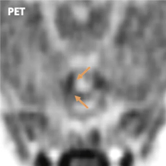
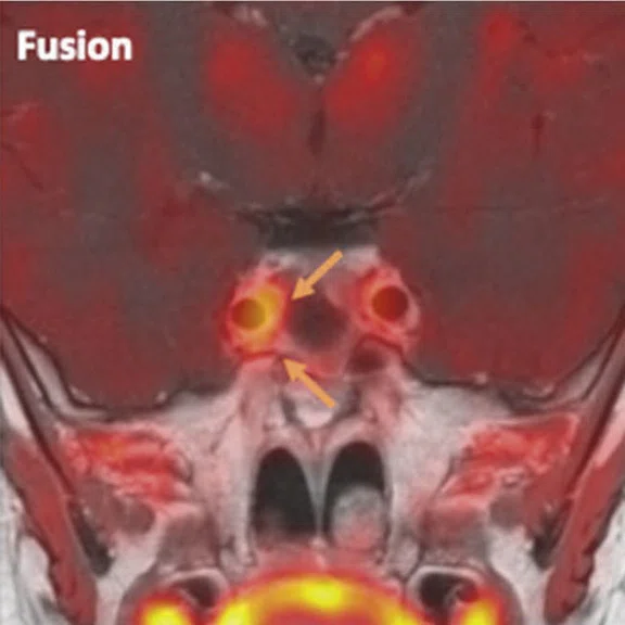
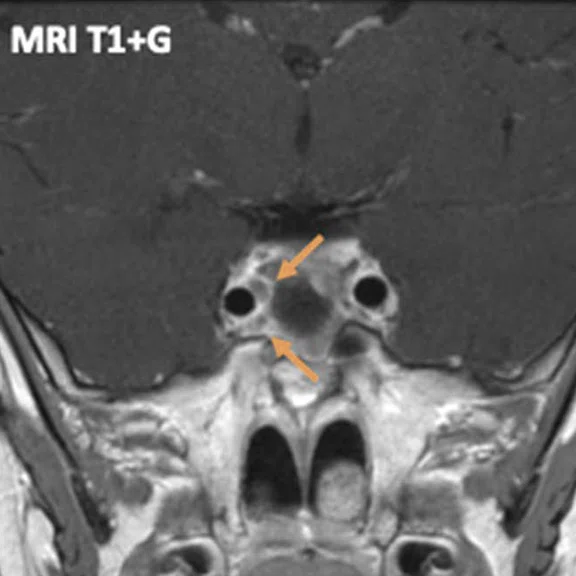
In our patient’s case, MR alone was repeatedly inconclusive with regards to differentiating a possible persistent or recurrent microadenoma from extensive post-operative scar tissue in the sella turcica. Using the hybrid PET/MR (SIGNA™ PET/MR, GE Healthcare) with [18F]FET, we could clearly see focal high uptake on the PET scan at the medial side of the right carotid artery, enabling us to delineate a 2-3 mm hypointense microadenoma from surrounding scar tissue on MR.
Opposed to MR only, [18F]FET PET/MR appeared to be excellent at delineating a recurrent pituitary microadenoma in this case. [18F]FET is a radiolabeled amino acid which enables the display of local higher metabolic activity in lesions with overexpression of particular types of amino acid transporters. Visualization of this focus of pathologically high metabolism provided by hybrid PET/MR imaging was clearly helpful in identifying this very small adenoma.
Discussion
In my line of research at Erasmus University Medical Center, we aim to demonstrate that the combination of PET/MR can improve the diagnosis and localization of disease, not only for pituitary pathology but also for other lesions in the brain and head and neck area. The hybrid PET/MR system enables us to produce some of the most highly detailed images of anatomy and molecular pathophysiology that are currently available. This allows us to study the molecular biology of diseases non-invasively, and helps us select the most effective therapeutic approach, and the patients most likely to benefit from treatment.
Figure 1.
In the case of a six-year-old with a recurrent pituitary microadenoma, using a hybrid PET/MR with [18F]FET enabled clear visualization of focal high uptake on the PET scan directly medial from the right carotid artery, thus enabling delineation of a 2-3 mm hypointense recurrent microadenoma from surrounding scar tissue on MR (arrows). (A) PET, (B) fused PET/MR and (C) MR images.
Our example case is unique since the application of PET/MR for the detection of microadenoma is not regularly performed, certainly not using [18F]FET. The sparse literature on [18F]FET in this context is limited to a case report of an incidental finding of a pituitary lesion and our example case report. To strengthen the scientific basis for this novel diagnostic approach, a clinical case series of approximately 20-30 patients who underwent [18F]FET PET/MR for localization of a pituitary microadenoma will be published.
This research was made feasible by using the latest generation of combined integrated PET/MR. Older generation PET scanners generally have an insufficient spatial resolution and sensitivity to enable the detection of the signal in such small lesions. Furthermore, rather than fusing PET/CT and diagnostic MR, the perfect 3D matching of images through integrated acquisition in a single bed position of the PET and MR images increased diagnostic confidence to a level not possible when fusing PET/CT with MR images.
In our clinical practice, the greatest value of the use of hybrid [18F]FET PET/MR for detection of pituitary microadenoma would be the shortening of the diagnostic process in patients with previously ambiguous results from conventional diagnostics. Currently, patients are referred for one or multiple MR exams and subsequent invasive inferior petrosal sinus sampling, and often many months go by until sufficient information for surgical intervention is acquired. By contrast with PET/MR, the patient would undergo a single 20- to 30-minute diagnostic scan, shortening the decision- making time regarding treatment to mere days when the multidisciplinary evaluation process is included.
Using [18F]FET PET/MR, we expect to considerably increase the detection rate of microadenoma compared to MR alone and, furthermore, expect better localization compared to inferior petrosal sinus sampling. This will considerably help the neurosurgeon plan a surgical resection of the offending lesion. Since we can clearly pinpoint the location of the microadenoma, the surgeon is able to resect only the affected side of the pituitary. This is important for the patients’ quality of life, as they can retain the rest of the organ, thus obviating the need for medical multi-hormone substitution therapy. In contrast, when the entire pituitary is resected, the patient loses that normal hormone production and must remain on multiple forms of sometimes complex hormone substitution therapies for the rest of their life.
In our opinion, [18F]FET PET/MR is a promising novel tool for diagnosing and guiding treatment of primary and recurrent pituitary microadenomas, especially if MR alone does not provide sufficient diagnostic certainty.


