A
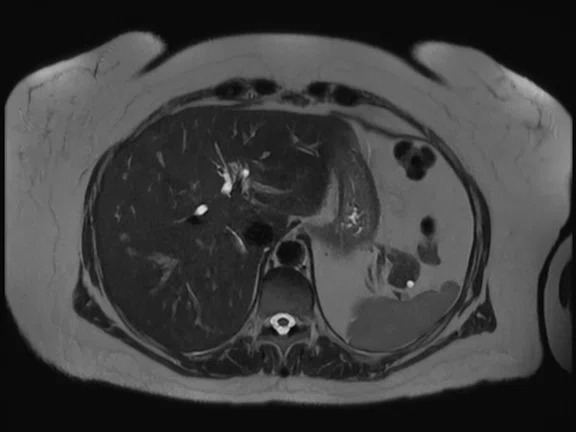
Figure 2.
MRCP exam on a healthy volunteer using the 21ch AIR™ MP Coil Large and 40ch PA Coil. (A) Axial T2 SSFSE, 0.9 x 1.3 x 4 mm, 13 sec.; (B) coronal T2 SSFSE, 0.7 x 1.3 x 2.5 mm, 12 sec.; (C) LAVA Star, 1.1 x 1.1 x 4.4 mm, free-breathing acquisition in 3:04 min.; and (D) 3D MRCP, 0.9 x 0.9 x 1.4 mm, respiratory triggered in 2:30 min.
B
Figure 2.
MRCP exam on a healthy volunteer using the 21ch AIR™ MP Coil Large and 40ch PA Coil. (A) Axial T2 SSFSE, 0.9 x 1.3 x 4 mm, 13 sec.; (B) coronal T2 SSFSE, 0.7 x 1.3 x 2.5 mm, 12 sec.; (C) LAVA Star, 1.1 x 1.1 x 4.4 mm, free-breathing acquisition in 3:04 min.; and (D) 3D MRCP, 0.9 x 0.9 x 1.4 mm, respiratory triggered in 2:30 min.
C
Figure 2.
MRCP exam on a healthy volunteer using the 21ch AIR™ MP Coil Large and 40ch PA Coil. (A) Axial T2 SSFSE, 0.9 x 1.3 x 4 mm, 13 sec.; (B) coronal T2 SSFSE, 0.7 x 1.3 x 2.5 mm, 12 sec.; (C) LAVA Star, 1.1 x 1.1 x 4.4 mm, free-breathing acquisition in 3:04 min.; and (D) 3D MRCP, 0.9 x 0.9 x 1.4 mm, respiratory triggered in 2:30 min.
D
Figure 2.
MRCP exam on a healthy volunteer using the 21ch AIR™ MP Coil Large and 40ch PA Coil. (A) Axial T2 SSFSE, 0.9 x 1.3 x 4 mm, 13 sec.; (B) coronal T2 SSFSE, 0.7 x 1.3 x 2.5 mm, 12 sec.; (C) LAVA Star, 1.1 x 1.1 x 4.4 mm, free-breathing acquisition in 3:04 min.; and (D) 3D MRCP, 0.9 x 0.9 x 1.4 mm, respiratory triggered in 2:30 min.
A
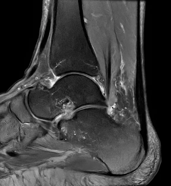
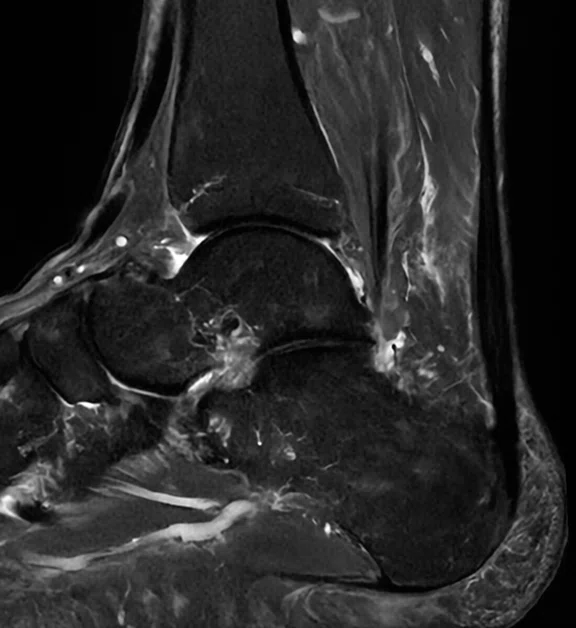
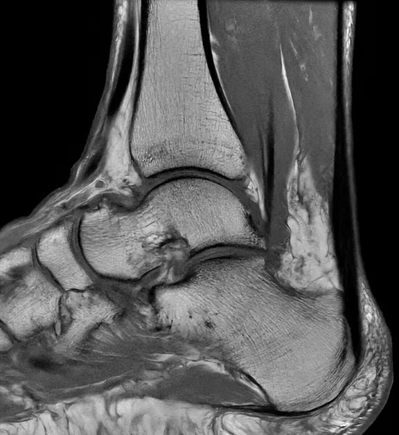
Figure 1.
Patient with foot pain and difficulty walking was referred for an MR exam. The 20ch AIR™ MP Coil Medium was used, providing a more comfortable exam for the patient because it better adapts to the anatomy and is lighter, putting less pressure on the patient’s painful joints/extremities. (A) Axial T2 FRFSE, 0.3 x 0.3 x 3.0 mm, 25 slices in 2:31 min.; (B) axial PD FatSat, 0.4 x 0.4 x 3.0 mm, 25 slices in 2:08 min.; (C) sagittal PD FatSat, 0.3 x 0.3 x 3.0 mm, 21 slices in 2:39 min.; (D) sagittal STIR, 0.4 x 0.4 x 3.0 mm, 21 slices in 2:56 min.; and (E) sagittal T1 FSE, 0.3 x 0.3 x 3.0 mm, 21 slices in 2:30 min.
B
Figure 1.
Patient with foot pain and difficulty walking was referred for an MR exam. The 20ch AIR™ MP Coil Medium was used, providing a more comfortable exam for the patient because it better adapts to the anatomy and is lighter, putting less pressure on the patient’s painful joints/extremities. (A) Axial T2 FRFSE, 0.3 x 0.3 x 3.0 mm, 25 slices in 2:31 min.; (B) axial PD FatSat, 0.4 x 0.4 x 3.0 mm, 25 slices in 2:08 min.; (C) sagittal PD FatSat, 0.3 x 0.3 x 3.0 mm, 21 slices in 2:39 min.; (D) sagittal STIR, 0.4 x 0.4 x 3.0 mm, 21 slices in 2:56 min.; and (E) sagittal T1 FSE, 0.3 x 0.3 x 3.0 mm, 21 slices in 2:30 min.
C
Figure 1.
Patient with foot pain and difficulty walking was referred for an MR exam. The 20ch AIR™ MP Coil Medium was used, providing a more comfortable exam for the patient because it better adapts to the anatomy and is lighter, putting less pressure on the patient’s painful joints/extremities. (A) Axial T2 FRFSE, 0.3 x 0.3 x 3.0 mm, 25 slices in 2:31 min.; (B) axial PD FatSat, 0.4 x 0.4 x 3.0 mm, 25 slices in 2:08 min.; (C) sagittal PD FatSat, 0.3 x 0.3 x 3.0 mm, 21 slices in 2:39 min.; (D) sagittal STIR, 0.4 x 0.4 x 3.0 mm, 21 slices in 2:56 min.; and (E) sagittal T1 FSE, 0.3 x 0.3 x 3.0 mm, 21 slices in 2:30 min.
D
Figure 1.
Patient with foot pain and difficulty walking was referred for an MR exam. The 20ch AIR™ MP Coil Medium was used, providing a more comfortable exam for the patient because it better adapts to the anatomy and is lighter, putting less pressure on the patient’s painful joints/extremities. (A) Axial T2 FRFSE, 0.3 x 0.3 x 3.0 mm, 25 slices in 2:31 min.; (B) axial PD FatSat, 0.4 x 0.4 x 3.0 mm, 25 slices in 2:08 min.; (C) sagittal PD FatSat, 0.3 x 0.3 x 3.0 mm, 21 slices in 2:39 min.; (D) sagittal STIR, 0.4 x 0.4 x 3.0 mm, 21 slices in 2:56 min.; and (E) sagittal T1 FSE, 0.3 x 0.3 x 3.0 mm, 21 slices in 2:30 min.
E
Figure 1.
Patient with foot pain and difficulty walking was referred for an MR exam. The 20ch AIR™ MP Coil Medium was used, providing a more comfortable exam for the patient because it better adapts to the anatomy and is lighter, putting less pressure on the patient’s painful joints/extremities. (A) Axial T2 FRFSE, 0.3 x 0.3 x 3.0 mm, 25 slices in 2:31 min.; (B) axial PD FatSat, 0.4 x 0.4 x 3.0 mm, 25 slices in 2:08 min.; (C) sagittal PD FatSat, 0.3 x 0.3 x 3.0 mm, 21 slices in 2:39 min.; (D) sagittal STIR, 0.4 x 0.4 x 3.0 mm, 21 slices in 2:56 min.; and (E) sagittal T1 FSE, 0.3 x 0.3 x 3.0 mm, 21 slices in 2:30 min.
result


PREVIOUS
${prev-page}
NEXT
${next-page}
Subscribe Now
Manage Subscription
FOLLOW US
Contact Us • Cookie Preferences • Privacy Policy • California Privacy PolicyDo Not Sell or Share My Personal Information • Terms & Conditions • Security
© 2024 GE HealthCare. GE is a trademark of General Electric Company. Used under trademark license.
IN PRACTICE
A continuum of MR upgrades transforms one magnet’s potential
A continuum of MR upgrades transforms one magnet’s potential
Aalborg University Hospital in Denmark installed its first MR system from GE HealthCare, the SIGNA™ HD 1.5T, in 2004. As the largest hospital in the North Denmark region, Aalborg University Hospital serves the area’s approximately 250,000 residents. Each year, the hospital’s 80 radiologists and 170 radiographers perform about 300,000 imaging exams across modalities (CT, MR, ultrasound, X-ray and interventional) and all types of anatomies for pediatric, adult and geriatric patients, as well as patients with complex medical conditions. Forty radiographers are dedicated to MR imaging.
Aalborg University Hospital in Denmark installed its first MR system from GE HealthCare, the SIGNA™ HD 1.5T, in 2004. As the largest hospital in the North Denmark region, Aalborg University Hospital serves the area’s approximately 250,000 residents. Each year, the hospital’s 80 radiologists and 170 radiographers perform about 300,000 imaging exams across modalities (CT, MR, ultrasound, X-ray and interventional) and all types of anatomies for pediatric, adult and geriatric patients, as well as patients with complex medical conditions. Forty radiographers are dedicated to MR imaging.
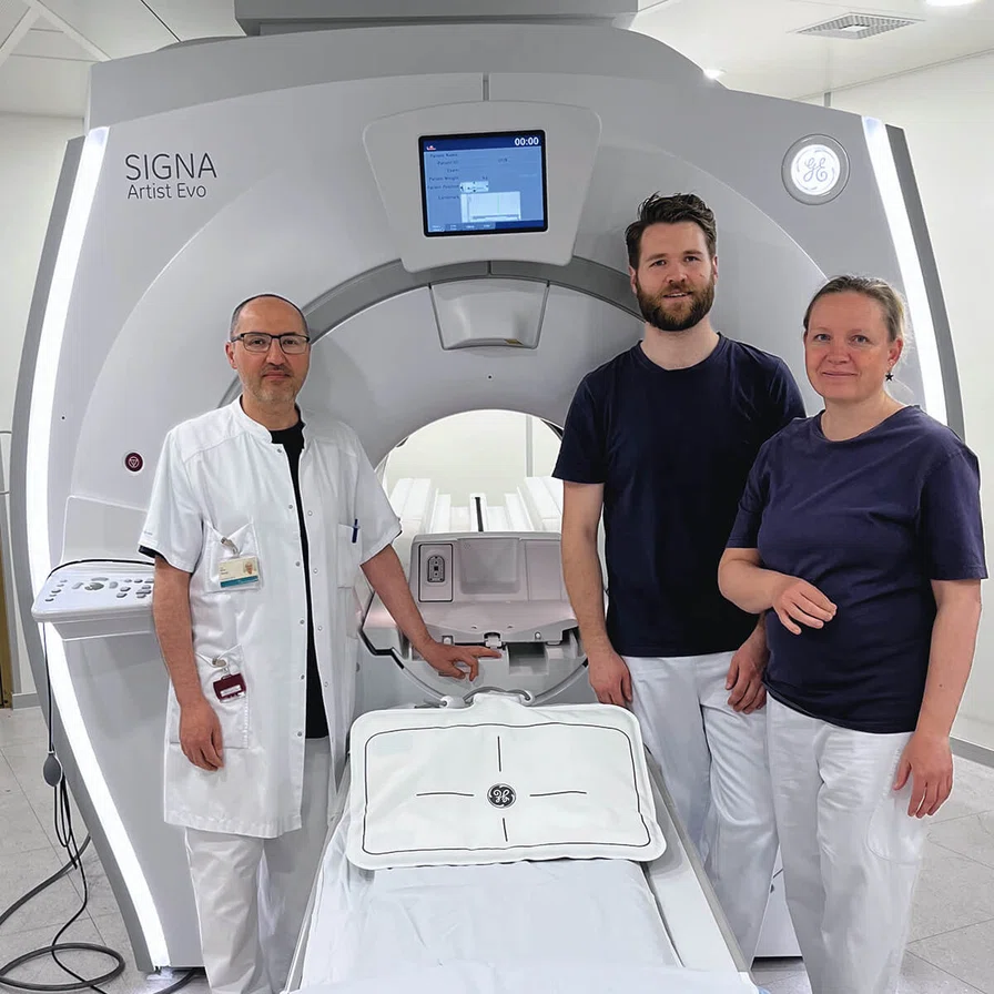
To keep up with the region’s demand for MR imaging, the hospital upgraded to the SIGNA™ Explorer 1.5T through the SIGNA™ Explorer Lift program in 2018. In the fall of 2022, the facility further improved its imaging capabilities by upgrading to another GE HealthCare system, the SIGNA™ Artist Evo 1.5T with AIR™ Recon DL and AIR™ Coils. The SIGNA™ Artist Evo is the latest innovation from GE HealthCare that widens the bore on legacy 1.5T MR systems to 70 cm, all while improving system performance to be on par with a state-of-the-art premium MR system.Aalborg University Hospital now has six GE HealthCare MR systems, including the first SIGNA™ Artist Evo in the European Union.
The hospital stayed with GE HealthCare because the state-of-the-art magnet remains current with GE HealthCare’s SIGNA™ Continuum™ program, giving Aalborg the option to upgrade hardware, electronics and applications. Plus, they were impressed by the quality and homogeneity of the magnet.
“As a radiologist, I’m very satisfied with the homogeneity of this magnet. This can have a significant impact on the accuracy and clarity of the images produced, so the homogeneity and magnet quality is critical here,” says Idris Abdulrahman Abdullah Akreyi, MD, musculoskeletal radiologist at Aalborg University Hospital.
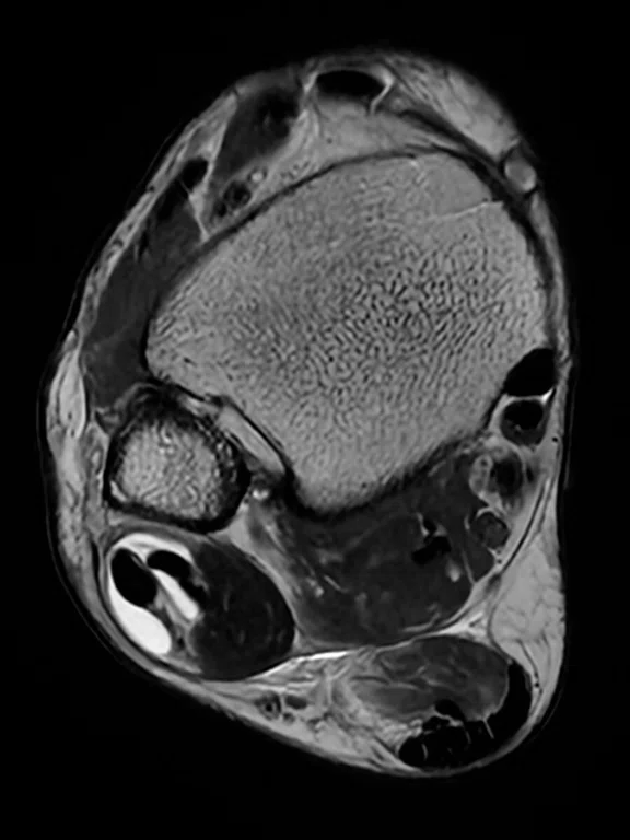
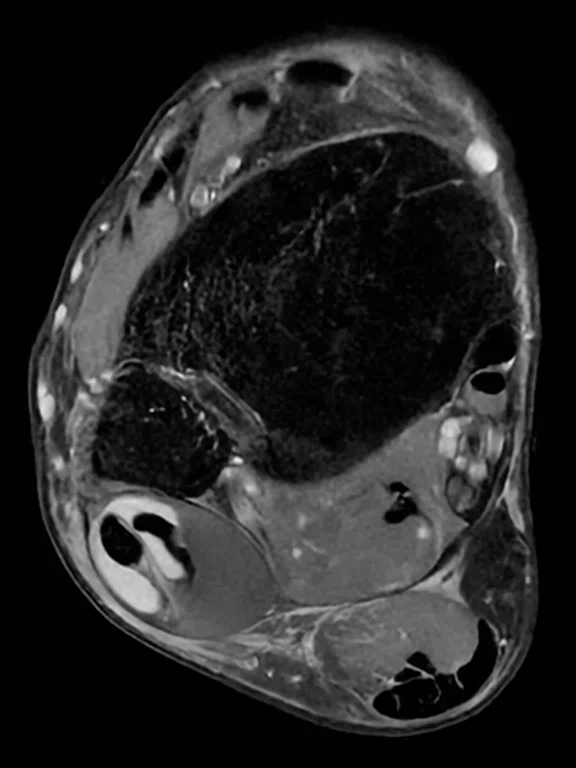
Figure 1.
Patient with foot pain and difficulty walking was referred for an MR exam. The 20ch AIR™ MP Coil Medium was used, providing a more comfortable exam for the patient because it better adapts to the anatomy and is lighter, putting less pressure on the patient’s painful joints/extremities. (A) Axial T2 FRFSE, 0.3 x 0.3 x 3.0 mm, 25 slices in 2:31 min.; (B) axial PD FatSat, 0.4 x 0.4 x 3.0 mm, 25 slices in 2:08 min.; (C) sagittal PD FatSat, 0.3 x 0.3 x 3.0 mm, 21 slices in 2:39 min.; (D) sagittal STIR, 0.4 x 0.4 x 3.0 mm, 21 slices in 2:56 min.; and (E) sagittal T1 FSE, 0.3 x 0.3 x 3.0 mm, 21 slices in 2:30 min.
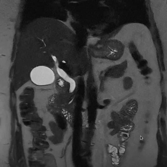
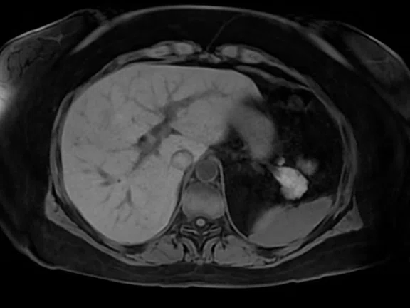
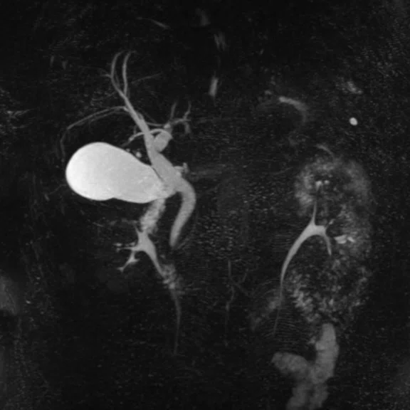
Figure 2.
MRCP exam on a healthy volunteer using the 21ch AIR™ MP Coil Large and 40ch PA Coil. (A) Axial T2 SSFSE, 0.9 x 1.3 x 4 mm, 13 sec.; (B) coronal T2 SSFSE, 0.7 x 1.3 x 2.5 mm, 12 sec.; (C) LAVA Star, 1.1 x 1.1 x 4.4 mm, free-breathing acquisition in 3:04 min.; and (D) 3D MRCP, 0.9 x 0.9 x 1.4 mm, respiratory triggered in 2:30 min.
An easy upgrade
To manage the upgrade, the hospital rescheduled existing appointments and added scan slots on Friday nights and Sunday afternoons. Having six GE HealthCare scanners that can handle all types of exams helped them stay flexible during the transition. “It’s not a big deal for our staff because they’re used to all the scanners, so that makes it easier,” says Louise Bach, MR radiographer at Aalborg University Hospital.
At Aalborg, upgrading the system to SIGNA™ Artist Evo saved time and eliminated the need to build out the MR suite, which reduced costs. Plus, it was an easier process with less paperwork for Bach, since she didn’t have to go through the procurement process. “We were already pleased with our GE HealthCare MR systems and our radiographers are used to operating these machines. So, it’s easy and very convenient to go with this upgrade path. But that’s not the only reason we upgraded. We also did it because we’re satisfied with the images and wanted to continue improving them with the latest advancements in MR imaging,” says Bach.
With the SIGNA™ Artist Evo, there are fewer constraints on imaging capabilities. “Now it’s possible for us to be a little more creative because we aren’t penalized if there’s not enough signal,” says Bach.
Overall, the SIGNA™ Artist Evo is helping Aalborg improve MR imaging across the board. “The SIGNA™ Artist Evo improves our diagnostic capabilities and increases efficiency while enhancing patient comfort and reducing cost,” says Dr. Akreyi.
This means Dr. Akreyi can make more confident diagnoses, which benefits clinicians and patients. “It means a lot for the patient’s outcome for me to be confident in my report. And the clinicians waiting for my conclusion will be also very satisfied,” he says.
Wide bore benefits
Now, with the wide-bore, the team at Aalborg can accommodate larger patients and provide a more comfortable exam for patients with all types of body habitus, as well as claustrophobic patients. “It’s less stressful to be in a wide bore than in a normal bore,” Bach says.
“The patient’s experience and improved patient comfort can reduce the need for sedation or other interventions, which can save a lot of time, cost and resources,” adds Dr. Akreyi.
When they do scan patients under anesthesia, the wide bore system Emakes it easier to accommodate the equipment needed for the procedure. “It’s really nice to have the wide bore for the tubing and equipment the patients need to have during the exam. It’s much easier to handle the anesthetized patient when there is more space around them during the exam. And it’s easier to watch the patients a bit more closely during the exam, such as seeing their chest move during respiration,” says Bach.
Improving image quality with AIR™ Recon DL and AIR™ Coils
At Aalborg University Hospital, AIR™ Recon DL has helped reduce scan times by approximately 50%. Bach anticipates AIR™ Recon DL will help improve image quality on routine, non-emergent cases and will improve scan times when speed is critical, such as strokes and spinal injuries. “We would definitely use it to get better image quality and resolution in some cases, such as fingers and toes. But we’ll also use it for emergency patients to get faster scan times,” she says.
AIR™ Recon DL also helps radiologists see complex anatomy and very small structures more clearly. “If the image quality is a little bit blurred or something isn’t clear, it’s difficult to describe and it will cost a lot of time for the radiologist. And even the diagnosis might not be correct. The SIGNA™ Artist Evo scanner with AIR™ Recon DL will have a dramatic impact on our outcomes as a radiologist and the whole radiology department in our hospital,” says Dr. Akreyi.
The SIGNA™ Artist Evo gives Aalborg University Hospital the ability to use the lightweight flexible blanket-like AIR™ Coils. Currently, the hospital has one 16-channel AIR™ Anterior Array (AA) Coil and two AIR™ Multi-Purpose (MP) Coils, Large (21-channel) and Medium (20-channel). Dr. Akreyi credits both the AIR™ Coils and AIR™ Recon DL with improving the level of detail in images, particularly small extremities. The SIGNA™ Artist Evo has been a game changer for these scans. “I’m amazed with the quality, accuracy, clarity and the diagnostic value in these images, especially in the fingers and the toes,” he says.
Recently, a referring physician contacted Dr. Akreyi in a panic because their patient’s finger scans taken at another hospital didn’t have clear image quality, which prevented the radiologist from delivering a confident diagnosis. He was happy to tell her they had a new scanner that would solve this issue for them. “I said, ‘I have good news for you.’ Because we have a better signal-to-noise ratio, we can dramatically reduce noise and improve image quality,” he says.
Bach says the AIR™ Coils improve both technologist productivity and workflow, as well as patient comfort. “The AIR™ Coils make it easier to position the patient more comfortably so they will lie still for the amount of time that we need. We can also get the coil close to the patient’s anatomy being scanned, improving the signal. And if you look at the workflow for the staff or the radiographers, it’s less weight to move around and fewer coil changes,” she says.
Many exams at Aalborg cover a large region, such as the entire femur from the knee to the hip joint, which was difficult to capture with the previous scanner. Now they can capture all of it more clearly in one scan. “With the AIR™ Coils, we can cover a large region and the whole femur, including the hip and the knee. It’s a great improvement in our daily practice,” says Dr. Akreyi.
The AIR™ Coils and AIR™ Recon DL work together to improve image quality and shorten scan times on the SIGNA™ Artist Evo compared to the previous system, especially in patient populations that have difficulty remaining motionless for an extended period of time, such as pediatrics, geriatrics and those with complex medical conditions. Aalborg is also planning to use the SIGNA™ Artist Evo to scan children without anesthesia, due to the shorter scan times.
“We’re all very satisfied with the quality of imaging and speed of the examinations, especially in these groups of patients with complex medical conditions and for those who can’t lie down without moving,” says Dr. Akreyi.
In addition, the system is easy to use, which makes the MR technologists more efficient and productive. “It’s very user friendly. I would say it’s plug and play. You don’t have to be a very skilled technologist to start using it,” Bach says.
The freedom to image clearly
The SIGNA™ Artist Evo has prompted the hospital to consider updating their protocols to maximize the new technologies and deliver the best image quality with thinner slices and scan time reductions.
“We used to think we couldn’t go lower than maybe 3 or 4 millimeters in slice thickness, or it’s going to take so long that the patient will move during the sequence,” says Bach. “But that’s not the case anymore. We have to reconsider our protocols and get used to thinking we can acquire 2 millimeter slices and can scan with a much smaller field of view.”


