C
Figure 1.
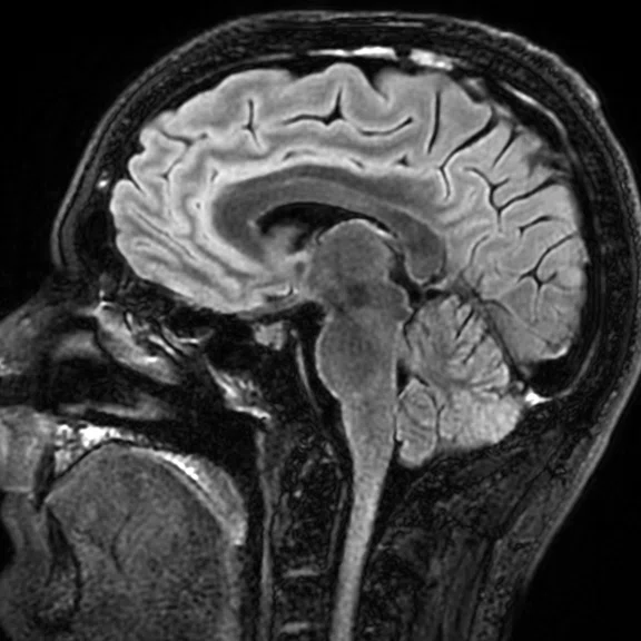
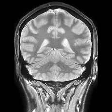
Figure 1. By using the combination of AIR™ Recon, AIR™ 48ch Head Coil and AIR x™, radiomed can perform a complete brain exam, including planning, in approximately 7 min. (A) Sagittal 3D T2 FLAIR Cube (long TR), 1 mm isotropic, 3:08 min. (B) Coronal T2* GRE, 5 mm, 54 sec. (C) Axial T2 PROPELLER, 5 mm, 1:22 min. (D) Axial DWI, 5 mm, 34 sec. (E) Axial T1 MEMP, 5 mm, 1:13 min.
D
Figure 1.
Figure 1. By using the combination of AIR™ Recon, AIR™ 48ch Head Coil and AIR x™, radiomed can perform a complete brain exam, including planning, in approximately 7 min. (A) Sagittal 3D T2 FLAIR Cube (long TR), 1 mm isotropic, 3:08 min. (B) Coronal T2* GRE, 5 mm, 54 sec. (C) Axial T2 PROPELLER, 5 mm, 1:22 min. (D) Axial DWI, 5 mm, 34 sec. (E) Axial T1 MEMP, 5 mm, 1:13 min.
E
Figure 1.
Figure 1. By using the combination of AIR™ Recon, AIR™ 48ch Head Coil and AIR x™, radiomed can perform a complete brain exam, including planning, in approximately 7 min. (A) Sagittal 3D T2 FLAIR Cube (long TR), 1 mm isotropic, 3:08 min. (B) Coronal T2* GRE, 5 mm, 54 sec. (C) Axial T2 PROPELLER, 5 mm, 1:22 min. (D) Axial DWI, 5 mm, 34 sec. (E) Axial T1 MEMP, 5 mm, 1:13 min.
A
Figure 1.
Figure 1. By using the combination of AIR™ Recon, AIR™ 48ch Head Coil and AIR x™, radiomed can perform a complete brain exam, including planning, in approximately 7 min. (A) Sagittal 3D T2 FLAIR Cube (long TR), 1 mm isotropic, 3:08 min. (B) Coronal T2* GRE, 5 mm, 54 sec. (C) Axial T2 PROPELLER, 5 mm, 1:22 min. (D) Axial DWI, 5 mm, 34 sec. (E) Axial T1 MEMP, 5 mm, 1:13 min.
B
Figure 1.
Figure 1. By using the combination of AIR™ Recon, AIR™ 48ch Head Coil and AIR x™, radiomed can perform a complete brain exam, including planning, in approximately 7 min. (A) Sagittal 3D T2 FLAIR Cube (long TR), 1 mm isotropic, 3:08 min. (B) Coronal T2* GRE, 5 mm, 54 sec. (C) Axial T2 PROPELLER, 5 mm, 1:22 min. (D) Axial DWI, 5 mm, 34 sec. (E) Axial T1 MEMP, 5 mm, 1:13 min.
result


PREVIOUS
${prev-page}
NEXT
${next-page}
Subscribe Now
Manage Subscription
FOLLOW US
Contact Us • Cookie Preferences • Privacy Policy • California Privacy PolicyDo Not Sell or Share My Personal Information • Terms & Conditions • Security
© 2024 GE HealthCare. GE is a trademark of General Electric Company. Used under trademark license.
IN PRACTICE
A trifecta of AIR technologies with data analytics delivers intelligent efficiency
A trifecta of AIR technologies with data analytics delivers intelligent efficiency
Increasing productivity and efficiency while maintaining or improving quality is an ongoing process at radiomed, a private practice with nine locations across west-central Germany. As reimbursements/remuneration for radiologists decrease, there is greater pressure to increase productivity.
Increasing productivity and efficiency while maintaining or improving quality is an ongoing process at radiomed, a private practice with nine locations across west-central Germany. As reimbursements/remuneration for radiologists decrease, there is greater pressure to increase productivity.
"We have to treat more patients in less time," says Christopher Ahlers, MD, Managing Partner at radiomed. "Our staff was already working to near capacity with about two patients per hour on some of the equipment."
The SIGNA™ Pioneer installed in 2015 at the practice’s Wiesbaden imaging center was upgraded in October 2019 to SIGNA™Works AIR™ Edition software, which includes AIR x™ and AIR™ Recon. In 2016, radiomed began collaborating with GE Healthcare on Imaging Insights/MR Excellence, one of GE’s Edison Applications that provides comprehensive and actionable insights based on data analytics.
With the upgrade to SIGNA™Works AIR™ Edition, Dr. Ahlers says it is a completely different machine. "The continued innovations on this system definitely add value over time. We are not looking only at scanner hardware or one MR system, rather we see this as a partnership where these innovations and solutions add value to our workflow."
AIR™ technologies
AIR x™ is one innovation that is changing neuro MR imaging at radiomed. More than 1,000 scans have been performed with AIR x™, an intelligent MR slice prescription using deep-learning algorithms to automatically detect and prescribe slices for neurological exams and deliver consistent, quantifiable results.
According to Dr. Ahlers and Maxi Wetzel, RT(R)(MR), Lead Technologist, AIR x™ delivers three key advantages: enhanced productivity, ease of use and reproducibility for longitudinal studies.
"Exams are a bit quicker because the time needed for prescription is less with AIR x™," Wetzel explains. "The slice prescription is completed when we open the first series, therefore, the first scan can be started earlier and the patient has less idle time in the scanner. For the less experienced technologist, the prescription time can take longer than the experienced user, and this is no longer the case. Plus, we don’t have to be as precise with patient positioning because it is already done with AIR x™."
Because the planning is faster, the total examination time for the patient is shorter, Wetzel adds.
"We can scan more patients per day due to the reduction in planning time. Patients also find the shorter time spent in the scanner a very pleasant benefit."
Maxi Wetzel
Consistency in prescription also impacts the radiologist when reading the study and comparing to prior exams. For Dr. Ahlers, the solution is most advantageous in patients with multiple sclerosis or neoplastic lesions, where the radiologist is looking for tiny lesions to compare changes in size or shape longitudinally across exams.
"Better reproducibility with automated prescription and planning facilitates our reading of the case, as we are more confident and can often read faster," adds Dr. Ahlers. "We don’t have to work around the images for the same appearance or obtain a precise measurement when comparing priors."
There is the option to make manual adjustments if needed. Yet Wetzel trusts AIR x™ and says it’s a marked improvement compared to READYBrain, an atlas-based approach that requires a special localization scan that takes additional time. In fact, she and her team rarely used READYBrain, whereas AIR x™ has been implemented into all of radiomed’s neuro protocols so it is used on every neuro MR exam, every time. Even the most experienced technologists at radiomed find that AIR x™ enhances their work.
In tandem with AIR x™, radiomed also benefits from AIR™ Recon, a reconstruction technique that reduces background noise and out-of-field-of-view artifacts for improved signal-to-noise ratio (SNR) and clearer, crisper images.
As with AIR x™, AIR™ Recon is always on for every MR exam on SIGNA™ Pioneer at radiomed.
"We see a significant increase in SNR and also a significant decrease – almost disappearing – of background noise," says Dr. Ahlers. "The images look more brilliant, especially the structures around important anatomy."
Also, radiomed can use the SNR boost to increase the scan matrix or spatial resolution, or reduce scan time.
Optimizing protocols is a continuous process at radiomed; it’s been no different after implementing SIGNA™Works AIR™ Edition and the AIR™ 48-channel Head Coil. Dr. Ahlers and his team worked with the GE applications team to optimize the neuro protocols utilizing the power of these three technologies.
The result is an approximately 7-minute complete brain exam. The higher SNR provided by AIR™ Recon and the AIR™ 48ch Head Coil enables shorter scan times, while AIR x™ shortens the technologist’s planning time to only 40 seconds (Figure 1).
C
D
E
Figure 1.
By using the combination of AIR™ Recon, AIR™ 48ch Head Coil and AIR x™, radiomed can perform a complete brain exam, including planning, in approximately 7 min. (A) Sagittal 3D T2 FLAIR Cube (long TR), 1 mm isotropic, 3:08 min. (B) Coronal T2* GRE, 5 mm, 54 sec. (C) Axial T2 PROPELLER, 5 mm, 1:22 min. (D) Axial DWI, 5 mm, 34 sec. (E) Axial T1 MEMP, 5 mm, 1:13 min.
Using data to improve MR performance
When radiomed began collaborating with GE on the Imaging Insights/MR Excellence program, the goal was to improve MR performance.
"What has happened since is beyond my expectations," Dr. Ahlers says. "We’ve been able to increase productivity while maintaining quality, and in some instances, improve quality."
Imaging Insights combines applied intelligence with data analytics to provide imaging managers with information on productivity and quality of care, including workflow, system utilization, protocols and referral patterns. The initial impact was staggering: the practice saw up to a 30 percent increase in productivity and increased MR exams from 130 to 170 per week across its fleet of MR scanners. As important, patient wait times for an exam also slightly decreased.
Data from Imaging Insights also quantitatively supports the team’s experience with AIR x™. With AIR x™, there are 70 percent fewer mouse clicks, reducing the average planning time for a neuro exam by 66 percent.
"Thanks to Imaging Insights, we were finally able to identify idle times of our MR technology and thus increase our cost efficiency while maintaining the quality of our imaging," Dr. Ahlers explains. "It has enabled us to optimize and standardize suboptimal scheduling and resource utilization, as well as scan protocols. The data-based analyses help us to make more informed business decisions for improved efficiency and patient care."
