A
Figure 4.
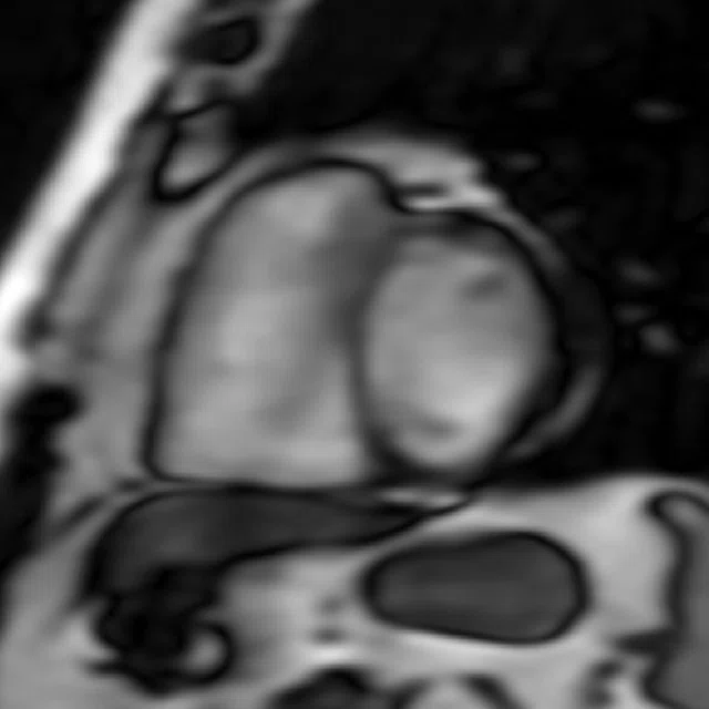
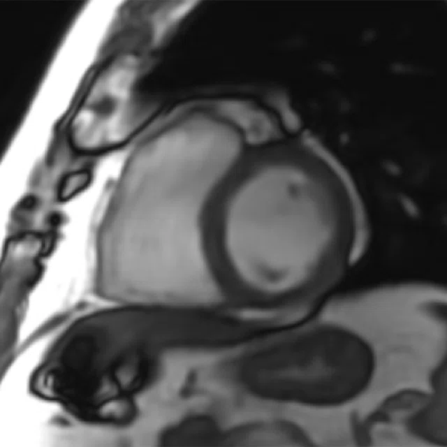
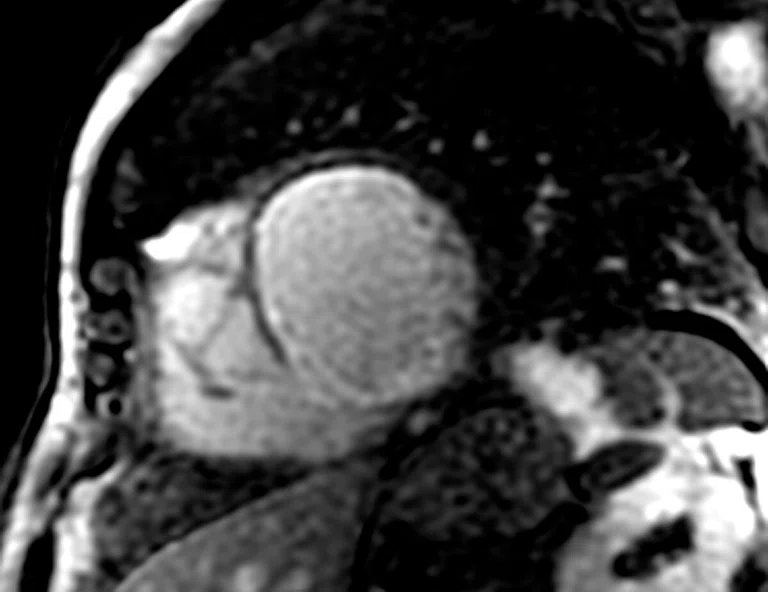
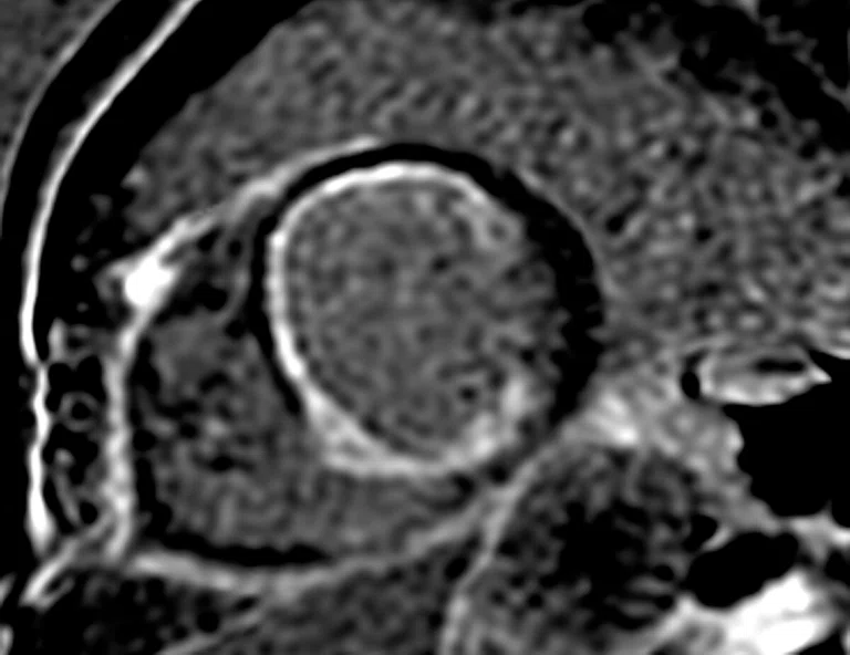
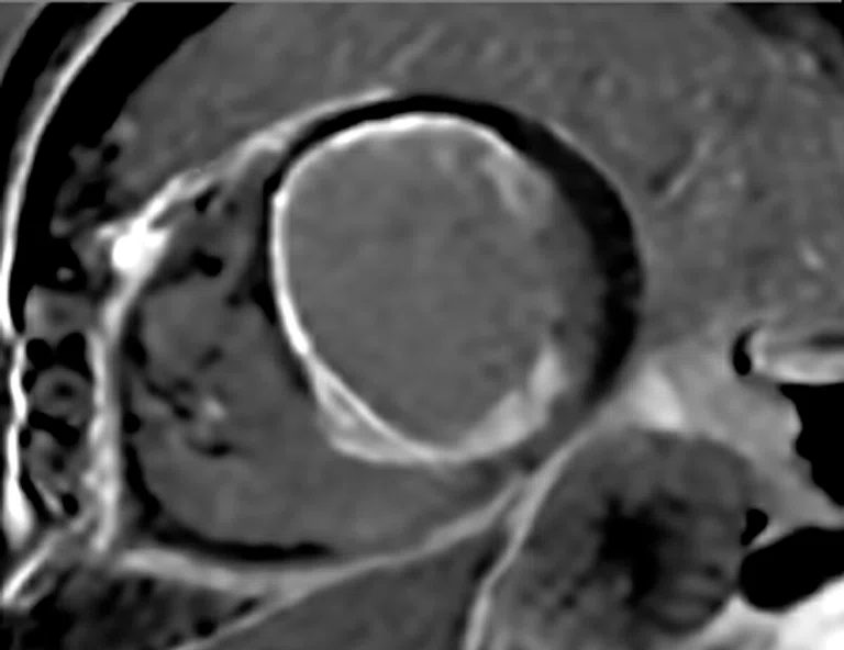
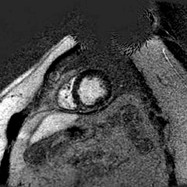
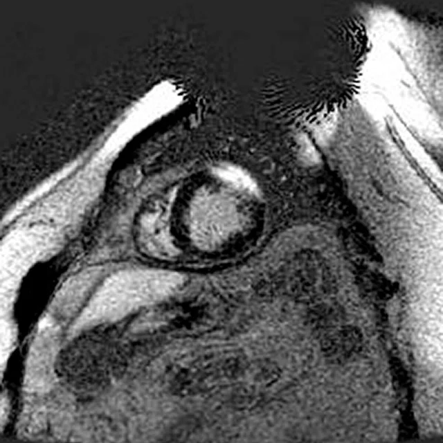
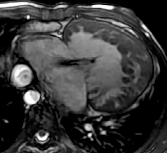
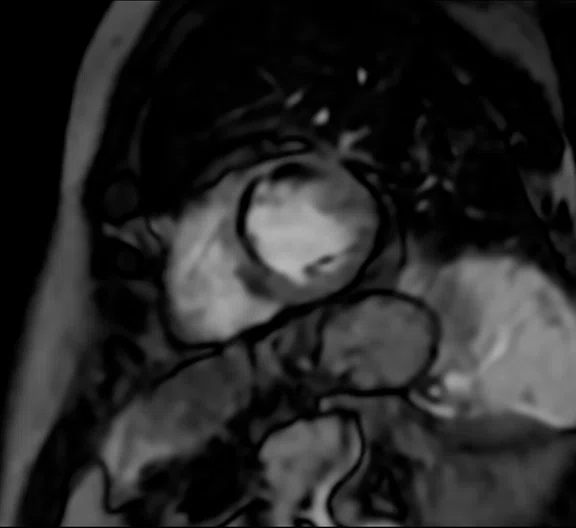
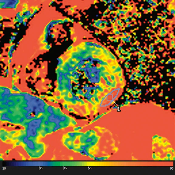
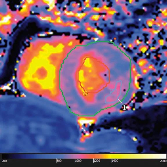
(A) Cine FIESTA provides high contrast between blood pool and myocardium as seen in this example on a patient with a mitral valve defect. (B) Short axis motion correction perfusion (rest) demonstrating a filling defect of the left ventricle. T1 and T2 Mapping in a COVID-19 myocarditis patient; (C) T2 map demonstrating 90% increased focal T2 compared to normal value and (D) MOLLI demonstrating a slight 7% increased focal native T1.
B
Figure 4.
(A) Cine FIESTA provides high contrast between blood pool and myocardium as seen in this example on a patient with a mitral valve defect. (B) Short axis motion correction perfusion (rest) demonstrating a filling defect of the left ventricle. T1 and T2 Mapping in a COVID-19 myocarditis patient; (C) T2 map demonstrating 90% increased focal T2 compared to normal value and (D) MOLLI demonstrating a slight 7% increased focal native T1.
1. Adam RD, Shambrook J, Flett AS. The Prognostic Role of Tissue Characterisation using Cardiovascular Magnetic Resonance in Heart Failure. Card Fail Rev. 2017 Nov;3(2):86-96.
2. Peterzan MA, Rider OJ, Anderson LJ. The Role of Cardiovascular Magnetic Resonance Imaging in Heart Failure. Card Fail Rev. 2016 Nov;2(2):115-122.
3. Yancy CW, Jessup M, Bozkurt B, et al. 2013 ACCF/AHA guideline for the management of heart failure: a report of the American College of Cardiology Foundation/American Heart Association Task Force on Practice Guidelines. J Am Coll Cardiol. 2013 Oct 15;62(16):e147-239.
4. Ponikowski P, Voors AA, Anker SD, et al. 2016 ESC Guidelines for the diagnosis and treatment of acute and chronic heart failure: The Task Force for the diagnosis and treatment of acute and chronic heart failure of the European Society of Cardiology (ESC) Developed with the special contribution of the Heart Failure Association (HFA) of the ESC. Eur Heart J. 2016 Jul 14;37(27):2129-2200.
5. National Institute for Health and Care Excellence Heart Failure Guidelines. Available at: www. nice.org.uk/guidance/conditions-and-diseases/cardiovascular-conditions/heart-failure. Accessed March 24, 2022.
6. Gulati M, Levy PD, Mukherjee D, et al. 2021 AHA/ACC/ASE/CHEST/SAEM/SCCT/SCMR Guideline for the Evaluation and Diagnosis of Chest Pain: A Report of the American College of Cardiology/ American Heart Association Joint Committee on Clinical Practice Guidelines. Circulation. 2021 Nov 30;144(22):e368-e454.
A
Figure 1.
DL Cine is designed to enable a free-breathing Cine acquisition in one heartbeat. (A) Conventional free-breathing, real-time FIESTA and (B) free-breathing, one-beat DL Cine acquisitions for heart function.
B
Figure 1.
DL Cine is designed to enable a free-breathing Cine acquisition in one heartbeat. (A) Conventional free-breathing, real-time FIESTA and (B) free-breathing, one-beat DL Cine acquisitions for heart function.
‡ Technology in development that represents ongoing research and development efforts. These technologies are not products and may never become products. Not for sale. Not cleared or approved by the U.S. FDA or any other global regulator for commercial availability.
‡ Technology in development that represents ongoing research and development efforts. These technologies are not products and may never become products. Not for sale. Not cleared or approved by the U.S. FDA or any other global regulator for commercial availability.
7. Sandino CM, Lai P, Vasanawala SS, Cheng JY. Accelerating cardiac cine MRI using a deep learningbased ESPIRiT reconstruction. Magn Reson Med. 2021 Jan;85(1):152-167.
8. Zucker EJ, Sandino CM, Kino A, Lai P, Vasanawala SS. Free-breathing Accelerated Cardiac MRI Using Deep Learning: Validation in Children and Young Adults. Radiology. 2021 Sep;300(3):539-548.
‡‡ 510(k) pending with the US FDA. Not yet CE marked. Not available for sale.
A
Figure 2.
Phase sensitive MDE acquisitions. (A) Single Shot PS MDE with TI set for myocardium, (B) Single Shot PS MDE with TI set for blood pool depicting the SNR challenges with this approach and (C) Single Shot PS MDE with TI set for blood pool with AIR™ Recon DL, helping overcome this challenge compared to (B).
B
Figure 2.
Phase sensitive MDE acquisitions. (A) Single Shot PS MDE with TI set for myocardium, (B) Single Shot PS MDE with TI set for blood pool depicting the SNR challenges with this approach and (C) Single Shot PS MDE with TI set for blood pool with AIR™ Recon DL, helping overcome this challenge compared to (B).
C
Figure 2.
Phase sensitive MDE acquisitions. (A) Single Shot PS MDE with TI set for myocardium, (B) Single Shot PS MDE with TI set for blood pool depicting the SNR challenges with this approach and (C) Single Shot PS MDE with TI set for blood pool with AIR™ Recon DL, helping overcome this challenge compared to (B).
A
Figure 3.
(A) Wideband MDE improves tissue suppression in the presence of MR-Conditional implants compared to (B) conventional MDE.
B
Figure 3.
(A) Wideband MDE improves tissue suppression in the presence of MR-Conditional implants compared to (B) conventional MDE.
10. Muscogiuri G, Martini C, Gatti M, et al. Feasibility of late gadolinium enhancement (LGE) in ischemic cardiomyopathy using 2D-multisegment LGE combined with artificial intelligence reconstruction deep learning noise reduction algorithm. Int J Cardiol. 2021 Nov 15;343:164-170.
9. Ferreira VM, Piechnik SK, Robson MD, Neubauer S, Karamitsos TD. Myocardial tissue characterization by magnetic resonance imaging: novel applications of T1 and T2 mapping. J Thorac Imaging. 2014 May;29(3):147-54.
C
Figure 4.
(A) Cine FIESTA provides high contrast between blood pool and myocardium as seen in this example on a patient with a mitral valve defect. (B) Short axis motion correction perfusion (rest) demonstrating a filling defect of the left ventricle. T1 and T2 Mapping in a COVID-19 myocarditis patient; (C) T2 map demonstrating 90% increased focal T2 compared to normal value and (D) MOLLI demonstrating a slight 7% increased focal native T1.
D
Figure 4.
(A) Cine FIESTA provides high contrast between blood pool and myocardium as seen in this example on a patient with a mitral valve defect. (B) Short axis motion correction perfusion (rest) demonstrating a filling defect of the left ventricle. T1 and T2 Mapping in a COVID-19 myocarditis patient; (C) T2 map demonstrating 90% increased focal T2 compared to normal value and (D) MOLLI demonstrating a slight 7% increased focal native T1.
result
9. Ferreira VM, Piechnik SK, Robson MD, Neubauer S, Karamitsos TD. Myocardial tissue characterization by magnetic resonance imaging: novel applications of T1 and T2 mapping. J Thorac Imaging. 2014 May;29(3):147-54.


PREVIOUS
${prev-page}
NEXT
${next-page}
Subscribe Now
Manage Subscription
FOLLOW US
Contact Us • Cookie Preferences • Privacy Policy • California Privacy PolicyDo Not Sell or Share My Personal Information • Terms & Conditions • Security
© 2024 GE HealthCare. GE is a trademark of General Electric Company. Used under trademark license.
TECH TRENDS
A wing-to-wing portfolio of cardiac sequences you can take to heart
A wing-to-wing portfolio of cardiac sequences you can take to heart
by Heide Harris, RT(R)(MR), MR Modality Leader, and Steve Lawson, RT(R)(MR), Global MR Clinical Marketing Manager, GE Healthcare
Cardiac MR (CMR) is considered the gold standard for the non-invasive evaluation of ischemic heart disease and heart failure.1 It accurately assesses cardiac anatomy and function and has the unique ability to identify pathological tissue characteristics of underlying disease.2 As a result of a large prognostic evidence base, the National Institute for Health and Care Excellence, as well as North American and European heart failure guidelines, now recommend CMR as the first-line imaging exam.3-5 Further, the 2021 AHA/ACC/ASE/ CHEST/SAEM/SCCT/SCMR guidelines for evaluating and diagnosing chest pain includes CMR with a Class 1 or Class 2a recommendation in all clinical scenarios, and is now always at least equivalent to other functional imaging tests (stress echo, MPS-SPECT, PET).6
AIR™ technologies benefitting CMR
AIR™ Coils – AIR™ Anterior Array (AA) Coil and AIR™ Multi-Purpose (MP) Coils – are GE Healthcare’s award-winning, extremely versatile, form-fitting and lightweight coils that are easy to position and comfortable for patients. Considering that patient obesity is a common challenge in CMR imaging, AIR™ Coils provide an advantage, as they allow the coil to be closer to the chest wall, providing deeper penetration and maximizing SNR. Technologists can simply drape them over the torso of a high BMI patient if needed. AIR™ Coils were designed with cardiac imaging in mind and have been optimized to provide better temporal resolution and improved image quality through shortened TRs.
Representing a new standard in coil innovation, AIR™ Coils enable a simplified, faster workflow while making the patient more comfortable. Other new features for workflow include AIR Touch™ to streamline patient positioning and coil selection on the user interface. When performing cardiac MR, the GE user interface allows free-breathing, real-time localization to ensure you scan in the right place while reserving the need to breathhold on the critical sequences like MDE later in the exam. This provides an opportunity for the patient to relax. The entire exam can be planned within seconds of starting it.
Wing-to-wing CMR portfolio
GE’s full wing-to-wing cardiac portfolio allows for a one-stop solution from plan to scan and includes partnerships with industry-leading cardiac post-processing companies, which is essential for cardiac analysis.
CMR protocols require a delicate balance between SNR, resolution and time. Suppression techniques often used in cardiac imaging come at the expense of signal. GE’s pioneering deep-learning- based reconstruction algorithm, AIR™ Recon DL, produces images with less noise, which may improve segmentation and perfusion analysis.
With the improved sharpness of AIR™ Recon DL, clinicians can now see smaller structures and obtain thinner slices with improved contrast-to-noise ratio (CNR) in a fast exam time. The higher SNR provided by AIR™ Recon DL is particularly useful with sequences that suffer from a lack of signal, such as single shot MDE or perfusion (time course).
Let’s take a deeper dive into the four main categories and sequences for cardiac MR exams: Function, morphology, flow and tissue characterization.
Function
In CMR assessments, Cine (2D FIESTA) is routinely used for qualitative and quantitative assessment of the heart’s function. With 2D FIESTA Cine, clinicians can visualize excellent tissue contrast between blood pool, myocardium and valves for qualitative assessment of valvular structure and anatomy (Figure 4). Quantitative LV and RV function values can be obtained with third-party post-processing tools such as cvi42®, suiteHEART® or Arterys®. Cine options include retrospective and prospective gating options that are ideal to use in challenging patients.
DL Cine‡, a technique pioneered by Stanford University, is a work-in-progress application from GE that is being developed to support rapid imaging. It is an approach that combines highly undersampled 2D Cine imaging with deep-learning reconstruction to leverage respiratory triggering to avoid breathholds for the patient.7-8 DL Cine includes automated segmentation‡ to easily and quickly capture quantitative metrics. DL Cine is being designed to enable free-breathing scanning that’s easier for the patient (Figure 1).
For strain/tissue tracking‡‡, third-party, post-processing products are being developed to analyze the acquired data from the FIESTA pulse sequences and facilitate quantifiable assessment of vessel wall motion.
Morphology
Black blood FSE enables visualization of the structural morphology of the heart. It includes double inversion recovery (IR) preparation, T1 and T2 weighting, and several fat suppression options, including triple IR. Black blood FSE sequences can be acquired in a conventional breathhold scan, or it can be acquired as a single shot (SSh) to accommodate more challenging patients who are unable to hold their breath. Regardless of the weighting or FatSat used, the black blood sequences are compatible with AIR™ Recon DL.
For a bright blood, whole-heart coverage, 3D Heart is available. It’s a non-contrast, free-breathing, 3D sequence. It can be either FIESTA based (at 1.5T) or GRE based (at 3.0T). 3D Heart allows for the assessment of anomalous coronaries, adult and pediatric congenital heart disease, and aorta and cardiac chambers. Because it’s a 3D sequence, it can easily be reformatted to help visualize small vessels or evaluate anatomical structures, particularly for exams where the anatomy is atypical or unique to the patient.
Flow
ViosWorks 4D Flow acquires a whole-heart functional exam in a non-gated, free-breathing acquisition. It is a highly accelerated acquisition that uses HyperKat reconstruction for any exam, from routine clinical scanning to complex anatomy.
2D Phase Contrast measures/quantifies blood flow with phase-contrast velocity encoding. In addition to quantitative blood flow and peak velocity measurements, it also provides the information clinicians need to evaluate the direction that the blood is flowing, which is critical in cases of valvular disease.
Tissue characterization
With the development of the late gadolinium enhancement (LGE) technique using an inversion-recovery T1-weighted sequence, it became possible to identify patterns and distribution of fibrosis and scar to differentiate ischemic and nonischemic cardiomyopathies. Additionally, further refinement of T2-weighted techniques allowed for the detection of myocardial inflammation and edema, which further established CMR as a tool for myocardial tissue characterization.9
Cardiac perfusion is utilized on patients with known or suspected coronary artery disease to determine if there are perfusion defects in the myocardium of the left ventricle that are caused by the narrowing of one or more of the coronary arteries. It can be performed with or without a stressing agent, although in some cases both approaches are used. The Time Course sequence is a robust motion-corrected acquisition for assessing the myocardium for perfusion defects. It utilizes an image registration algorithm to compensate the misalignments of respiratory motion and residual cardiac motion at both 1.5T and 3.0T. Time Course perfusion is also compatible with AIR™ Recon DL.
When it comes to the myocardial delayed enhanced (MDE) sequences, also known as LGE, it’s all about having the “right tool in the toolbox” for the patient’s exam. MDE sequences are designed to suppress the signal in the myocardium with an IR pulse approximately 10 minutes after contrast injection, called the peak enhancement time. The inversion time (TI) is determined by Cine IR or look-locker and acquires multiple inversion times to identify which sufficiently suppresses the myocardial tissue. The MDE sequences use a wideband inversion pulse to provide improved image quality and homogeneity in the myocardial tissue against inversion inaccuracies caused by MR-Conditional cardiovascular implanted electronic devices (Figure 3).
Figure 2.
Phase sensitive MDE acquisitions. (A) Single Shot PS MDE with TI set for myocardium, (B) Single Shot PS MDE with TI set for blood pool depicting the SNR challenges with this approach and (C) Single Shot PS MDE with TI set for blood pool with AIR™ Recon DL, helping overcome this challenge compared to (B).
GE offers diverse, free-breathing or high-resolution MDE sequence options to meet every patient’s needs. Each option provides uniform suppression of healthy myocardium and excellent CNR.
The 2D segmented MDE was the original sequence and was once considered the gold standard. It is a single-slice, breathhold sequence that offered higher resolution slices. However, it was a longer sequence (~15 seconds per slice), which made it more challenging for patients. As technology advanced, a single shot version (SSh MDE) was introduced to provide a whole-heart, free-breathing acquisition in seconds. Although both 2D MDE and SSh MDE are compatible with AIR™ Recon DL, scan times of SSh MDE are three times shorter compared to segmented MDE.10
The MDE sequences are time sensitive. Additional Cine IR scans can be acquired beyond the peak enhancement time to reevaluate the TI time, however, that adds more scanning time. For a better solution, a phase-sensitive MDE (PS MDE) is less sensitive to the selected TI value, which provides a wider range of TIs wherein myocardial nulling is effective. It can, therefore, reduce the number of repeat MDE scans. PS MDE allows the flexibility in TI for dark blood pool; adding AIR™ Recon DL overcomes SNR challenges and allows for the use of a rapid single-shot acquisition (Figure 2).
MDE can be acquired as a 3D, whole-heart, free-breathing navigated sequence, providing the added advantage of reformatting into any plane.
Recently, parametric T1-mapping and T2-mapping techniques opened a new frontier for CMR to explore tissue characteristics in more quantitative terms.9 These novel mapping strategies offer the promise of objective assessment of myocardial tissue properties, providing an absolute quantitative measure rather than just a qualitative or semi-quantitative evaluation. They are primarily used to assess trauma, edema or inflammatory disease such as myocarditis (Figure 4).
Figure 4.
(A) Cine FIESTA provides high contrast between blood pool and myocardium as seen in this example on a patient with a mitral valve defect. (B) Short axis motion correction perfusion (rest) demonstrating a filling defect of the left ventricle. T1 and T2 Mapping in a COVID-19 myocarditis patient; (C) T2 map demonstrating 90% increased focal T2 compared to normal value and (D) MOLLI demonstrating a slight 7% increased focal native T1.
GE offers two versions of T1 mapping: MOLLI (MOdified Look-Locker Inversion recovery) and SmarT1MAP. Both provide quantitative assessment of T1 relaxation time of myocardium and have integrated motion correction; however, they are acquired differently. The MOLLI sequence starts with an inversion pulse and acquires multiple inversion times to calculate the apparent T1 (or T1*). SmarT1MAP uses a saturation recovery 90° pulse acquired with various prep times and calculates the true T1 of the tissues. Both MOLLI and SmarT1MAP can be inline post-processed on the MR console automatically or after the acquisition.
T2 mapping provides quantitative assessment of the myocardium T2 relaxation time. The primary applications of T2 mapping are in diffuse inflammation (myocarditis) and potential detection of edema in response to ischemic injury.
T2* mapping provides the quantitative measurements to assess iron overload. Iron can build up in the heart due to hereditary hemochromatosis. Patients with thalassemia also have high levels of iron in their heart tissue.
The power of quantification
Acquiring the data from the MR scanner is only one part of the job when it comes to cardiac imaging. Having the right tools to quantify the data is another. While there is cardiac analysis software available on the MR console (e.g., CardioMaps, T2*, reformats), GE has integrated industry-leading cardiac MR post-processing applications to deliver more in-depth analysis to complete the SIGNA™Works wing-to-wing cardiac solution, including cvi42® (Circle Cardiovascular Imaging), suiteHEART® (NeoSoft) and ViosWorks (Arterys®).
The simple and easy-to-use suiteHEART® provides fully automated processed segmentation for function, flow and tissue characterization. This comprehensive solution includes time course, delayed enhancement, StarMap and Patent Foramen Ovale (PFO). With the Integrated Virtual™ Fellow, it speeds up CMR reporting with a deep-learning (DL) assistant to organize and analyze exams for operational efficiency improvement.
cvi42® features a highly automated workflow with advanced function (including mass and volume), 4D flow, tissue characterization, perfusion, tissue mapping modules and DL-based contour detection. It offers structured reporting to improve report turnaround times and has a fully customizable interface to adapt to user preferences.ViosWorks 4D Flow acquires a whole-heart functional exam in a non-gated, free-breathing acquisition that provides anatomical, functional and quantitative blood flow data. It is a highly accelerated acquisition that uses HyperKat reconstruction to enable the visualization of the direction and speed of blood flow. HyperKat takes advantage of data sparsity and correlates it spatially and temporally and also adapts temporally to increase the accuracy of reconstruction.
With ViosWorks 4D Flow on a GE MR system, users can acquire a <2 mm³ resolution whole-heart in under 8 minutes. It’s simple and fast workflow automates the editable segmentation for LV and RV workup with results validated and compared to expert consensus data. Scar quantification and perfusion analysis are both accelerated with AI, while the quantitative MDE and semi-quantitative perfusion tools provide a more objective analysis.
For nearly any cardiac indication, GE provides a CMR sequence that allows for a one-stop solution from plan to scan. In partnership with industry-leading post-processing applications, clinicians can obtain the quantitative analyses they need to manage cardiac patients in a less invasive way. Adding GE’s DLbased AIR™ Recon DL to CMR sequences further increases SNR and reduces scan times, providing more detail for the clinician, while using AIR™ AA Coils enhances patient comfort.
References
Gulati M, Levy PD, Mukherjee D, et al. 2021 AHA/ACC/ASE/CHEST/SAEM/SCCT/SCMR Guideline for the Evaluation and Diagnosis of Chest Pain: A Report of the American College of Cardiology/ American Heart Association Joint Committee on Clinical Practice Guidelines. Circulation. 2021 Nov 30;144(22):e368-e454.


