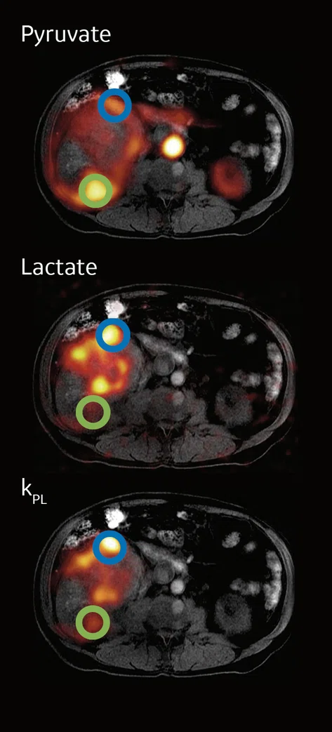
Figure 1.
Hyperpolarized 13C in a subject with kidney cancer. Circles represent areas of high (blue) and low (green) metabolism.
A
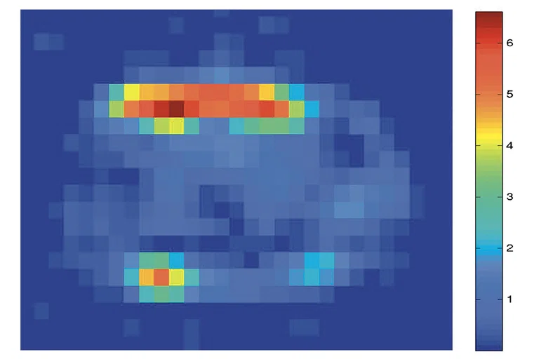
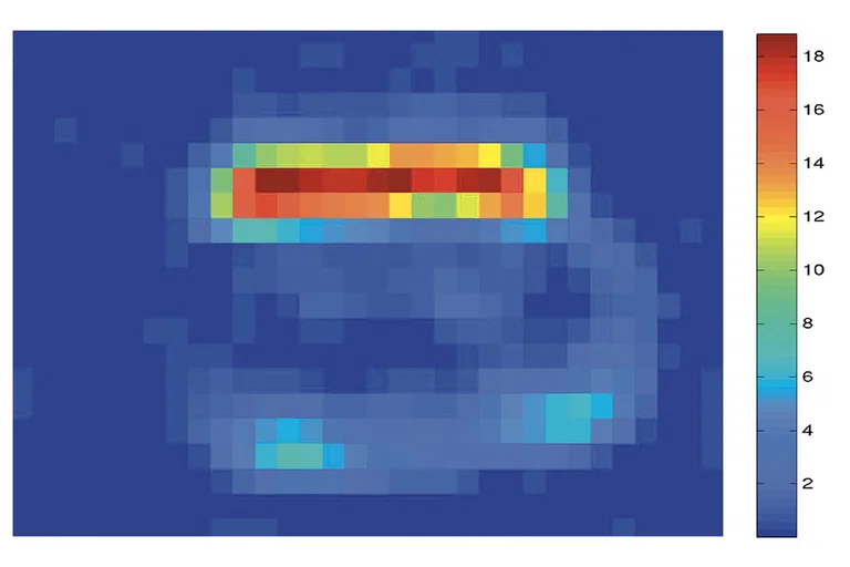
Figure 4.
SNR improvement in 13C imaging with a 16ch coil. (A) Prior 8ch coil, 4 anterior + posterior and (B) 16ch anterior coil only.B
Figure 4.
SNR improvement in 13C imaging with a 16ch coil. (A) Prior 8ch coil, 4 anterior + posterior and (B) 16ch anterior coil only.
‡ Technology in development that represents ongoing research and development efforts. These technologies are not products and may never become products. Not for sale. Not cleared or approved by the US FDA or any other global regulator for commercial availability.
‡ Technology in development that represents ongoing research and development efforts. These technologies are not products and may never become products. Not for sale. Not cleared or approved by the US FDA or any other global regulator for commercial availability.
1. Stødkilde-Jørgensen H, Laustsen C, Hansen ESS, et al. Pilot Study Experiences With Hyperpolarized [1-13C]pyruvate MRI in Pancreatic Cancer Patients. J Magn Reson Imaging, (2020); 51:961-963.
2. Gallagher FA, Woitek R, McLean MA, et al. Imaging breast cancer using hyperpolarized carbon-13 MRI. Proc Natl Acad Sci U S A. 2020 Jan 28;117(4):2092-2098.
3. Woitek R, McLean MA, Gill AB, et al. Hyperpolarized 13C MRI of Tumor Metabolism Demonstrates Early Metabolic Response to Neoadjuvant Chemotherapy in Breast Cancer. Radiol Imaging Cancer. 2020 Jul 31;2(4):e200017.
4. Woitek R, McLean MA, Ursprung S, et al. Hyperpolarized Carbon-13 MRI for Early Response Assessment of Neoadjuvant Chemotherapy in Breast Cancer Patients.
Cancer Res. 2021 Dec 1;81(23):6004-6017.
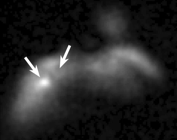
A
Figure 2.
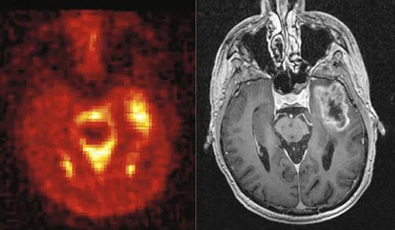
(A) 23Na imaging in a subject with melanoma metastases and (B) 23Na imaging in a subject with glioma. 23Na signal is elevated within tumors, especially in the presence of necrosis, as seen in the central non-enhancing glioma core.
B
Figure 2.
(A) 23Na imaging in a subject with melanoma metastases and (B) 23Na imaging in a subject with glioma. 23Na signal is elevated within tumors, especially in the presence of necrosis, as seen in the central non-enhancing glioma core.
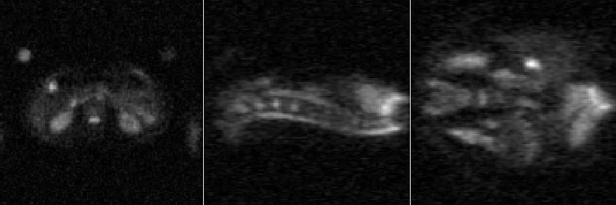
Figure 3.
Healthy control, abdominal imaging with 23Na with sagittal and coronal reconstructions illustrating strong homogenous signal across a 50 cm extent.
5. Bøgh N, Laustsen C, Hansen ESS, Tankisi H, Bertelsen LB, Blicher JU. Imaging Neurodegenerative Metabolism in Amyotrophic Lateral Sclerosis with Hyperpolarized [1-13C]pyruvate MRI. Tomography. 2022 Jun 14;8(3):1570-1577.
6. Gray LR, Tompkins SC, Taylor EB. Regulation of pyruvate metabolism and human disease. Cell Mol Life Sci. 2014 Jul;71(14):2577-604.
7. Joergensen SH, Hansen ESS, Bøgh N, et al. Detection of increased pyruvate dehydrogenase flux in the human heart during adenosine stress test using hyperpolarized [1-13C]pyruvate cardiovascular magnetic resonance imaging. J Cardiovasc Magn Reson 24, 34 (2022). https://doi.org/10.1186/s12968-022-00860-6
8. Bøgh N, Hansen ESS, Mariager CØ, Bertelsen LB, Ringgaard S, Laustsen C. Cardiac pH-Imaging With Hyperpolarized MRI. Front Cardiovasc Med. 2020 Nov 5;7:603674.
9. Kaggie JD, Lanz T, McLean MA, Riemer F, Schulte RF, Benjamin AJV, Kessler DA, Sun C, Gilbert FJ, Graves MJ, Gallagher FA. Combined 23 Na and 13 C imaging at 3.0 Tesla using a single-tuned large FOV birdcage coil. Magn Reson Med. 2021 Sep;86(3):1734-1745.
10. Rao MR,Robb FJ, Taracila V, Zhang W, Wild JM. Highly-Flexible Highly-Decoupled 1H RF Coils for MR Imaging of the lungs as utilized within a 129Xe Transmit-Receive RF Coil at 1.5T. Proceedings of the 2022 Joint Annual Meeting of the ISMRM-ESMRMB. Abstract 4102. Available at www.ismrm.org/22m.
‡ Technology in development that represents ongoing research and development efforts. These technologies are not products and may never become products. Not for sale. Not cleared or approved by the US FDA or any other global regulator for commercial availability.
‡‡ For research use only. Not cleared for clinical use in the US or any other country.
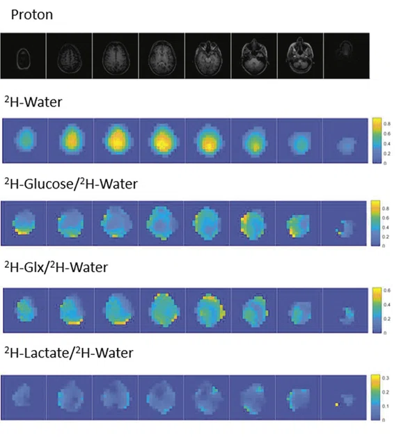
Figure 5.
Deuterium metabolic imaging. Color bars represent signal ratio to the water peak.


result
PREVIOUS
${prev-page}
NEXT
${next-page}
Subscribe Now
Manage Subscription
FOLLOW US
Contact Us • Cookie Preferences • Privacy Policy • California Privacy PolicyDo Not Sell or Share My Personal Information • Terms & Conditions • Security
© 2024 GE HealthCare. GE is a trademark of General Electric Company. Used under trademark license.
SPOTLIGHT
Advancing our knowledge of disease using multinuclear spectroscopy and imaging
Advancing our knowledge of disease using multinuclear spectroscopy and imaging
by Christoffer Laustsen, Dr. Med., PhD, Chair, Professor of Translational MRI, Aarhus University, Jutland, Denmark, and Joshua D. Kaggie, PhD, Senior Research Assistant, Mary A. McLean, PhD, Senior Research Associate, Ferdia A. Gallagher, PhD, FRCR FRCP, Professor of Translational Imaging and CRUK Senior Cancer Research Fellow, and Martin J. Graves, PhD, Professor of MR Physics, University of Cambridge, Cambridge, UK
Multinuclear spectroscopy and imaging (MNSI)‡ is a developing technique that enables an assessment of biological and metabolic changes in various diseases throughout the body using a range of non-proton (1H) nuclei. Key areas of MNSI research are currently neuroimaging (neurodegenerative diseases, stroke and traumatic brain injury), cardiac/pulmonary, musculoskeletal and oncology.
Multinuclear spectroscopy and imaging (MNSI)‡ is a developing technique that enables an assessment of biological and metabolic changes in various diseases throughout the body using a range of non-proton (1H) nuclei. Key areas of MNSI research are currently neuroimaging (neurodegenerative diseases, stroke and traumatic brain injury), cardiac/pulmonary, musculoskeletal and oncology.
Advances in MR hardware and software, such as multi-channel radiofrequency coils and high-performance gradients that increase signal together with ultra-fast pulse sequences, allow MNSI to capture metabolism in vivo,‡ providing a personalized and precise evaluation of disease in an individual.
Three nuclei are the subject of collaborative research between the University of Cambridge/Cancer Research UK Cambridge Institute and Aarhus University and Aarhus University Hospital: hyperpolarized carbon-13 (HP 13C), sodium (23Na) and deuterium (2H).
Oncology applications
The majority of clinical research applications for MNSI in the field of oncology are focused on tumor grading, progression and therapy response. For example, metabolism has been shown to vary across different types of tumors. Post-therapy, MNSI may help determine whether the metabolism is increasing or decreasing, informing the oncologist regarding the success of a particular therapy at an early stage.
Diagnosis and staging
Standard 1H MR has an established role in detecting cancer. The team have been exploring whether HP 13C can help stratify how aggressive a tumor is, which could provide important information to the oncologist for determining how to manage these patients.
The use of HP 13C is an area of active research in a range of cancers including prostate cancer, where many men have less aggressive forms of the disease that may not require immediate treatment and where active surveillance may be more appropriate. If HP 13C can provide this capability, it would better inform patient management. This same approach could also be used in cases of kidney, liver, pancreatic, breast and brain cancers, which all present with a range of aggressiveness across lesion types. Aarhus researchers are investigating if adding MNSI to a standard MR exam may help determine which lesions are malignant and, of those, which ones are more aggressive.
HP 13C may be useful in earlier detection of pancreatic cancer. The pancreas generates a significant amount of alanine in normal cells and, therefore, the pancreas may provide a fuel source for tumor development.1 Pre-cancerous pancreatic tumors can progress from inflammation to disease over the course of years or decades. Currently, it is not clear when that transition occurs, and understanding lesion aggressiveness by using MNSI is a potential application that could make a difference in this deadly cancer.
Treatment response
HP 13C research is currently focused on pyruvate as a general metabolic biomarker, as it metabolizes into high levels of lactate within aggressive cancers. The team are also investigating the use of HP 13C fumarate, which is metabolized into malate in areas where there is active, or very recent, cell death or necrosis. This may provide a slightly different insight into the effectiveness of therapy. The presence of a signal from malate could be a good indication that the therapy is effectively killing the tumor. 23Na MNSI has similar potential for treatment response monitoring with higher levels of sodium in areas with cell death than it is within the active tumor cells.
HP 13C is being used by the team in Aarhus to examine treatment response within the first week of therapy. Breast tumors demonstrate heterogeneity both within and between tumors, leading to variability in treatment response. In Cambridge, both 23Na and HP 13C are being used to examine breast cancer treatment response and further studies using 2H MNSI are planned following administration of oral 2H-labeled glucose.
Cambridge research have shown that HP 13C imaging in breast cancer has the capability to identify more aggressive cancers, which may be useful in stratifying disease.2 HP 13C has also been shown to provide an early response assessment in triple-negative breast cancer patients undergoing neoadjuvant chemotherapy.3,4
Neuro applications
In neurodegenerative diseases, 2H is being used to help identify areas of impaired glycolysis, or areas with a lack of glucose or oxygen supply, that have been identified in many brain diseases and injuries, such as Alzheimer’s disease, stroke and traumatic brain injury.
In Aarhus, several research efforts are examining neurodegenerative diseases and the long-term effects of COVID. Even in the presence of an intact blood-brain barrier, HP 13C allows brain metabolism to be probed. As an example, in one amyotrophic lateral sclerosis (ALS) study, subjects with symptoms that predominantly affected one side of the body showed an increased metabolism in the part of the brain controlling that side of the body. The group showed an increase in the production of lactate and bicarbonate in that area of the brain, demonstrating activation of the region responsible for the symptoms.5
Cardiac applications
There is currently some debate as to whether FDG-PET predicts myocardial viability. Pyruvate is the end-product of glycolysis and an important molecule for human metabolism so it could provide additional information in these patients. Disruption of pyruvate metabolism can cause mild or severe disease, depending on the location or severity of the disruption.6
Standard 1H MR is useful in the evaluation of cardiovascular function, particularly in the developing field of quantitative myocardial perfusion imaging using gadolinium (Gd)-based contrast agents. HP 13C could be an alternative to Gd-based perfusion imaging.7 In cases of chronic heart disease, decreased tissue viability can be visualized with HP 13C. MNSI can also be used to evaluate cardiac reserve and the cardiac response to treatment by imaging it at rest and following stress.
As shown in a published study, HP 13C MNSI accurately shows myocardial acid-base disturbances, which are commonly found in congestive heart failure and compromised cardiopulmonary function patients. It is possible that HP 13C MNSI with HP 13C pyruvate, may be useful in selecting heart failure patients for novel therapies or for detecting the localization, severity and reversibility of ischemia in coronary heart disease.8
Why MNSI
Like standard 1H MR, MNSI does not involve ionizing radiation. There are many instances where clinicians avoid PET and/or CT due to radioactivity, particularly in pediatric and young adult imaging, and in patients with genetic or chronic diseases that require regular imaging.
As mentioned above, the potential to identify patients benefiting from cancer treatment early in their therapy cycle is advantageous because it can reduce both the high cost of treatment and toxicity to the body in cases where there is no benefit. Additionally, there is the potential to obtain more temporal information about the lesion or disease using HP 13C, 23Na and 2H methods because these can be used for serial imaging.
MNSI can be utilized across the spectrum of commercially available field strengths, from 1.5T to 7.0T. HP 13C is ideal at 3.0T. Non-hyperpolarized nuclei, like 3Na and 2H, benefit from the use of ultra-high-field MR, e.g., 7.0T. While these nuclei can be used at 3.0T, with 7.0T the higher signal provides better sensitivity and, for 2H, better separation of the spectral peaks from different metabolites.
MNSI hardware and software
For MNSI, multinuclear coils are often developed by the institution and, in some cases, in collaboration with an MR or coil manufacturer. For example, Cambridge are using an MNSI breast coil (RAPID Biomedical GmgH, Rimpar, Germany). It is important to remember 1H coils are used in addition to other nuclei (e.g., carbon) coils in MNSI MR imaging. This might require scanning with an additional separate coil, or enabling an integrated multiple-frequency coil. The breast coil operates at two frequencies for 23Na and 1H, and can compete with clinical coils for quality and image acceleration. While it is possible to acquire data at both frequencies simultaneously, this is still a very limited field of research due to additional hardware complications.
At Cambridge, the team worked with RAPID Biomedical to develop and demonstrate the feasibility of a 23Na birdcage coil with a craniocaudal field of view (FOV) up to 48 cm to image a large FOV that is comparable to conventional hydrogen (1H) imaging. When used with an 8-channel receive coil, the team obtained a two-fold increase in 23Na SNR.9
One coil that shows promise in providing the 1H imaging required for MNSI is similar in design to GE Healthcare’s AIR™ Anterior Array (AA) Coil, although modifications are required to perform at 13C frequency. The advantage of this coil design, which is similar to a blanket and is placed over the patient, is that it can accommodate people of various sizes and shapes, which is a limiting factor with rigid coil designs, and it easily allows for the use of carbon or other nuclei coils. GE is collaborating with Cambridge to develop a 3.0T 13C 16-channel AIR™ Coil‡, enabling the potential for whole-body, multinuclear imaging as well as the inclusion of larger-sized patients for MNSI research. Further, the University of Sheffield published findings that regular 1H AIR™ Coils have a unique low-frequency rejection, enabling their use in multinuclear transmitters without additional carbon-blocking circuitry.10
Similarly, GE has supported MNSI research by developing or modifying pulse sequences for imaging of these different nuclei, such as 13C, 23Na, phosphorous (15P) and 2H. The MNSI Research Pack‡‡ from GE provides flexibility for users, including reading in arbitrary radiofrequency pulses and gradient waveforms; allowing for semi-automated prescan optimization using a Bloch-Siegert shift off resonance pulse; reading in lists with flip angles, TRs, TEs, RF phases and rotations; and enabling changes for gating, SSFP, etc. Current plug-ins available within the MNSI research pack to institutions with a GE research agreement include: MNSI Prescan, IDEAL Spiral CSI, SPSP Spiral Imaging, EPSI, MR Parameter Mapping and 3D radial UTE (waveforms and reconstruction).
Conclusion
The ability to image different nuclei is a promising area of research that may help clinicians detect various diseases and inform treatment pathways. Continued industry-academia collaboration will further advance development of system hardware and software necessary to move MNSI further into clinical use.


