A
Figure 2.
Comparison of female pelvis exams: (A-D) patient 1 was scanned with the prior software (PX26) and (E-H) patient 2 was scanned with SIGNA™Works AIR™ Edition, which includes AIR™ Recon and Flexible NPW. Note the higher resolution, smaller FOV imaging and shorter scan time provided with AIR™ Recon (see chart below).
B
Figure 2.
Comparison of female pelvis exams: (A-D) patient 1 was scanned with the prior software (PX26) and (E-H) patient 2 was scanned with SIGNA™Works AIR™ Edition, which includes AIR™ Recon and Flexible NPW. Note the higher resolution, smaller FOV imaging and shorter scan time provided with AIR™ Recon (see chart below).
C
Figure 2.
Comparison of female pelvis exams: (A-D) patient 1 was scanned with the prior software (PX26) and (E-H) patient 2 was scanned with SIGNA™Works AIR™ Edition, which includes AIR™ Recon and Flexible NPW. Note the higher resolution, smaller FOV imaging and shorter scan time provided with AIR™ Recon (see chart below).
D
Figure 2.
Comparison of female pelvis exams: (A-D) patient 1 was scanned with the prior software (PX26) and (E-H) patient 2 was scanned with SIGNA™Works AIR™ Edition, which includes AIR™ Recon and Flexible NPW. Note the higher resolution, smaller FOV imaging and shorter scan time provided with AIR™ Recon (see chart below).
E
Figure 2.
Comparison of female pelvis exams: (A-D) patient 1 was scanned with the prior software (PX26) and (E-H) patient 2 was scanned with SIGNA™Works AIR™ Edition, which includes AIR™ Recon and Flexible NPW. Note the higher resolution, smaller FOV imaging and shorter scan time provided with AIR™ Recon (see chart below).
F
Figure 2.
Comparison of female pelvis exams: (A-D) patient 1 was scanned with the prior software (PX26) and (E-H) patient 2 was scanned with SIGNA™Works AIR™ Edition, which includes AIR™ Recon and Flexible NPW. Note the higher resolution, smaller FOV imaging and shorter scan time provided with AIR™ Recon (see chart below).
G
Figure 2.
Comparison of female pelvis exams: (A-D) patient 1 was scanned with the prior software (PX26) and (E-H) patient 2 was scanned with SIGNA™Works AIR™ Edition, which includes AIR™ Recon and Flexible NPW. Note the higher resolution, smaller FOV imaging and shorter scan time provided with AIR™ Recon (see chart below).
H
Figure 2.
Comparison of female pelvis exams: (A-D) patient 1 was scanned with the prior software (PX26) and (E-H) patient 2 was scanned with SIGNA™Works AIR™ Edition, which includes AIR™ Recon and Flexible NPW. Note the higher resolution, smaller FOV imaging and shorter scan time provided with AIR™ Recon (see chart below).
A
Figure 3.
Comparison images of an ankle exam; (A-D) on the prior software (PX26) all using NEX 2 and (E-H) SIGNA™Works AIR™ Edition all using NEX 1 except (E) using NEX 1.5. Total scan time reduction was 45 percent, from 31 min. to 17 min., with AIR™ Recon (see chart below).
B
Figure 3.
Comparison images of an ankle exam; (A-D) on the prior software (PX26) all using NEX 2 and (E-H) SIGNA™Works AIR™ Edition all using NEX 1 except (E) using NEX 1.5. Total scan time reduction was 45 percent, from 31 min. to 17 min., with AIR™ Recon (see chart below).
C
Figure 3.
Comparison images of an ankle exam; (A-D) on the prior software (PX26) all using NEX 2 and (E-H) SIGNA™Works AIR™ Edition all using NEX 1 except (E) using NEX 1.5. Total scan time reduction was 45 percent, from 31 min. to 17 min., with AIR™ Recon (see chart below).
D
Figure 3.
Comparison images of an ankle exam; (A-D) on the prior software (PX26) all using NEX 2 and (E-H) SIGNA™Works AIR™ Edition all using NEX 1 except (E) using NEX 1.5. Total scan time reduction was 45 percent, from 31 min. to 17 min., with AIR™ Recon (see chart below).
E
Figure 3.
Comparison images of an ankle exam; (A-D) on the prior software (PX26) all using NEX 2 and (E-H) SIGNA™Works AIR™ Edition all using NEX 1 except (E) using NEX 1.5. Total scan time reduction was 45 percent, from 31 min. to 17 min., with AIR™ Recon (see chart below).
F
Figure 3.
Comparison images of an ankle exam; (A-D) on the prior software (PX26) all using NEX 2 and (E-H) SIGNA™Works AIR™ Edition all using NEX 1 except (E) using NEX 1.5. Total scan time reduction was 45 percent, from 31 min. to 17 min., with AIR™ Recon (see chart below).
G
Figure 3.
Comparison images of an ankle exam; (A-D) on the prior software (PX26) all using NEX 2 and (E-H) SIGNA™Works AIR™ Edition all using NEX 1 except (E) using NEX 1.5. Total scan time reduction was 45 percent, from 31 min. to 17 min., with AIR™ Recon (see chart below).
H
Figure 3.
Comparison images of an ankle exam; (A-D) on the prior software (PX26) all using NEX 2 and (E-H) SIGNA™Works AIR™ Edition all using NEX 1 except (E) using NEX 1.5. Total scan time reduction was 45 percent, from 31 min. to 17 min., with AIR™ Recon (see chart below).
A
Figure 4.
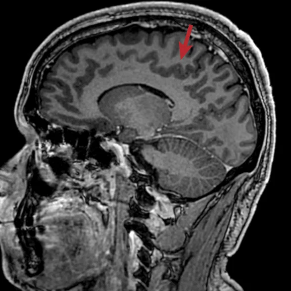
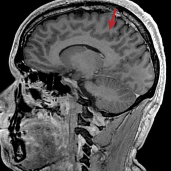
MP-RAGE delivers better gray/white matter differentiation in a post-contrast sequence (arrows). (A) Sagittal MP-RAGE and (B) sagittal 3D BRAVO.
B
Figure 4.
MP-RAGE delivers better gray/white matter differentiation in a post-contrast sequence (arrows). (A) Sagittal MP-RAGE and (B) sagittal 3D BRAVO.
A
Figure 5.
3D MRCP exam with FOCUS, HyperSense, HyperCube and AIR™ Recon. (A) Axial T2 FatSat PROPELLER RTr, (B, C) coronal T2 SSFSE navigated and (D, E) volume-rendered reformats.
B
Figure 5.
3D MRCP exam with FOCUS, HyperSense, HyperCube and AIR™ Recon. (A) Axial T2 FatSat PROPELLER RTr, (B, C) coronal T2 SSFSE navigated and (D, E) volume-rendered reformats.
C
Figure 5.
3D MRCP exam with FOCUS, HyperSense, HyperCube and AIR™ Recon. (A) Axial T2 FatSat PROPELLER RTr, (B, C) coronal T2 SSFSE navigated and (D, E) volume-rendered reformats.
D
Figure 5.
3D MRCP exam with FOCUS, HyperSense, HyperCube and AIR™ Recon. (A) Axial T2 FatSat PROPELLER RTr, (B, C) coronal T2 SSFSE navigated and (D, E) volume-rendered reformats.
E
Figure 5.
3D MRCP exam with FOCUS, HyperSense, HyperCube and AIR™ Recon. (A) Axial T2 FatSat PROPELLER RTr, (B, C) coronal T2 SSFSE navigated and (D, E) volume-rendered reformats.
result


PREVIOUS
${prev-page}
NEXT
${next-page}
Subscribe Now
Manage Subscription
FOLLOW US
Contact Us • Cookie Preferences • Privacy Policy • California Privacy PolicyDo Not Sell or Share My Personal Information • Terms & Conditions • Security
© 2024 GE HealthCare. GE is a trademark of General Electric Company. Used under trademark license.
IN PRACTICE
AIR Recon: a game changer at Al Qassimi Hospital
AIR Recon: a game changer at Al Qassimi Hospital
In 2016, the United Arab Emirates (UAE) Ministry of Health announced the establishment of Unison – the first joint project of its kind in the field of medical imaging between the public and private sectors – between Abu Dhabi International Medical Services Company (ADI), a leading healthcare solutions provider, and GE Healthcare. The partnership helps facilitate and accelerate access to quality medical services throughout the UAE.
In 2016, the United Arab Emirates (UAE) Ministry of Health announced the establishment of Unison – the first joint project of its kind in the field of medical imaging between the public and private sectors – between Abu Dhabi International Medical Services Company (ADI), a leading healthcare solutions provider, and GE Healthcare. The partnership helps facilitate and accelerate access to quality medical services throughout the UAE.
"Since the forming of Unison, we have worked toward enhancing the operational efficiency of radiology departments throughout the Ministry of Health hospital network in the UAE," says Hatem Abou El Abbass Ghonim, PhD, Professor, Senior Consultant Radiologist, Al Qassimi Hospital, Sharjah, and Medical Director, Unison CI in PPP, UAE Ministry of Health and Prevention (MOHaP). "Strengthening radiology competencies is crucial to boosting the speed, quality and reliability of diagnostic services to help deliver better patient outcomes. Intelligent workflow applications powered by AIR™ automate the scan process to drive consistency. When paired with advanced imaging applications, AIR™ Recon, AIR Touch™ and AIR™ Coils deliver simply better versatility and productivity gains along with industry-leading image quality."
As part of the Unison collaboration, Al Qassimi Hospital acquired a SIGNA™ Pioneer system. Al Qassimi Hospital is one of the largest MOHaP hospitals in Sharjah UAE, providing efficient healthcare services across multiple disciplines and specializations.
In January 2020, the hospital’s SIGNA™ Pioneer was upgraded to the SIGNA™ Works AIR™ Edition software, which includes AIR™ Recon, AIR Touch™ and AIR™ Coils.
AIR Touch™ accelerates the scanning process through automated coil selection and landmarking. With the anatomical-based protocol optimization, the technologist does not have to place the center of the coil over the center of the anatomy to be scanned. AIR Touch™ optimizes the protocol parameters with a single touch, delivering on average a 59 percent productivity gain from plan-to-scan at Al Qassimi Hospital. It has also helped the hospital reduce repeating a sequence when the coil selection does not match the anatomy being scanned.
The use of AIR™ Coils has made scanning workflow even more streamlined. Now, the technologist just places the AIR™ Coil on the patient, without measuring or centering, and the coil and AIR Touch™ work in tandem to automatically select the anatomy to be scanned.
A new reconstruction algorithm, AIR™ Recon, fundamentally shifts the balance between image quality and scan time. It reduces background noise and out of field-of-view (FOV) artifacts and improves SNR for clearer, crisper images and shorter scan times, reducing the potential need for rescans.
"AIR™ Recon is a key solution that offers dramatic improvement over existing image reconstruction techniques. We are now able to scan patients with shorter scan times and maintain high-resolution results, which is one of my priorities. The ability to save time in a routine exam is essential."
Professor Hatem Abou El Abbass Ghonim
Mohyi Eldin Fahmy, MD, Consultant Radiologist, Head of Medical Imaging Department, Al Qassimi Hospital, has regularly seen the impact of AIR™ Recon on his clinical routine because it is compatible with all pulse sequences. In addition to increased SNR, he can also obtain smaller FOVs and utilize Flexible No Phase Wrap (NPW) for even more scan time savings.
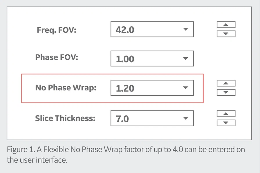
Prior to the SIGNA™Works AIR™ Edition upgrade, when the NPW imaging option was turned on, the phase matrix would only double and the NEX would be reduced by half. After the upgrade, NPW allows the user to adjust the amount of oversampling along phase-encoding direction to avoid wrapping artifacts. In addition, NEX is no longer confusingly coupled with the NPW option.
A female pelvis case scanned with the upgrade to SIGNA™Works AIR™ Edition was compared to a similar patient scanned with the prior software (PX26). With the new software, the total exam time was 10 percent shorter and the FOV went from 24 to 18 cm. The use of AIR™ Recon also enabled higher resolution images in lower scan times (Figure 2).
Figure 2.
Comparison of female pelvis exams: (A-D) patient 1 was scanned with the prior software (PX26) and (E-H) patient 2 was scanned with SIGNA™Works AIR™ Edition, which includes AIR™ Recon and Flexible NPW. Note the higher resolution, smaller FOV imaging and shorter scan time provided with AIR™ Recon (see chart below).
In another case involving an ankle exam, the technologist achieved a 45 percent scan time reduction between the prior software version and SIGNA™Works AIR™ Edition utilizing AIR™ Recon (Figure 3).
"AIR™ from GE Healthcare packs innovations that deliver versatility, productivity and consistent quality, empowering any technologist to produce images with remarkable clarity," Dr. Fahmy says. "From new applications to enhancements to existing technologies, the new software release is just simply better."
One of the new applications is 3D MP-RAGE, a 3D gradient echo pulse sequence designed for neuro imaging that uses sequential view ordering and adjustable recovery time to further improve contrast-to-noise ratios, as well as reduce ringing artifacts. In a comparison of MP-RAGE in SIGNA™Works AIR™ Edition and BRAVO in the previous software, Dr. Fahmy found that MP-RAGE delivered better gray/white matter differentiation in a post-contrast sequence (Figure 4).
Applying SIGNA™Works AIR™ Edition in their daily workflow is crucial. Dr. Fahmy explains, "The advantage of having this technology enables us to scan a 3D MRCP using HyperCube and HyperSense that deliver the freedom to scan with a focused FOV and faster scan time with higher resolution," (Figure 5).
"The AIR™ advanced technologies are game-changers in MR that strengthen clinical power and enable radiologists to achieve improved workflow, enhanced patient comfort and reduced exam times," Professor Ghonim adds. "With the introduction of these advanced solutions, Al Qassimi Hospital will further strengthen its position as one of the leading centers in the region offering world-class diagnostic services."


