1. Guzik TJ, Mohiddin SA, Dimarco A, et al. COVID-19 and the cardiovascular system: implications for risk assessment, diagnosis, and treatment options. Cardiovasc Res. 2020 Aug 1;116(10):1666-1687.
2. Kelle S, Bucciarelli-Ducci C, Judd RM, et al. Society for Cardiovascular Magnetic Resonance (SCMR) recommended CMR protocols for scanning patients with active or convalescent phase COVID-19 infection. J Cardiovasc Magn Reson 22, 61 (2020).
2. Kelle S, Bucciarelli-Ducci C, Judd RM, et al. Society for Cardiovascular Magnetic Resonance (SCMR) recommended CMR protocols for scanning patients with active or convalescent phase COVID-19 infection. J Cardiovasc Magn Reson 22, 61 (2020).
2. Kelle S, Bucciarelli-Ducci C, Judd RM, et al. Society for Cardiovascular Magnetic Resonance (SCMR) recommended CMR protocols for scanning patients with active or convalescent phase COVID-19 infection. J Cardiovasc Magn Reson 22, 61 (2020).
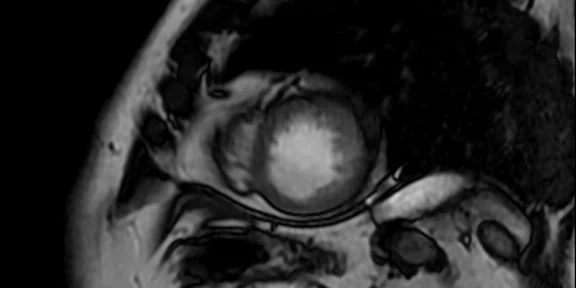
A
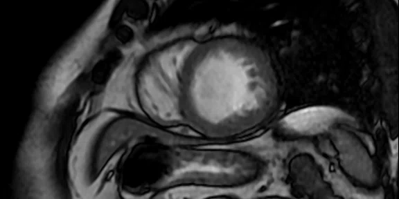
Figure 1.
(A-D) Short axis FIESTA post-contrast, 1.9 x 1.6 x 8 mm, 3:08 min.
B
Figure 1.
(A-D) Short axis FIESTA post-contrast, 1.9 x 1.6 x 8 mm, 3:08 min.
C
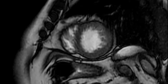
Figure 1.
(A-D) Short axis FIESTA post-contrast, 1.9 x 1.6 x 8 mm, 3:08 min.
D
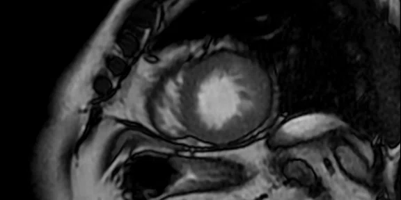
Figure 1.
(A-D) Short axis FIESTA post-contrast, 1.9 x 1.6 x 8 mm, 3:08 min.
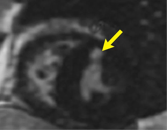
A
Figure 2.
(A) Short axis SS PS MDE, 1.9 x 2.8 x 8.0 mm, 15 slices; (B, C) 2 CAV (2D MDE Radial), A B C 1.7 x 2.3 x 8 mm, 3 slices.
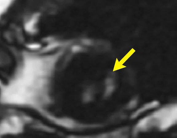
B
Figure 2.
(A) Short axis SS PS MDE, 1.9 x 2.8 x 8.0 mm, 15 slices; (B, C) 2 CAV (2D MDE Radial), A B C 1.7 x 2.3 x 8 mm, 3 slices.
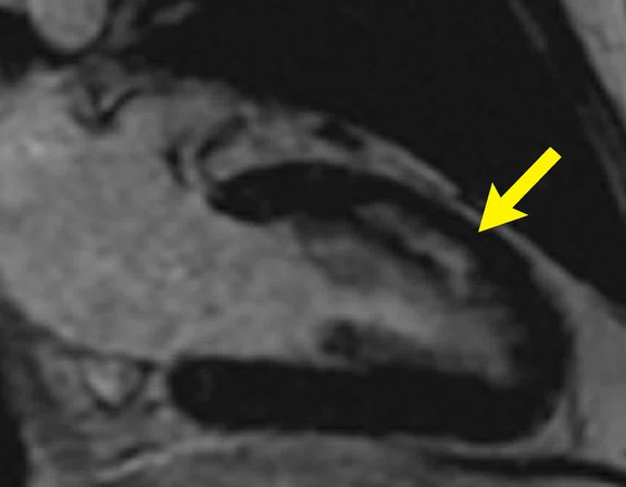
C
Figure 2.
(A) Short axis SS PS MDE, 1.9 x 2.8 x 8.0 mm, 15 slices; (B, C) 2 CAV (2D MDE Radial), A B C 1.7 x 2.3 x 8 mm, 3 slices.
result


PREVIOUS
${prev-page}
NEXT
${next-page}
Subscribe Now
Manage Subscription
FOLLOW US
Contact Us • Cookie Preferences • Privacy Policy • California Privacy PolicyDo Not Sell or Share My Personal Information • Terms & Conditions • Security
© 2024 GE HealthCare. GE is a trademark of General Electric Company. Used under trademark license.
SPOTLIGHT
Assessing COVID-19 cardiac-related disease
Assessing COVID-19 cardiac-related disease
by Olivier Vignaux, MD, PhD, radiologist, American Hospital of Paris, France
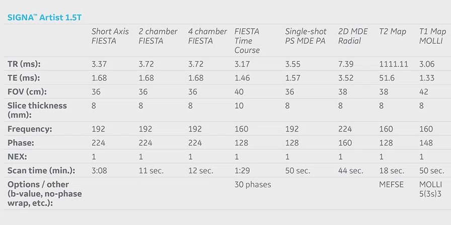
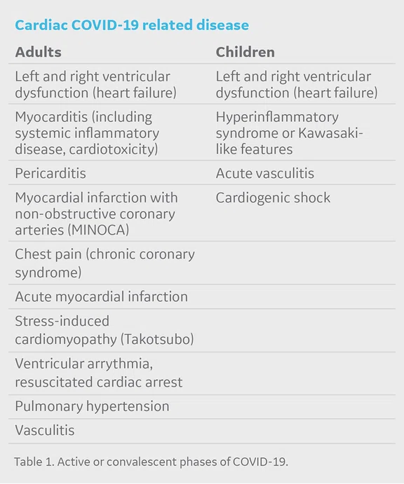
The novel coronavirus disease 2019, caused by SARS-CoV-2 (COVID-19), not only impacts the respiratory system, it is also responsible for major complications of the cardiovascular system including hypertension, acute cardiac injury and myocarditis1. The mechanisms involved as well as the impacts on this system are not yet fully understood, and many studies are underway on these subjects. Based on recent literature2, and according to guidelines released by the Society for Cardiovascular Magnetic Resonance (SCMR) for cardiac MR (CMR) imaging of COVID-19 patients, the expected CMR indications in patients with active or convalescent phase of COVID-19 are presented in Table 1.
The SCMR released specific CMR recommendations for scanning patients with known or suspected COVID-19 disease, including those recovering from the disease2. As patients with COVID-19 often could have pulmonary difficulties and cannot take long breath-holds, a very short protocol (> 15 minutes, minimum) is suggested. A longer protocol (> 30 minutes, desirable) is proposed if the patient’s functional status allows a scan time of around 30 minutes.
Diagnosis of cardiac involvement in COVID-19 related disease can include the use of CMR. In our facility, the improved signal of the AIR™ Anterior Array (AA) Coil enables the acquisition of excellent image quality to depict myocardial involvement related to SARS-CoV-2. In this patient case, MR imaging exhibited intramyocardial delayed enhancement due to direct myocarditis and/or myocardial infarction with non-obstructive coronary arteries (MINOCA, microvascular impairment).
Patient history
A 46-year-old male weighing 78 kg (172 lb.) positive for COVID-19 (PCR test) with chest pain and an increase in troponin levels was referred to CMR to evaluate presence of myocardial infarction.
Results
Coronary angiography exhibited only moderate stenosis of LAD. MR appearance in favor of latero-basal, anterior middle and antero-latero-apical intra-myocardial lesions. These lesions suggest an inflammatory and/or microcirculatory involvement rather than macro-circulatory infarctions (intra-myocardial site, non-systematized bifocal involvement, absence of hypokinesis).
Discussion
Determining cardiac involvement in COVID-19 patients is crucial for prognosis, e.g., risk of sudden cardiac death. The use of CMR can identify COVID-19 patients with possible cardiac injury and help predict progression of disease-related complications. With the AIR™ AA Coil, we are able to obtain high-quality cardiac images to assess these patients using both the minimal and desired protocols as recommended by the SCMR.


