A
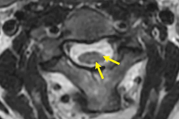
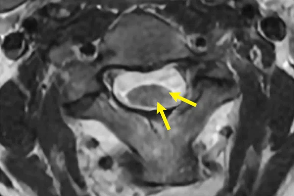
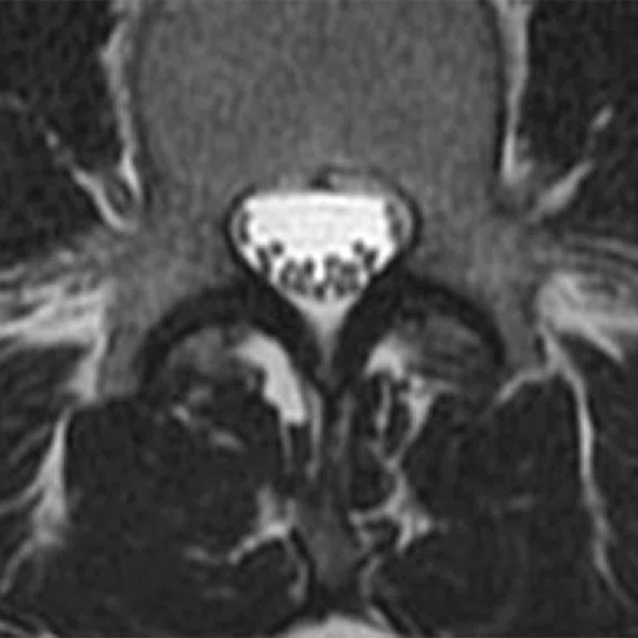
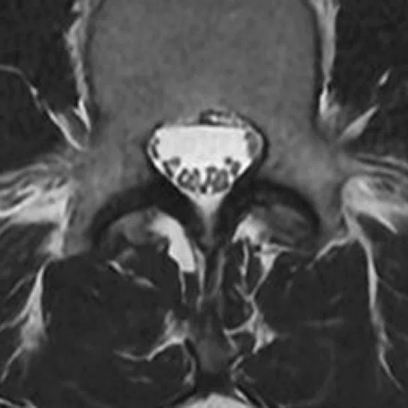
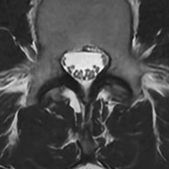
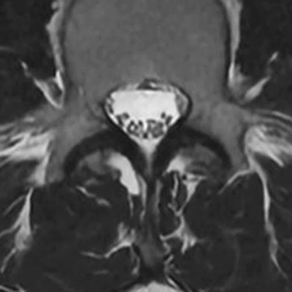
Figure 2.
AIR™ Recon DL improves image quality and maintains lesion conspicuity. (A) Conventional and (B) AIR™ Recon DL reconstructed images.
B
Figure 2.
AIR™ Recon DL improves image quality and maintains lesion conspicuity. (A) Conventional and (B) AIR™ Recon DL reconstructed images.
‡Not yet CE marked for 1.5T. Not available for sale in all regions.
A
Figure 1.
A multi-rater evaluation study presented at RSNA 2019 reported that the AIR™ Recon DL images were rated higher with a significant improvement in both SNR and anatomic structure. Y-axis is the mean of the difference between AIR™ Recon DL and the conventional image.
B
Figure 1.
A multi-rater evaluation study presented at RSNA 2019 reported that the AIR™ Recon DL images were rated higher with a significant improvement in both SNR and anatomic structure. Y-axis is the mean of the difference between AIR™ Recon DL and the conventional image.
A-1
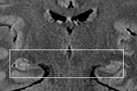
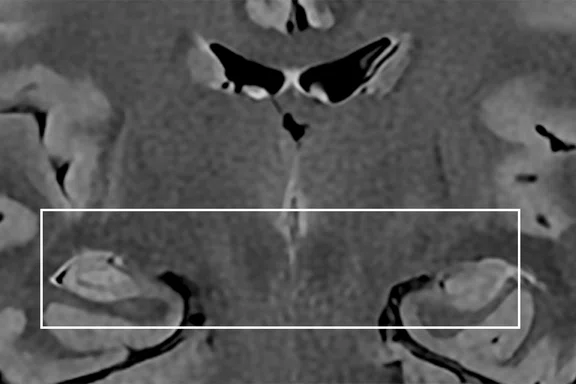
Figure 3.
AIR™ Recon DL is useful for denoising small structures such as the hippocampus. (A) Conventional and (B) AIR™ Recon DL reconstructed images.
Images courtesy of Columbia University Medical Center.
B-1
Figure 3.
AIR™ Recon DL is useful for denoising small structures such as the hippocampus. (A) Conventional and (B) AIR™ Recon DL reconstructed images.
Images courtesy of Columbia University Medical Center.
A-2
Figure 3.
AIR™ Recon DL is useful for denoising small structures such as the hippocampus. (A) Conventional and (B) AIR™ Recon DL reconstructed images.
Images courtesy of Columbia University Medical Center.
B-2
Figure 3.
AIR™ Recon DL is useful for denoising small structures such as the hippocampus. (A) Conventional and (B) AIR™ Recon DL reconstructed images.
Images courtesy of Columbia University Medical Center.
A-1
Figure 4.
In these inverted T2 images, the AIR™ Recon DL zoomed images show sharper detail, which assists the radiologist when evaluating small structures. (A) Conventional and (B) AIR™ Recon DL reconstructed images.
Images courtesy of Columbia University Medical Center.
A-2
Figure 4.
In these inverted T2 images, the AIR™ Recon DL zoomed images show sharper detail, which assists the radiologist when evaluating small structures. (A) Conventional and (B) AIR™ Recon DL reconstructed images.
Images courtesy of Columbia University Medical Center.
B-1
Figure 4.
In these inverted T2 images, the AIR™ Recon DL zoomed images show sharper detail, which assists the radiologist when evaluating small structures. (A) Conventional and (B) AIR™ Recon DL reconstructed images.
Images courtesy of Columbia University Medical Center.
B-2
Figure 4.
In these inverted T2 images, the AIR™ Recon DL zoomed images show sharper detail, which assists the radiologist when evaluating small structures. (A) Conventional and (B) AIR™ Recon DL reconstructed images.
Images courtesy of Columbia University Medical Center.
A
Figure 5.
In addition to improving image quality, AIR™ Recon DL can also be used for faster imaging, including reducing NEX. (A) Original image, 2 NEX, 2:17 min. (B) AIR™ Recon DL low, 2 NEX, 2:17 min. (C) AIR™ Recon DL medium, 2 NEX, 2:17 min. (D) AIR™ Recon DL high, 1 NEX, 1:12 min.
B
Figure 5.
In addition to improving image quality, AIR™ Recon DL can also be used for faster imaging, including reducing NEX. (A) Original image, 2 NEX, 2:17 min. (B) AIR™ Recon DL low, 2 NEX, 2:17 min. (C) AIR™ Recon DL medium, 2 NEX, 2:17 min. (D) AIR™ Recon DL high, 1 NEX, 1:12 min.
C
Figure 5.
In addition to improving image quality, AIR™ Recon DL can also be used for faster imaging, including reducing NEX. (A) Original image, 2 NEX, 2:17 min. (B) AIR™ Recon DL low, 2 NEX, 2:17 min. (C) AIR™ Recon DL medium, 2 NEX, 2:17 min. (D) AIR™ Recon DL high, 1 NEX, 1:12 min.
D
Figure 5.
In addition to improving image quality, AIR™ Recon DL can also be used for faster imaging, including reducing NEX. (A) Original image, 2 NEX, 2:17 min. (B) AIR™ Recon DL low, 2 NEX, 2:17 min. (C) AIR™ Recon DL medium, 2 NEX, 2:17 min. (D) AIR™ Recon DL high, 1 NEX, 1:12 min.
1. Villanueva-Meyer J, Shin D, Li Y, et al. Denoising MR Images of the Cervical Spine: Multi-Reader Assessment of a Deep Learning Approach. Radiological Society of North America 2019 Scientific Assembly and Annual Meeting, December 1 - December 6, 2019, Chicago IL. archive.rsna.org/2019/19018947.html. Accessed October 7, 2020.
A
Figure 4.
In these inverted T2 images, the AIR™ Recon DL zoomed images show sharper detail, which assists the radiologist when evaluating small structures.(A) Conventional and (B) AIR™ Recon DL reconstructed images. Images courtesy of Columbia University Medical Center.
Figure 4.
In these inverted T2 images, the AIR™ Recon DL zoomed images show sharper detail, which assists the radiologist when evaluating small structures.(A) Conventional and (B) AIR™ Recon DL reconstructed images. Images courtesy of Columbia University Medical Center.
B
Figure 4.
In these inverted T2 images, the AIR™ Recon DL zoomed images show sharper detail, which assists the radiologist when evaluating small structures.(A) Conventional and (B) AIR™ Recon DL reconstructed images. Images courtesy of Columbia University Medical Center.
Figure 4.
In these inverted T2 images, the AIR™ Recon DL zoomed images show sharper detail, which assists the radiologist when evaluating small structures.(A) Conventional and (B) AIR™ Recon DL reconstructed images. Images courtesy of Columbia University Medical Center.
result


PREVIOUS
${prev-page}
NEXT
${next-page}
Subscribe Now
Manage Subscription
FOLLOW US
Contact Us • Cookie Preferences • Privacy Policy • California Privacy PolicyDo Not Sell or Share My Personal Information • Terms & Conditions • Security
© 2024 GE HealthCare. GE is a trademark of General Electric Company. Used under trademark license.
IN PRACTICE
Elevating neuroimaging with AIR Recon DL
Elevating neuroimaging with AIR Recon DL
Based on an ASNR 2020 webinar by Javier Villanueva-Meyer, MD, Assistant Professor, Neuroradiology, University of California, San Francisco
A patient with suspected compressive myelopathy is transferred to a tertiary institution for an advanced neurological evaluation. Unfortunately, the quality of the patient’s MR exam from the other facility limits the neuroradiologist’s interpretation of the existing exam. A repeat scan is requested and the results are slightly better, though still suboptimal. This is a scenario that often occurs at institutions across the world, where a lack of consistency and uniformity across imaging providers can lead to duplicate or unnecessary imaging exams and increase healthcare costs.
A patient with suspected compressive myelopathy is transferred to a tertiary institution for an advanced neurological evaluation. Unfortunately, the quality of the patient’s MR exam from the other facility limits the neuroradiologist’s interpretation of the existing exam. A repeat scan is requested and the results are slightly better, though still suboptimal. This is a scenario that often occurs at institutions across the world, where a lack of consistency and uniformity across imaging providers can lead to duplicate or unnecessary imaging exams and increase healthcare costs.
The reality is that adequate neuroradiological diagnosis often hinges on identifying subtle changes or lesions in small anatomies – such as the hippocampus or sella turcica – as well as determining tumor margins for surgical planning. While technological advances in MR imaging have enhanced neuroimaging capabilities, image quality is also dependent upon patient compliance. Patients may be clinically unstable or suffer from a neurodegenerative disease that makes it difficult to control movement and therefore remain still for the duration of an MR exam. A suboptimal exam can make evaluating small structures a difficult – and sometimes nearly impossible – task.
Enter AIR™ Recon DL‡, GE Healthcare’s novel MR image reconstruction method that uses a deep learning-based convolutional neural network to reconstruct MR images at 3.0T and 1.5T. It delivers both SNR improvement and image sharpening for higher spatial resolution and improved image detail.
"When we look at the enhanced AIR™ Recon DL image side-by-side with the conventional image, we can really see a significant improvement," says Javier Villanueva-Meyer, MD, Assistant Professor of Neuroradiology at the University of California, San Francisco (UCSF).
As with many new reconstruction techniques that enhance image quality, a key question is whether lesion conspicuity is maintained. At RSNA 2019, Dr. Villanueva-Meyer and colleagues presented a multi-rater evaluation study1 on the difference between conventional and AIR™ Recon DL reconstructed images of the cervical spine. The raters were asked to evaluate signal-to-noise ratio (SNR), anatomic structure definition, diagnostic certainty, overall image quality and artifacts. In each category except artifacts, where the rating was nearly equivalent, the AIR™ Recon DL images were rated higher with a significant improvement in both SNR and anatomic structure.
"We can use this tool to speed imaging up and actually use that to add another facet of value to the MR exam. Similarly, we can use the shortened scan time to reduce the possibility of patient motion and therefore improve diagnostic confidence."
Dr. Javier Villanueva-Meyer
For example, rather than utilize a T2 Flex scan that takes 2:40 minutes, Dr. Villanueva-Meyer suggests a T2 STIR that takes 1:51 minutes with AIR™ Recon DL and is similar in quality to the longer T2 Flex scan. Another example involves reducing NEX from 2 to 1 to cut scan time nearly in half while using AIR™ Recon DL to enhance the image – in some cases better than the original 2 NEX image – at a fraction of the scan time.
Implementing AIR™ Recon DL can be tailored to a specific institution’s preferences, as well. Dr. Villanueva-Meyer advises that if the initial enhancement or change in image quality is perhaps too much or something the clinicians aren’t accustomed to, the SNR improvement factor can be reduced and then gradually increased as the radiologists gain more experience with it.
"There is broad utility for using AIR™ Recon DL in diagnosis," Dr. Villanueva-Meyer adds. "Whether it is scanning the unstable patient, reducing scan time, reducing the artifacts from motion that occur, or bringing up the image quality and SNR, there’s opportunity for improving throughput on an MR system."
For some institutions, the additional time savings afforded by AIR™ Recon DL can also facilitate research activities by increasing available scanner time.
"It was important to ensure that we could still see pathology and we were happy to find that AIR™ Recon DL maintained lesion conspicuity in a non-inferiority analysis," he adds.
"One clinical scenario that we are often asked by surgeons is whether or not there’s involvement of the cavernous sinus in pituitary tumors," Dr. Villanueva-Meyer explains. "Another is to evaluate for mesial temporal sclerosis. Sometimes we have difficulty seeing these small structures unless we have a truly optimal exam. This is where technology like AIR™ Recon DL can be really helpful in the diagnosis by making the anatomy a little bit more clear."
The image clarity and sharper detail provided by AIR™ Recon DL is noticeable when zooming in to view these small structures, such as in the hippocampus (Figures 3 and 4) or when evaluating small structures like the anatomy near the vertex of the brain, the brain CSF interface or within the subarachnoid space. It’s also helpful when evaluating tumor features and lesion margins, as well as areas of edema.
"I want to clearly see that the infiltrative margins are maintained (in brain tumor cases) or have the ability to identify texture differences, which is helpful for making the distinction between true vasogenic edema and infiltrative edema," says Dr. Villanueva-Meyer.
AIR™ Recon DL is also helpful for enhancing images of the skull base in orbital exams to provide sharper detail within the interorbital contents, or imaging of the extracranial vessels, where images often have relatively low SNR.
"I’m really excited about applying this tool to cut the time for some sequences while maintaining diagnostic quality and, therefore, allowing more time for investigational sequences within the allotted scan time."
Dr. Javier Villanueva-Meyer
Conversely, Dr. Villanueava-Meyer adds there’s a safety perspective as well, where facilities can speed up imaging and decrease exposure between patients and staff during the current pandemic setting.


