1. McGee KP, Campeau NG, Witte RJ, et al. Evaluation of a New, Highly Flexible Radiofrequency Coil for MR Simulation of Patients Undergoing External Beam Radiation Therapy. J Clin Med. 2022 Oct 11;11(20):5984.
2. Scherman J, Af Wetterstedt S, Persson E, Olsson LE, Jamtheim Gustafsson C. Geometric impact and dose estimation of on-patient placement of a lightweight receiver coil in a clinical magnetic resonance imaging-only radiotherapy workflow for prostate cancer. Phys Imaging Radiat Oncol. 2023 Mar 24;26:100433.
2. Scherman J, Af Wetterstedt S, Persson E, Olsson LE, Jamtheim Gustafsson C. Geometric impact and dose estimation of on-patient placement of a lightweight receiver coil in a clinical magnetic resonance imaging-only radiotherapy workflow for prostate cancer. Phys Imaging Radiat Oncol. 2023 Mar 24;26:100433.
result


PREVIOUS
${prev-page}
NEXT
${next-page}
Subscribe Now
Manage Subscription
FOLLOW US
Contact Us • Cookie Preferences • Privacy Policy • California Privacy PolicyDo Not Sell or Share My Personal Information • Terms & Conditions • Security
© 2024 GE HealthCare. GE is a trademark of General Electric Company. Used under trademark license.
News
First global installation of AIR™ RT Suite at Radium Hospital in Norway
First global installation of AIR™ RT Suite at Radium Hospital in Norway
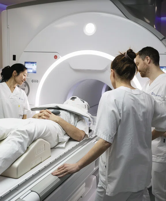
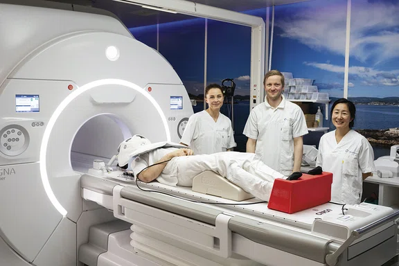
Radium Hospital, part of Oslo University Hospital in Norway, is the first hospital to implement GE HealthCare’s AIR™ RT Suite, featuring the Universal Couchtop™ MRI Overlay (CIVCO Medical Solutions, Kalona, IA) and 32-channel AIR™ Open Coil. As a leading cancer center, Radium began using MR for radiation therapy planning in 2006 for brachytherapy and gynecological treatments.
The 32-channel AIR™ Open Coil is designed for higher signal-to-noise ratio (SNR) and is compatible with all SIGNA™ wide bore scanners. It is easy to set up, requires less training for technologists and offers wide compatibility with patient immobilization devices.
Radium has been conducting initial set-up and quality assurance imaging on volunteers. According to Edmund Reitan, lead MRI radiographer, the AIR™ Open Coil takes significantly less time to set up than a conventional MR coil. Since the coil is flexible, it conforms well to the person’s head and fixation mask.
Reitan adds, “The geometric factor of the MR coil seems to be comparable to other head and neck coils, which enables us to use parallel imaging in the same way as we normally would in a diagnostic Head & Neck MR coil. This has not been the case with other MR coil set-ups for RT with fixation masks.”
“Image quality so far is superior to what we see from conventional MR coil set-ups,” says Knut Håkon Hole, senior MRI radiologist. “It provides better SNR and more homogeneous signal in difficult areas like the anterior neck directly below the chin.”
“We are looking forward to implementing and exploring the potential benefits of using the AIR™ RT-system in RT planning,” adds Line Nilsen, PhD, radiation physicist, Radium Hospital.
DOWNLOAD ARTICLE HERE


