A
Figure 2.
(A) Patient with severe arrhythmia scanned with a short axis FIESTA Cine with multiple breathholds resulting in motion artifacts, 2 x 2.3 x 8 mm, 1:45 min. for a complete short axis stack. (B) Same patient scanned in one heartbeat with a short axis Sonic DL™ Cardiac Cine with a single breathhold, 2.2 x 2.2 x 8 mm, 24 sec. for a complete short axis stack. Images courtesy of Fairfax Radiological Consultants.
B
Figure 2.
(A) Patient with severe arrhythmia scanned with a short axis FIESTA Cine with multiple breathholds resulting in motion artifacts, 2 x 2.3 x 8 mm, 1:45 min. for a complete short axis stack. (B) Same patient scanned in one heartbeat with a short axis Sonic DL™ Cardiac Cine with a single breathhold, 2.2 x 2.2 x 8 mm, 24 sec. for a complete short axis stack. Images courtesy of Fairfax Radiological Consultants.
A
Figure 1.
Same patient as featured on front cover. 4 chamber acquisition with Sonic DL™ Cardiac Cine, 1.4 x 1.4 x 8 mm, 7 sec. per slice, 60 cardiac phases with a 23 ms temporal resolution per phase. Images courtesy of Stanford University.
B
Figure 1.
Same patient as featured on front cover. 4 chamber acquisition with Sonic DL™ Cardiac Cine, 1.4 x 1.4 x 8 mm, 7 sec. per slice, 60 cardiac phases with a 23 ms temporal resolution per phase. Images courtesy of Stanford University.
A
Figure 3.
Sonic DL™ is designed to deliver higher spatial resolution imaging in a shorter scan time. (A) Short axis FIESTA Cine with multiple breathholds, 2 x 2 x 8 mm, 2:10 min. for a complete short axis stack compared to (B) a short axis Sonic DL™ Cardiac Cine in fewer breathholds 1.8 x 1.2 x 8 mm, 1:18 min. for a complete short axis stack.
B
Figure 3.
Sonic DL™ is designed to deliver higher spatial resolution imaging in a shorter scan time. (A) Short axis FIESTA Cine with multiple breathholds, 2 x 2 x 8 mm, 2:10 min. for a complete short axis stack compared to (B) a short axis Sonic DL™ Cardiac Cine in fewer breathholds 1.8 x 1.2 x 8 mm, 1:18 min. for a complete short axis stack.
A
Figure 4.
Sonic DL™ Cardiac Cine is designed to enable high-quality, free-breathing cardiac acquisitions. (A) Short axis FIESTA Cine with multiple breathholds, 2 x 2 x 8 mm, 2:10 min. for a complete short axis stack compared to (B) a short axis Sonic DL™ Cardiac Cine with free-breathing and respiratory gating, 2 x 2.2 x 8 mm, 51 sec. for a complete short axis stack.
B
Figure 4.
Sonic DL™ Cardiac Cine is designed to enable high-quality, free-breathing cardiac acquisitions. (A) Short axis FIESTA Cine with multiple breathholds, 2 x 2 x 8 mm, 2:10 min. for a complete short axis stack compared to (B) a short axis Sonic DL™ Cardiac Cine with free-breathing and respiratory gating, 2 x 2.2 x 8 mm, 51 sec. for a complete short axis stack.
‡ At time of printing, 510(k) pending at US FDA. Not yet CE marked. Not available for sale.
IMV 2022 MR Market Outlook Report. Available at www.imv.com.
1. IMV 2022 MR Market Outlook Report. Available at www.imv.com.
2. Sandino CM, Lai P, Vasanawala SS, Cheng JY. Accelerating cardiac cine MRI using a deep learning-based ESPIRiT reconstruction. Magn Reson Med. 2021;85(1):152-167.
3. Lai P, Brau A. “Improving cardiac cine MRI on 3T using 2D kt accelerated auto-calibrating parallel imaging.” Journal of Cardiovascular Magnetic Resonance 16.1 (2014): 1-2.
4. Zucker EJ, Sandino CM, Kino A, Lai P, Vasanawala SS. Free-breathing Accelerated Cardiac MRI Using Deep Learning: Validation in Children and Young Adults. Radiology. 2021;300(3):539-548.
5. Orii M, Sone M, Osaki T, et al. Reliability of respiratory-gated real-time two-dimensional cine incorporating deep learning reconstruction for the assessment of ventricular function in an adult population. Int J Cardiovasc Imaging. 2023;39(5):1001-1011.


result
PREVIOUS
${prev-page}
NEXT
${next-page}
Subscribe Now
Manage Subscription
FOLLOW US
Contact Us • Cookie Preferences • Privacy Policy • California Privacy PolicyDo Not Sell or Share My Personal Information • Terms & Conditions • Security
© 2024 GE HealthCare. GE is a trademark of General Electric Company. Used under trademark license.
TECH TRENDS
Life-speed cardiac imaging with Sonic DL
Life-speed cardiac imaging with Sonic DL
Artificial Intelligence (AI) is a powerful tool to simplify workflow, increase productivity and improve diagnostic confidence by helping obtain consistent, high-quality images. GE HealthCare is committed to continually offering AI features that address these important customer needs.
Artificial Intelligence (AI) is a powerful tool to simplify workflow, increase productivity and improve diagnostic confidence by helping obtain consistent, high-quality images. GE HealthCare is committed to continually offering AI features that address these important customer needs.
It is well known in MR that there exists a fundamental trade-off between signal-to-noise ratio (SNR), resolution and scan times. GE HealthCare developed AIR™ Recon DL to increase SNR and image sharpness, which can then be utilized to improve resolution and decrease scan times. Reduced scan times are achieved by optimizing parameters such as bandwidth, NEX and parallel imaging factor to make the most out of the additional SNR.
Acceleration techniques, such as ARC, ASSET and HyperSense (compressed sensing), have also been used to speed up the scan in order to reduce scan times.
SENSE-based methods require accurate estimation of coil sensitivity maps from calibration data. When the field-of-view (FOV) is smaller than the subject, the overlapping anatomies can lead to discontinuities in the coil sensitivity maps and cause residual ghosting in the reconstructed images. K-space parallel imaging approaches are robust to anatomy overlaps and allow for acceleration with a reduced FOV. Compressed sensing, such as HyperSense, has enabled rapid cine acquisitions with a single breathhold or while the patient is free-breathing, however, it requires careful hand-tuning of the relative weights assigned to data consistency and regularization terms, otherwise there is the potential to introduce aliasing artifacts. Additionally, the use of conventional acceleration techniques to enable faster scanning has led to noisier and lower image quality. Now, there is a better way to accelerate MR imaging.
The better way is Sonic DL™‡, a deep-learning-based image acquisition acceleration technique. Sonic DL™ uses a deep-learning engine to optimize every voxel by leveraging 6.5 million operations on the raw data, delivering astonishing new levels of acceleration. Because Sonic DL™ is not based on conventional acceleration techniques, it does not have the same limitations.
Sonic DL™ is GE HealthCare’s most rapid MR acceleration technique yet, designed to enable significantly shorter scan times without impacting diagnostic value. It can achieve rapid results with up to an 83% reduction in scan time versus a fully sampled Cine acquistion. And, it captures rapidly moving anatomies, such as the heart, without extending scan time.

A
B
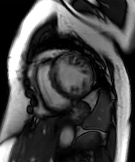
Figure 1.
Same patient as featured on front cover. 4 chamber acquisition with Sonic DL™ Cardiac Cine, 1.4 x 1.4 x 8 mm, 7 sec. per slice, 60 cardiac phases with a 23 ms temporal resolution per phase. Images courtesy of Stanford University.
Understanding the potential impact of Sonic DL™, GE HealthCare will first introduce Sonic DL™ Cardiac Cine to address the most challenging MR cases, cardiac cine, and enable MR imaging of patients who couldn’t be effectively imaged before, namely those with arrythmias and those who cannot hold their breath. Future generations of this solution are expected to be compatible with 2D and 3D sequences across all clinical areas, as well as expected to be combined with AIR™ Recon DL for image quality improvement.
Sonic DL™ Cardiac Cine is designed to achieve high frame rates in cardiac imaging and enable exceptionally fast scans, from minutes to seconds, minimizing the need for repetitive patient breathholds. The fast scan times of Sonic DL™ may help simplify challenging and lengthy procedures and help accommodate sick and uncooperative patients who cannot tolerate long exam times. To accomplish this, GE HealthCare is building on the success of AIR™ Recon DL and the company’s experience using deep-learning algorithms to enhance MR imaging.
The need for fast, robust cardiac MR (CMR) imaging is growing. According to IMV, 23% of all sites with at least one MR system perform CMR (excluding MRAs), an increase of 5 percentage points over 2021 data. Vascular and cardiovascular MRAs increased 13 percentage points to 81% of all sites.1
A
B
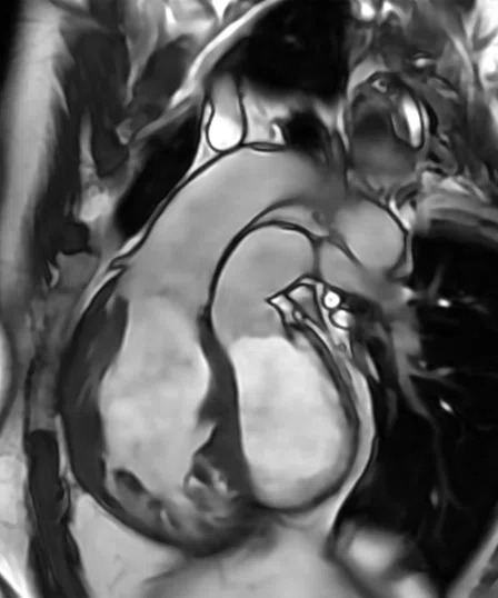
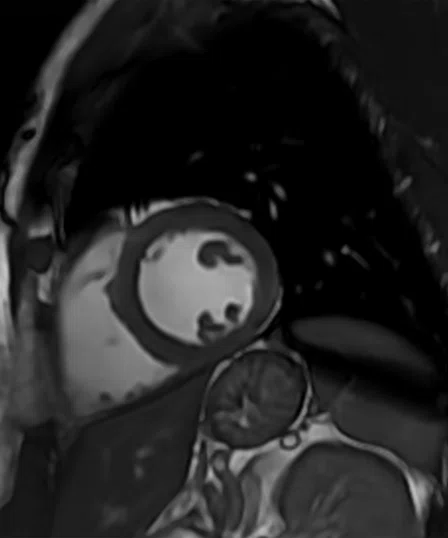
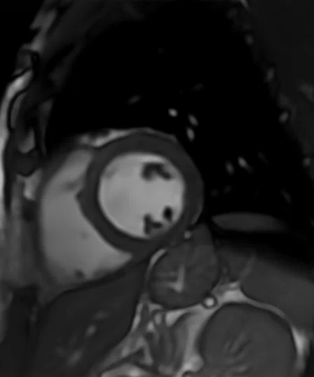
Figure 2.
(A) Patient with severe arrhythmia scanned with a short axis FIESTA Cine with multiple breathholds resulting in motion artifacts, 2 x 2.3 x 8 mm, 1:45 min. for a complete short axis stack. (B) Same patient scanned in one heartbeat with a short axis Sonic DL™ Cardiac Cine with a single breathhold, 2.2 x 2.2 x 8 mm, 24 sec. for a complete short axis stack. Images courtesy of Fairfax Radiological Consultants.
The development story
The idea behind the development of Sonic DL™ is to turn MR scan times from minutes to seconds. The concept began with a collaboration between GE HealthCare’s Applied Sciences Laboratory in Menlo Park, CA and Stanford University. Sandino et al. described the evaluation of a novel combined parallel imaging and deep-learning (DL) based reconstruction framework for accelerating cardiac cine MR acquisitions. The authors demonstrated the feasibility to use this novel prototype to capture a rapid single heartbeat per slice in a 2D cardiac cine scan.2
“We had an early prototype from the initial collaboration with Stanford,” explains Xucheng Zhu, Lead MRI AI Scientist at GE HealthCare. “We knew there would be a big opportunity in cardiac MR because it is one of most challenging and time-consuming patient exams.”
Zhu then worked to further develop the initial prototype into a more mature solution that could be used in a multi-center research study. As the solution evolved, it underwent further testing and external evaluation. Near the end of 2021, the solution moved into the product development stage under the guidance of Shiv Kaushik, Senior Applications Engineer at GE HealthCare, and his team.
“At this point, we had retired all the risks and performed a full evaluation of the solution, so it was ready to go into product development,” Kaushik says.
Zhu and Kaushik worked together to harmonize the pulse sequence, reconstruction and deep-learning model to optimize image quality. This is the point where Sonic DL™ Cardiac Cine began to take shape, with the DL-based reconstruction specially tuned and developed for a cardiac cine exam.
The sampling pattern originated from a variable density (VD) k-t sampling pattern from previous GE HealthCare development work3 that was then customized and optimized for Sonic DL™.
“A DL-reconstruction enhancement improved the results,” adds Zhu. “We had several different options for sampling patterns, so we tested and compared them, selecting the optimal sampling ordering and sampling density. So each step along the way, we fine-tuned the solution while in the product pipeline.”
A
B
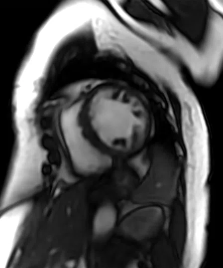
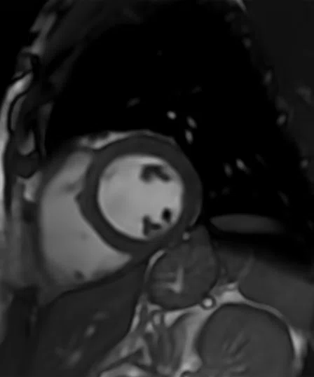
Figure 3.
Sonic DL™ is designed to deliver higher spatial resolution imaging in a shorter scan time. (A) Short axis FIESTA Cine with multiple breathholds, 2 x 2 x 8 mm, 2:10 min. for a complete short axis stack compared to (B) a short axis Sonic DL™ Cardiac Cine in fewer breathholds 1.8 x 1.2 x 8 mm, 1:18 min. for a complete short axis stack.
The development team lead by Kaushik and Zhu worked with GE HealthCare’s Digital engineering team, using the Edison™ AI Workbench and its Orchestrator framework, for model development. The next step was to pre-process the training data and train the model, which was then integrated into the product.
“There were multiple subsystems and multiple developers all handling different components,” says Kaushik. “We had a rigorous rhythm throughout the product development cycle to test all corner cases and retire all technical risks to ensure the product met our expectations.”
The development team worked with GE HealthCare’s Clinical Applications team to scan volunteers on different GE HealthCare MR systems with different coils. There were frequent — either weekly or biweekly — image quality reviews. Based on these results, the development team could quickly deploy changes, test and then iterate.
The solution blends DL with conventional MR techniques to optimize the acquisition and reconstruction. Zhu explains that as a data-driven approach, DL overcomes the limitations posed by conventional MR to accelerate the scan, delivering a robust solution that for the first time is designed to allow more efficient imaging of cardiac patients who previously had difficulty undergoing a CMR exam, namely those with arrhythmia. Sonic DL™ was designed to reconstruct images that match the acquired data by using an unrolled convolutional neural network with data consistency updates.
“Sonic DL™ Cardiac Cine impacts scan time reduction while AIR™ Recon DL impacts image quality,” adds Kaushik. “Separately, they do two very different things, but when used together, which is our goal, they deliver a robust, fast and high-quality cardiac exam across all cardiac patient populations.”
A
B
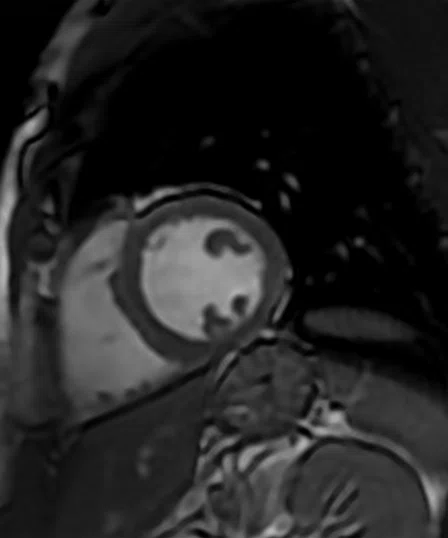
Figure 4.
Sonic DL™ Cardiac Cine is designed to enable high-quality, free-breathing cardiac acquisitions. (A) Short axis FIESTA Cine with multiple breathholds, 2 x 2 x 8 mm, 2:10 min. for a complete short axis stack compared to (B) a short axis Sonic DL™ Cardiac Cine with free-breathing and respiratory gating, 2 x 2.2 x 8 mm, 51 sec. for a complete short axis stack.
While conventional acceleration techniques typically delivered a factor of 2, Sonic DL™ is designed to deliver up to a factor of 12, enabling an acquisition in a single heartbeat per slice, making the patient more comfortable and without the need to undergo multiple breathholds to obtain a diagnostic cardiac exam.
With several academic institutions involved in the evaluation and validation process, there has already been positive external feedback regarding the potential of Sonic DL™ Cardiac Cine to deliver the image quality and rapid scanning needed in cardiac MR.4,5 Sonic DL™ Cardiac Cine will be available in a future MR software release.
Editor's Note:
Shiv Kaushik and Xucheng Zhu acknowledge the contributions made by the Sonic DL™ development team to bring this solution from prototype to product:
Ke Li, PhD, Applications Engineer (pulse sequence development) Kavitha Manickam, PhD, Senior Software Engineer (reconstruction development) Kailash Saravanan, MS, Software Engineer (reconstruction development) Eman Ali, Lead Clinical Development Specialist, MR (applications development) Sheila Washburn, MS, Digital Product Manager, MR Intelligent Scanner