1. Etkin CD, Springer BD. The American Joint Replacement Registry-the first 5 years. Arthroplast Today. 2017;3(2):67-69. Published 2017 Mar 14. doi:10.1016/j.artd.2017.02.002.
2. Bayliss LE, Culliford D, Monk AP, Glyn-Jones S, Prieto-Alhambra D, Judge A, Cooper C, Carr AJ, Arden NK, Beard DJ, Price AJ. The effect of patient age at intervention on risk of implant revision after total replacement of the hip or knee: a population-based cohort study. Lancet. 2017 Apr 8;389(10077):1424-1430. doi: 10.1016/S0140-6736(17)30059-4. Epub 2017 Feb 14. Erratum in: Lancet. 2017 Apr 8;389(10077):1398. PMID: 28209371; PMCID: PMC5522532.
3. AAOS: Volume of Primary and Revision Total Joint Replacement in U.S. Projected to Grow by Triple Digits. Orthopedic Design & Technology, March 2018. Available at: https://www.odtmag.com/contents/view_breaking-news/2018-03-09/volume-of-primary-and-revision-total-joint-replacement-in-us-projected-to-grow-by-triple-digits/.
4. How Many Spinal Fusions are Performed Each Year in the United States? iData Research, May 2018. Available at: https://idataresearch.com/how-many-instrumented-spinal-fusions-are-performed-each-year-in-the-united-states/.
5. Guerini H, Morvan G, Judet T, et al. IRM des prothèses de hanche. La Hanche, 2019. Proceedings of the 46th congress of the Societe D’Imagerie Musculo-squelettique.
‡Not yet CE marked. Not available for sale in all regions.
‡‡For research use only. Not cleared for clinical use in the USA or any other country.
‡‡‡Features that are part of SIGNA™ 7.0T but not cleared for clinical use in the USA or any other country.
A
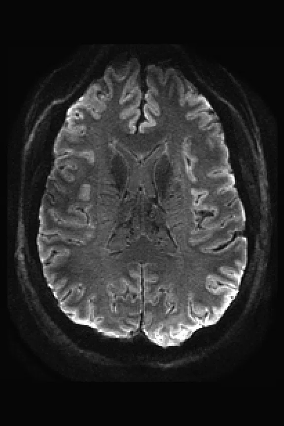
Figure 1.
(A) Mean diffusion-weighted image and the (C) track density image can be acquired with 0.8 mm slices with exquisite detail. The middle image (B) is the directionally encoded color equivalent to FA for CSD model.
B
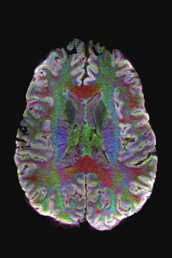
Figure 1.
(A) Mean diffusion-weighted image and the (C) track density image can be acquired with 0.8 mm slices with exquisite detail. The middle image (B) is the directionally encoded color equivalent to FA for CSD model.
C
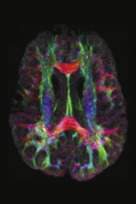
Figure 1.
(A) Mean diffusion-weighted image and the (C) track density image can be acquired with 0.8 mm slices with exquisite detail. The middle image (B) is the directionally encoded color equivalent to FA for CSD model.
A
Figure 2.
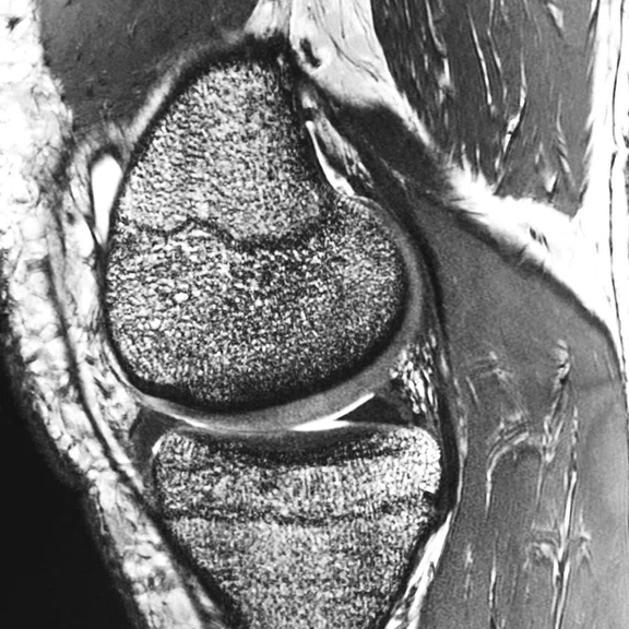
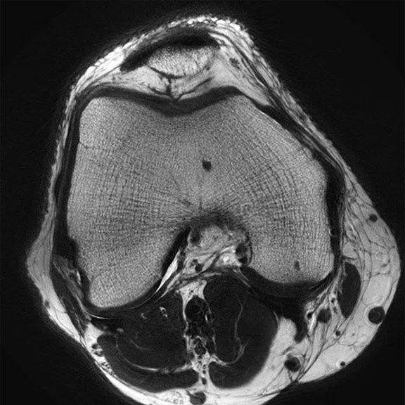
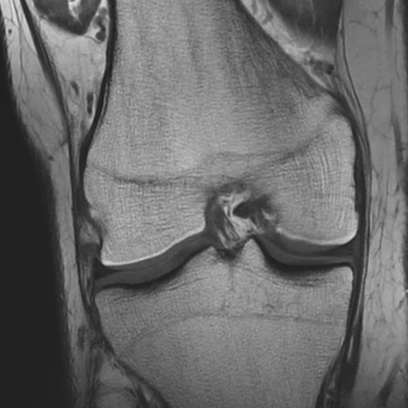
In addition to brain imaging, the SIGNA™ 7.0T is an ideal system for extremity imaging. (A) FIESTA-C, 150µm x 150µm x 1.2 mm, (B) 2D FSE PROPELLER, 293µm x 293µm x 2 mm, and (C) T1 FSE, 312µm x 312µm x 3 mm.
B
Figure 2.
In addition to brain imaging, the SIGNA™ 7.0T is an ideal system for extremity imaging. (A) FIESTA-C, 150µm x 150µm x 1.2 mm, (B) 2D FSE PROPELLER, 293µm x 293µm x 2 mm, and (C) T1 FSE, 312µm x 312µm x 3 mm.
C
Figure 2.
In addition to brain imaging, the SIGNA™ 7.0T is an ideal system for extremity imaging. (A) FIESTA-C, 150µm x 150µm x 1.2 mm, (B) 2D FSE PROPELLER, 293µm x 293µm x 2 mm, and (C) T1 FSE, 312µm x 312µm x 3 mm.
‡‡‡Features that are part of SIGNA™ 7.0T but not cleared for clinical use in the USA or any other country.
A
Figure 3.
With higher field strength, multinuclear spectroscopy is enabled on the SIGNA™ 7.0T. Shown here is a sodium (23Na) acquisition in a subject with glioblastoma multiforme. With multi echo acquisition, we are able to collect an estimate of total sodium and intra- and extra-cellular sodium in the subject. (A) T1 post-contrast, (B) total 23Na, (C) extracellular 23Na and (D) intracellular 23Na.
B
Figure 3.
With higher field strength, multinuclear spectroscopy is enabled on the SIGNA™ 7.0T. Shown here is a sodium (23Na) acquisition in a subject with glioblastoma multiforme. With multi echo acquisition, we are able to collect an estimate of total sodium and intra- and extra-cellular sodium in the subject. (A) T1 post-contrast, (B) total 23Na, (C) extracellular 23Na and (D) intracellular 23Na.
D
Figure 3.
With higher field strength, multinuclear spectroscopy is enabled on the SIGNA™ 7.0T. Shown here is a sodium (23Na) acquisition in a subject with glioblastoma multiforme. With multi echo acquisition, we are able to collect an estimate of total sodium and intra- and extra-cellular sodium in the subject. (A) T1 post-contrast, (B) total 23Na, (C) extracellular 23Na and (D) intracellular 23Na.
C
Figure 3.
With higher field strength, multinuclear spectroscopy is enabled on the SIGNA™ 7.0T. Shown here is a sodium (23Na) acquisition in a subject with glioblastoma multiforme. With multi echo acquisition, we are able to collect an estimate of total sodium and intra- and extra-cellular sodium in the subject. (A) T1 post-contrast, (B) total 23Na, (C) extracellular 23Na and (D) intracellular 23Na.
result
PREVIOUS
${prev-page}
NEXT
${next-page}
Subscribe Now
Manage Subscription
FOLLOW US
contact us • privacy policy • terms & conditions
© 2023 GE HealthCare. GE is a trademark of the General Electric Company used under trademark license.


IN PRACTICE
Scientific discovery meets clinical translation at 7.0T
Scientific discovery meets clinical translation at 7.0T
The promise of ultra-high-field MR imaging for neuro and extremity imaging applications took a significant step forward with the US FDA 510(k) clearance of the SIGNA™ 7.0T‡ from GE Healthcare. The new system brings together proven clinical technologies along with powerful hardware advancements to further propel MR imaging capabilities.
The promise of ultra-high-field MR imaging for neuro and extremity imaging applications took a significant step forward with the US FDA 510(k) clearance of the SIGNA™ 7.0T‡ from GE Healthcare. The new system brings together proven clinical technologies along with powerful hardware advancements to further propel MR imaging capabilities.
Designed to overcome the limitations of many of today’s clinical MR systems, SIGNA™ 7.0T is a powerful new platform for advancing neurological research and clinical translation. Unveiled during the virtual ISMRM 2020 meeting in August, the SIGNA™ 7.0T will be familiar to many GE MR users with the SIGNA™Works productivity platform and Total Digital Imaging (TDI). It brings together the best of GE’s high-strength advanced MR technology: the company’s new and most powerful gradient coil technology, UltraG, the SuperG gradient amplifiers and gradient drivers from the SIGNA™ Premier 3.0T, and the zero boil-off actively shielded 7.0T magnet from the Discovery™ MR950‡‡ research-only 7.0T system.
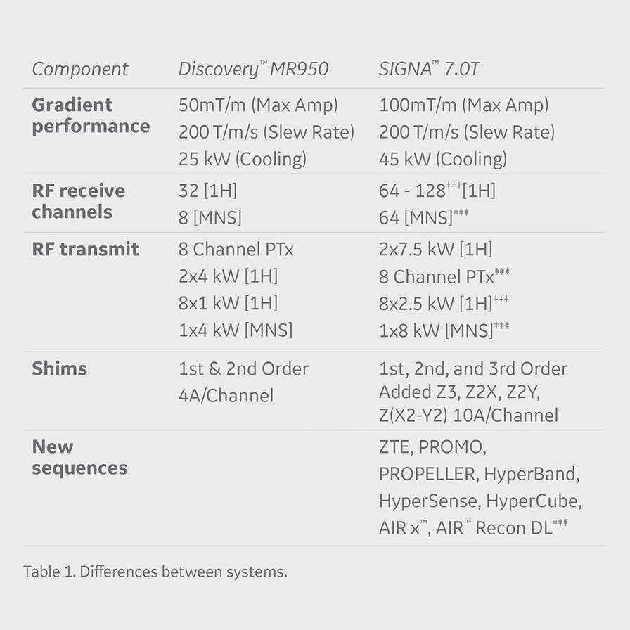
For the past five years, Vincent A. Magnotta, PhD, Professor of Radiology, Division of Neurology, Professor of Psychiatry and Professor of Biomedical Engineering, has conducted research on the Discovery™ MR950, an investigational pre-cursor to the SIGNA™ 7.0T system. According to Dr. Magnotta, the SIGNA™ 7.0T has many of the same capabilities and overall architecture as the Discovery™ MR950 yet it delivers several layers of improvements (Table 1).
"The additional RF capabilities of the SIGNA™ 7.0T are key to improving image quality and enabling multi-nuclear capabilities."
Dr. Vincent A. Magnotta
The RF architecture is designed to provide the power and robustness needed at 7.0T while also enabling new research applications‡‡‡.
UltraG gradients are designed to address the needs of diffusion and higher field strength, as well as overcome the T2 reduction in white matter that occurs at higher field strengths. The hollow, water-cooled conductor provides a sustained 45 kW heat dissipation and enables ultra-low eddy currents, torque and force balancing to minimize the magnet gradient interaction.
At higher resolution, it is necessary to account for distortion resulting from motion, adds Dr. Magnotta.
"As we move to higher resolution imaging, we need ways to account for and minimize even the smallest motion," he explains. "Applications such as ZTE and PROPELLER are relatively immune to patient motion."
These sequences are part of the SIGNA™Works AIR™ Edition, a broad library of applications that have been implemented clinically on GE’s 3.0T and 1.5T MR systems.
Using the ZTE-based T1-weighted sequence, Silenz, Dr. Magnotta and his team were able to significantly reduce blood flow artifacts. They also noticed improvements in image quality near metal in phantom scans with these techniques.
High-resolution diffusion imaging, including higher b-values, is another area where a 7.0T field strength is expected to yield impressive results (Figure 1).
High field strengths also improve MR spectroscopy applications, including scanning almost all the metabolites as well as gamma-aminobutyric acid (GABA) editing. Proton spectroscopy is an important application at 7.0T that requires accurate localization due to the finite RF pulse bandwidth. GE has addressed this with the development of semi-LASER, where adiabatic localization pulses for high localization accuracy for all metabolites.
Since the SIGNA™ 7.0T will include SIGNA™Works AIR™ Edition, users will have access to the AIR™ family of products, such as AIR x™ for reliably and reproducibly positioned scan planes, and advanced techniques such as HyperBand, HyperSense and HyperCube.
Advanced research capabilities of SIGNA 7.0T‡‡‡
In addition to offering an array of powerful clinical applications, the SIGNA™ 7.0T system further enables advanced research capabilities. By providing a multitude of uses, researchers and clinicians can extract higher information content of 7.0T images to address basic and scientific questions, make scientific breakthroughs and undertake clinical translational research. One system meets the needs of two communities.
The 64-channel TDI is scalable to 128 channels for use in both proton and multi-nuclear applications. In research mode, the open architecture supports third-party coil integration.
The research capabilities of SIGNA™ 7.0T are designed to enable multi-nuclear imaging capabilities, such as phosphorus spectroscopy, where the additional power delivers an SNR boost at metabolites that may be distal to the center frequency, or in sodium imaging with a multiecho acquisition to be able to estimate total sodium and intra and extra cellular sodium in the subject (Figure 3).
Figure 3.
With higher field strength, multinuclear spectroscopy is enabled on the SIGNA™ 7.0T. Shown here is a sodium (23Na) acquisition in a subject with glioblastoma multiforme. With multi echo acquisition, we are able to collect an estimate of total sodium and intra- and extra-cellular sodium in the subject. (A) T1 post-contrast, (B) total 23Na, (C) extracellular 23Na and (D) intracellular 23Na.
AIR™ Recon DL, a pioneering, deep-learning-based reconstruction algorithm that improves SNR and image sharpness, enabling shorter scan times, will be available with the research-only capabilities.


