T2 SSFSE, DWI and dynamic LAVA depict a rapidly enhancing liver lesion. Images courtesy of Hospital Universitario Quirón Salud, Madrid, Spain
T2 SSFSE, DWI and dynamic LAVA depict a rapidly enhancing liver lesion. Images courtesy of Hospital Universitario Quirón Salud, Madrid, Spain
Pre-contrast enhancement. Images courtesy of Hospital Universitario Quirón Salud, Madrid, Spain
Arterial phase. Images courtesy of Hospital Universitario Quirón Salud, Madrid, Spain
Portal phase. Images courtesy of Hospital Universitario Quirón Salud, Madrid, Spain
Late phase. Images courtesy of Hospital Universitario Quirón Salud, Madrid, Spain
Patient with Crohn’s disease demonstrating post-contrast enhancement of the terminal ileum. MRA was obtained in the same exam during the early arterial phase of a DISCO LAVA sequence. Images courtesy of Hospital Universitario Quirón Salud, Madrid, Spain
Patient with Crohn’s disease demonstrating post-contrast enhancement of the terminal ileum. MRA was obtained in the same exam during the early arterial phase of a DISCO LAVA sequence. Images courtesy of Hospital Universitario Quirón Salud, Madrid, Spain
Delayed phase axial LAVA acquisition 15 min. post Gd-EOB-DTPA. Scan time was 0:20 min. Images courtesy of Osaka University Hospital, Osaka, Japan
Delayed phase axial LAVA acquisition 15 min. post Gd-EOB-DTPA. Scan time was 0:20 min. Images courtesy of Osaka University Hospital, Osaka, Japan
Coronal MPR of an axial LAVA acquisition. Images courtesy of Osaka University Hospital, Osaka, Japan


result
PREVIOUS
${prev-page}
NEXT
${next-page}
Subscribe Now
Manage Subscription
FOLLOW US
Contact Us • Cookie Preferences • Privacy Policy • California Privacy PolicyDo Not Sell or Share My Personal Information • Terms & Conditions • Security
© 2024 GE HealthCare. GE is a trademark of General Electric Company. Used under trademark license.
SPOTLIGHT
SIGNA™Works AIR™ Edition gallery:
Abdominal imaging
SIGNA™Works AIR™ Edition gallery:
Abdominal imaging
All images in this gallery were acquired using AIR™


T2 SSFSE, DWI and dynamic LAVA depict a rapidly enhancing liver lesion. Images courtesy of Hospital Universitario Quirón Salud, Madrid, Spain

Pre-contrast enhancement

Arterial phase

Portal phase

Late phase

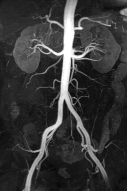
Patient with Crohn’s disease demonstrating post-contrast enhancement of the terminal ileum. MRA was obtained in the same exam during the early arterial phase of a DISCO LAVA sequence. Images courtesy of Hospital Universitario Quirón Salud, Madrid, Spain


Delayed phase axial LAVA acquisition 15 min. post Gd-EOB-DTPA. Scan time was 0:20 min. Images courtesy of Osaka University Hospital, Osaka, Japan

Coronal MPR of an axial LAVA acquisition. Images courtesy of Osaka University Hospital, Osaka, Japan