Figure 4.
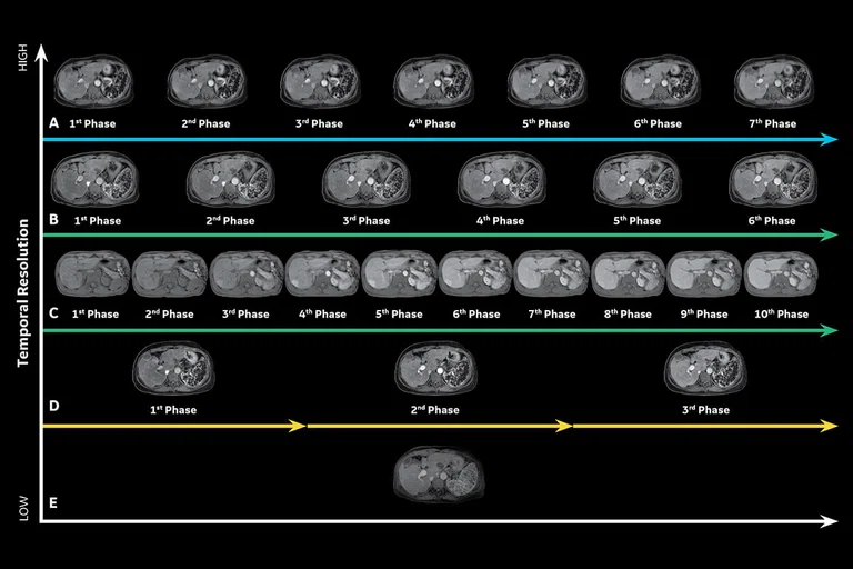
GE’s complete 3D dynamic abdominal MR portfolio. (A) DISCO with HyperSense, 1 x 1.6 x 3.6 mm, temporal resolution 3 sec., scan time 20 sec.; (B) DISCO, 1 x 1.6 x 3.6 mm, temporal resolution 4 sec., scan time 24 sec.; (C) DISCO Star, 1.2 x 1.2 x 2 mm, temporal resolution 6 sec., scan time 24 sec.; (D) LAVA or LAVA Flex with HyperSense, 1 x 1.6 x 3.6 mm, scan time 8 sec. x 3; (E) LAVA or LAVA Flex, 1 x 2 x 5 mm, scan time 14 sec. Images courtesy of Yamanashi Medical Center, Japan.
A
Figure 3.
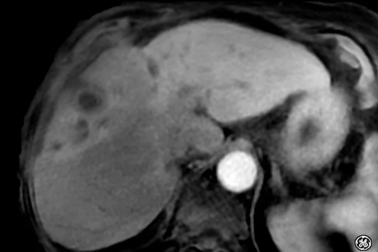
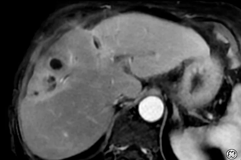
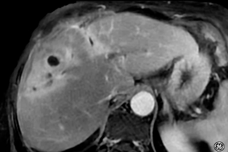
Same patient as Figure 2, Nov. 2020 free-breathing 3D dynamic liver exam. (A-D) Dynamic DISCO Star acquisition in the (A) pre, (B) early arterial, (C) arterial and (D) portal phases, 1.2 x 1.2 x 2 mm, 17 sec. (E) LAVA Star delayed acquisition, 1.2 x 1.2 x 2 mm, 3:21 min. Images courtesy of Acibadem Maslak Hospital, Istanbul, Turkey.
B
Figure 3.
Same patient as Figure 2, Nov. 2020 free-breathing 3D dynamic liver exam. (A-D) Dynamic DISCO Star acquisition in the (A) pre, (B) early arterial, (C) arterial and (D) portal phases, 1.2 x 1.2 x 2 mm, 17 sec. (E) LAVA Star delayed acquisition, 1.2 x 1.2 x 2 mm, 3:21 min. Images courtesy of Acibadem Maslak Hospital, Istanbul, Turkey.
C
Figure 3.
Same patient as Figure 2, Nov. 2020 free-breathing 3D dynamic liver exam. (A-D) Dynamic DISCO Star acquisition in the (A) pre, (B) early arterial, (C) arterial and (D) portal phases, 1.2 x 1.2 x 2 mm, 17 sec. (E) LAVA Star delayed acquisition, 1.2 x 1.2 x 2 mm, 3:21 min. Images courtesy of Acibadem Maslak Hospital, Istanbul, Turkey.
D
Figure 3.
Same patient as Figure 2, Nov. 2020 free-breathing 3D dynamic liver exam. (A-D) Dynamic DISCO Star acquisition in the (A) pre, (B) early arterial, (C) arterial and (D) portal phases, 1.2 x 1.2 x 2 mm, 17 sec. (E) LAVA Star delayed acquisition, 1.2 x 1.2 x 2 mm, 3:21 min. Images courtesy of Acibadem Maslak Hospital, Istanbul, Turkey.
E
Figure 3.
Same patient as Figure 2, Nov. 2020 free-breathing 3D dynamic liver exam. (A-D) Dynamic DISCO Star acquisition in the (A) pre, (B) early arterial, (C) arterial and (D) portal phases, 1.2 x 1.2 x 2 mm, 17 sec. (E) LAVA Star delayed acquisition, 1.2 x 1.2 x 2 mm, 3:21 min. Images courtesy of Acibadem Maslak Hospital, Istanbul, Turkey.
A
Figure 1.
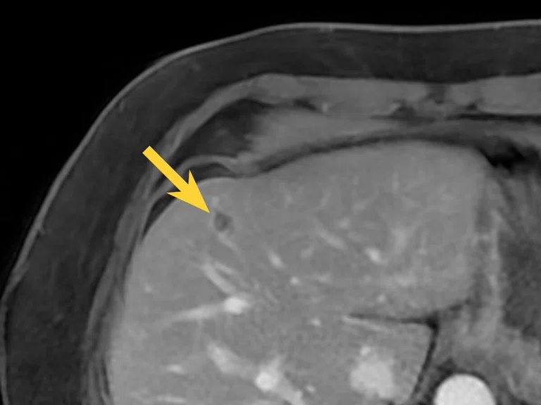
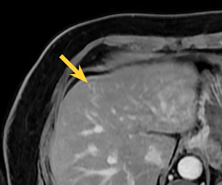
Due to motion during breath-hold, a lesion (arrow) is only visible on (A) LAVA Star compared to (B) LAVA.
B
Figure 1.
Due to motion during breath-hold, a lesion (arrow) is only visible on (A) LAVA Star compared to (B) LAVA.
A
Figure 2.
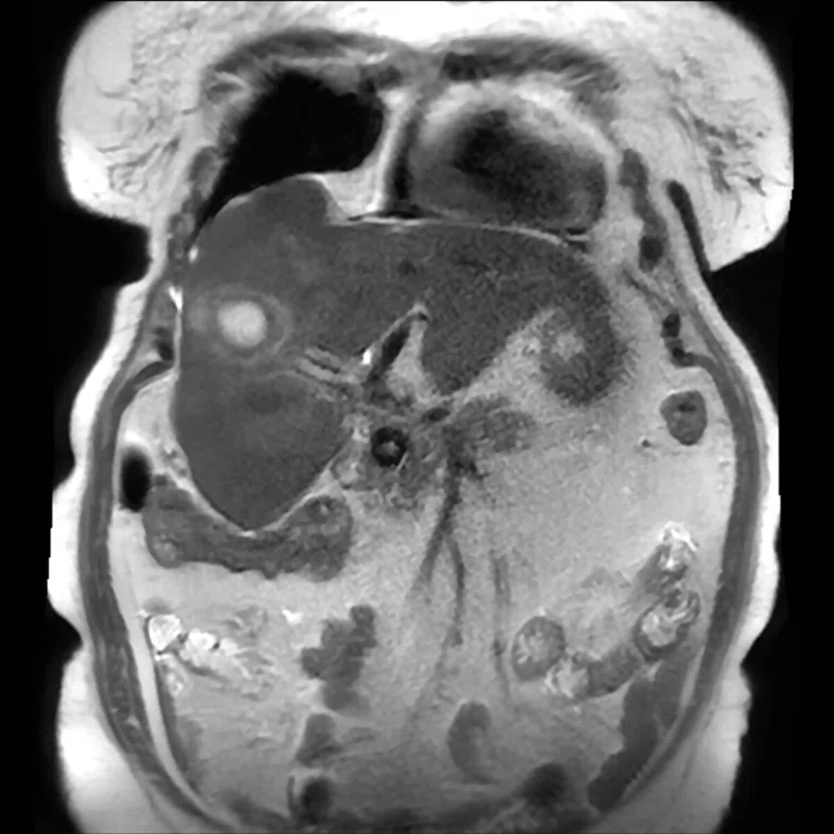
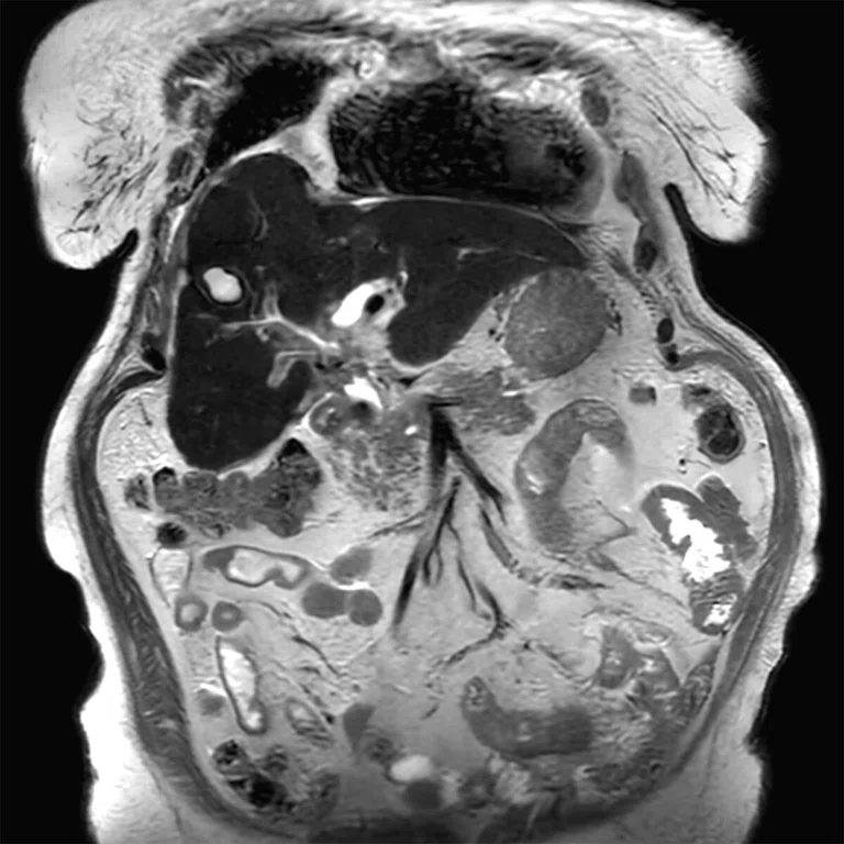
An 80-year-old female with a gallbladder tumor was scanned in Sept. 2020 and came back for a follow-up exam to evaluate R-lobe anterior abscesses after undergoing transarterial radioembolization in Nov. 2020. In between these two exams, SIGNA™Works AIR™ IQ Edition was installed, which includes AIR™ Recon DL and DISCO Star. (A) Sept. 2020 T2 SSFSE, (B) Nov. 2020 T2 SSFSE with AIR™ Recon DL, (C) Sept. 2020 DISCO with Auto Navigator and (D) Nov. 2020 DISCO Star. Images courtesy of Acibadem Maslak Hospital, Istanbul, Turkey.
B
Figure 2.
An 80-year-old female with a gallbladder tumor was scanned in Sept. 2020 and came back for a follow-up exam to evaluate R-lobe anterior abscesses after undergoing transarterial radioembolization in Nov. 2020. In between these two exams, SIGNA™Works AIR™ IQ Edition was installed, which includes AIR™ Recon DL and DISCO Star. (A) Sept. 2020 T2 SSFSE, (B) Nov. 2020 T2 SSFSE with AIR™ Recon DL, (C) Sept. 2020 DISCO with Auto Navigator and (D) Nov. 2020 DISCO Star. Images courtesy of Acibadem Maslak Hospital, Istanbul, Turkey.
C
Figure 2.
An 80-year-old female with a gallbladder tumor was scanned in Sept. 2020 and came back for a follow-up exam to evaluate R-lobe anterior abscesses after undergoing transarterial radioembolization in Nov. 2020. In between these two exams, SIGNA™Works AIR™ IQ Edition was installed, which includes AIR™ Recon DL and DISCO Star. (A) Sept. 2020 T2 SSFSE, (B) Nov. 2020 T2 SSFSE with AIR™ Recon DL, (C) Sept. 2020 DISCO with Auto Navigator and (D) Nov. 2020 DISCO Star. Images courtesy of Acibadem Maslak Hospital, Istanbul, Turkey.
D
Figure 2.
An 80-year-old female with a gallbladder tumor was scanned in Sept. 2020 and came back for a follow-up exam to evaluate R-lobe anterior abscesses after undergoing transarterial radioembolization in Nov. 2020. In between these two exams, SIGNA™Works AIR™ IQ Edition was installed, which includes AIR™ Recon DL and DISCO Star. (A) Sept. 2020 T2 SSFSE, (B) Nov. 2020 T2 SSFSE with AIR™ Recon DL, (C) Sept. 2020 DISCO with Auto Navigator and (D) Nov. 2020 DISCO Star. Images courtesy of Acibadem Maslak Hospital, Istanbul, Turkey.
2. Andre JB, Bresnahan BW, Mossa-Basha M, Hoff MN, Smith CP, Anzai Y, Cohen WA. Toward Quantifying the Prevalence, Severity, and Cost Associated With Patient Motion During Clinical MR Examinations. J Am Coll Radiol. 2015 Jul;12(7):689-95. doi: 10.1016/j.jacr.2015.03.007. Epub 2015 May 9. PMID: 25963225.
1. IMV 2020 MR Market Outlook Report.
result


PREVIOUS
${prev-page}
NEXT
${next-page}
Subscribe Now
Manage Subscription
FOLLOW US
Contact Us • Cookie Preferences • Privacy Policy • California Privacy PolicyDo Not Sell or Share My Personal Information • Terms & Conditions • Security
© 2024 GE HealthCare. GE is a trademark of General Electric Company. Used under trademark license.
TECH TRENDS
When timing is everything: 3D dynamic T1 scans in body imaging
When timing is everything: 3D dynamic T1 scans in body imaging
by Steve Lawson, RT(R)(MR), Global MR Clinical Marketing Manager and Heide Harris, RT(R)(MR), Global Product Marketing Director, MR Applications & Visualization, GE Healthcare
Due to its superior ability to image soft tissue in multiple planes, dynamic 3D imaging of the body is a critical exam in the diagnostic work-up of many patients as it helps differentiate between benign and malignant lesions, and provides information used for staging cancer. Dynamic contrast enhanced (DCE) imaging is derived from the multiple serial images that are collected after the injection of contrast.
Dynamic MR is often used for evaluating solid tumors and blood vessels as well as identifying tumor enhancement. Often, patients will first undergo a CT or ultrasound. If a lesion is depicted, then MR is used to examine the different characteristics of the lesion based on how it enhances. For example, is the liver lesion primary cancer, metastatic cancer or a hemangioma? How is the disease advancing? Dynamic MR can help provide these answers.
Further, some diseases may be reversible or, at a minimum, stopped from advancing. An important example is fatty liver disease, a progressive disease that leads to inflammation and tissue stiffness followed by scarring (cirrhosis) and eventually liver failure or hepatocellular carcinoma. If the condition is caught during the earlier stages of inflammation and liver stiffness, then it may be reversible with drug therapy.
Prostate MR has become one of the fastest growing procedures1 as it’s a critical exam for diagnosing and monitoring cancer progression, particularly if the patient’s care plan involves active surveillance for low-risk cancers or surgical intervention. For breast cancer, the dynamic 3D sequence is one of the most critical for evaluating the extent of disease in women as a follow up to abnormal findings from a mammogram. Throughout the rest of the abdomen – pancreas, kidneys, bladder, adrenal glands and pelvis - contrast is used to differentiate lesions at multiple points in time to determine tissue characterization.
Each area in the body has a specific time sequence to acquire the data. For instance, liver imaging should capture the early, mid and late arterial phase, followed by portal then delayed. The most critical piece is the timing of the bolus arrival because the technologist has only one opportunity to acquire the dynamic scan. If a patient breathes or moves during the series, the study may be deemed undiagnostic and they would have to be called back for another exam.
So let’s take a look at the different abdominal dynamic 3D MR imaging options available from GE Healthcare today.
LAVA and LAVA Flex
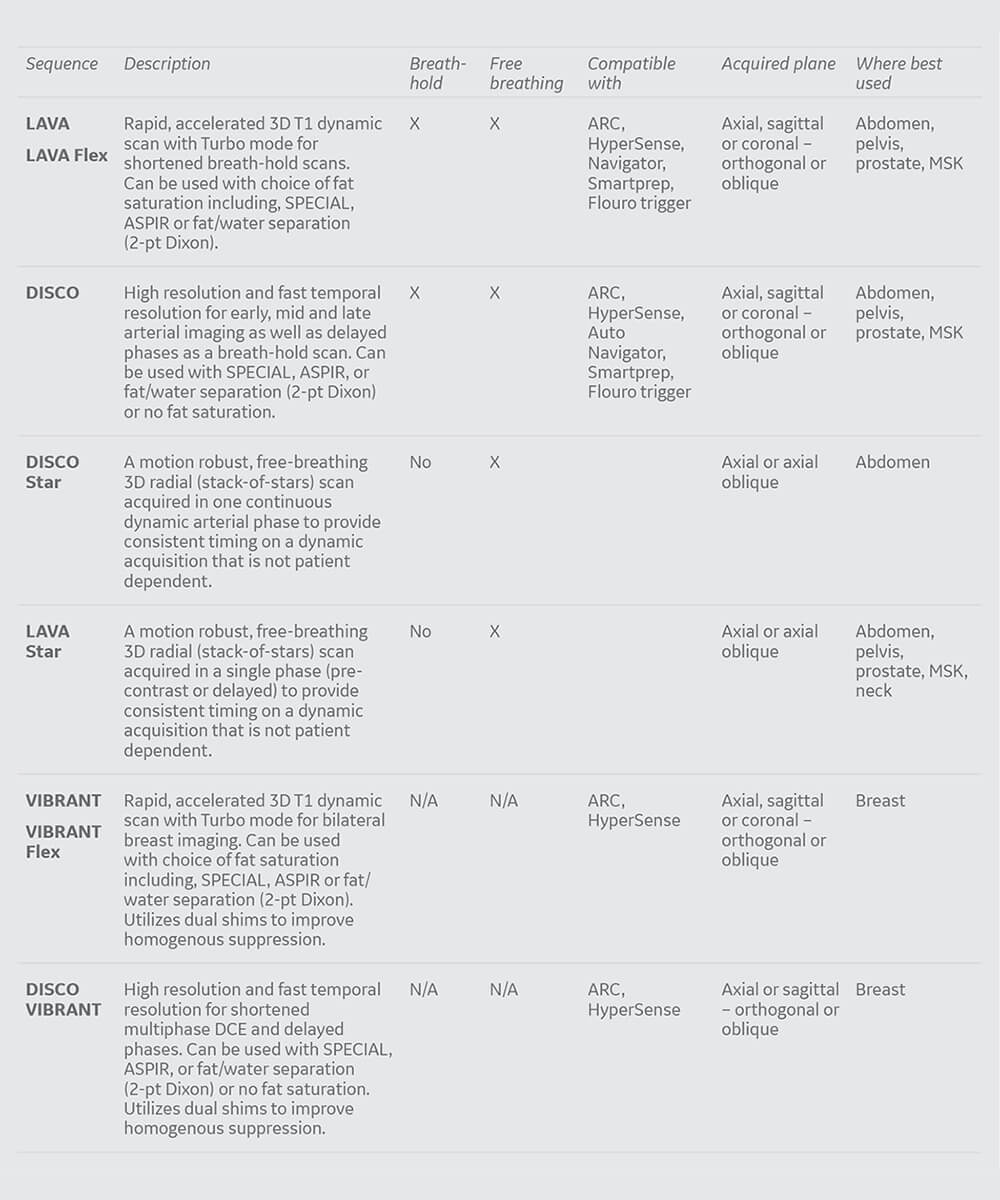
One of the original dynamic 3D T1 sequences is LAVA. LAVA is a rapid, accelerated dynamic scan that uses SPECIAL as a fat saturation technique, which is important because it suppresses the background tissues for better visualization of the contrast uptake in the anatomy. Another dynamic sequence that can be used is LAVA Flex. Like LAVA, it is a rapid, accelerated dynamic scan that uses a fat-water separation technique added to the core acquisition for robust, homogenous suppression of the tissue. Both of these can use Auto Navigator, ARC and HyperSense to further reduce scan times, which is important due to breath-hold requirements. They can be enhanced with Turbo mode in order to further shorten scan times. Typically, LAVA and LAVA Flex scans are approximately 15-19 seconds per phase.
Figure 2.
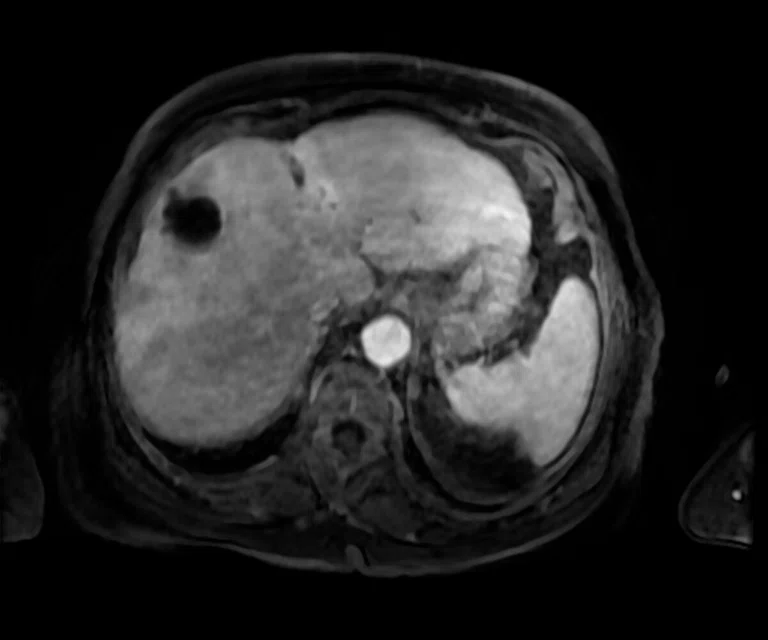
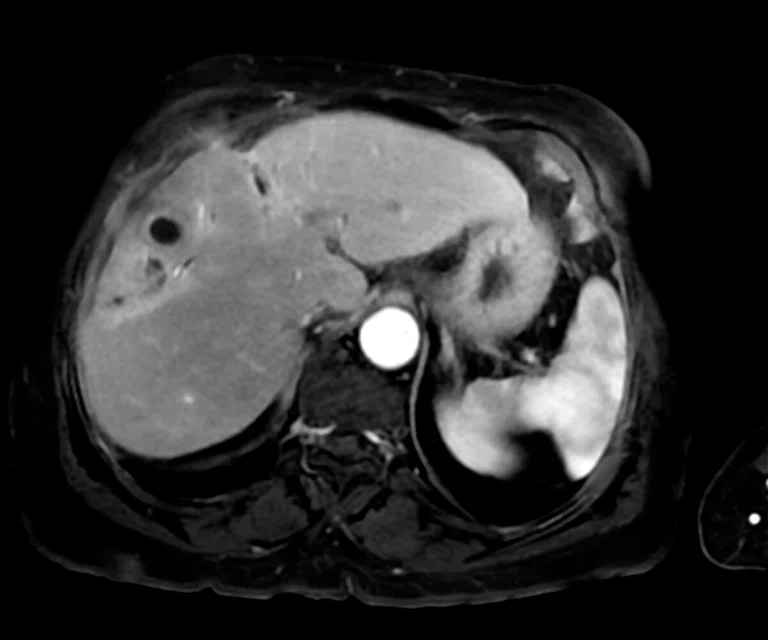
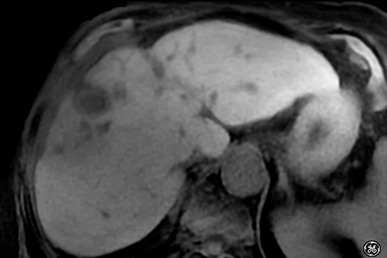
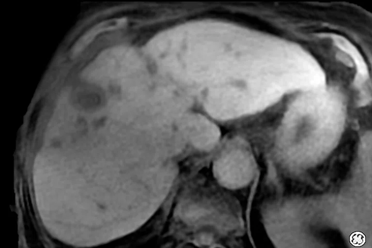
An 80-year-old female with a gallbladder tumor was scanned in Sept. 2020 and came back for a follow-up exam to evaluate R-lobe anterior abscesses after undergoing transarterial radioembolization in Nov. 2020. In between these two exams, SIGNA™Works AIR™ IQ Edition was installed, which includes AIR™ Recon DL and DISCO Star. (A) Sept. 2020 T2 SSFSE, (B) Nov. 2020 T2 SSFSE with AIR™ Recon DL, (C) Sept. 2020 DISCO with Auto Navigator and (D) Nov. 2020 DISCO Star. Images courtesy of Acibadem Maslak Hospital, Istanbul, Turkey.
DISCO
For acquisitions that require more temporal phases in a faster scan time without sacrificing resolution, DISCO is the dynamic scan of choice. With its ultrafast temporal resolution, DISCO acquires multiple phases as low as 3 seconds per phase with the ability to acquire more time points for kinetic information from a single scan. It’s the key sequence for obtaining Ktrans information (GE’s GenIQ) as it requires a temporal phase of 6 seconds or less. Ktrans measures the accumulation of a gadolinium-based contrast agent in the extravascular-extracellular space. DISCO is also flexible in terms of fat suppression techniques, as it can use SPECIAL, Flex or no suppression at all.
DISCO Star and LAVA Star
In the SIGNA™Works AIR™ IQ Edition software release, we’ve taken dynamic MR a step further with a complete free-breathing 3D T1 sequence. For patients who are not able to hold their breath, such as pediatrics, elderly or those who are critically ill, DISCO Star offers a solution to acquire a multi-phase, dynamic sequence that is motion robust and free-breathing for better patient compliance. This 3D radial (stack-of-stars) sequence acquires one continuous dynamic arterial phase for consistent, worry-free timing of the contrast arrival, which will vary from patient to patient depending on age, pathology and condition. As previously mentioned, timing is critical for assessing lesions. Because of how DISCO Star acquires data, the potential to miss the bolus is minimized, and it improves the consistency of contrast arrival regardless of the patient’s condition.
Though technically not a dynamic sequence, LAVA Star is critical for the pre-contrast or delayed sequences after the contrast has washed out. It provides a consistent, worry-free image quality regardless of the patient’s condition or ability to breath-hold. LAVA Star compensates for motion without the need for monitoring the patient’s breathing with Auto Navigator tracking or an external bellows to trigger the acquisition based on their breathing.
VIBRANT, VIBRANT Flex and DISCO VIBRANT
The dynamic sequence is one of the most important in breast imaging. VIBRANT is a dedicated sequence for T1-weighted DCE imaging of simultaneous, high-definition, fat-suppressed bilateral breast imaging in either the axial or sagittal plane. At times, fat saturation can be a challenge as the breast is surrounded by air in the lungs. An alternative to fat saturation is VIBRANT Flex. Much like LAVA Flex, it provides robust suppression of the tissue around a particularly challenging area to scan. DISCO VIBRANT brings the benefit of DISCO for excellent spatial and temporal resolution imaging to the breast along with the ability to gather more time points in a single dynamic volume.
Figure 3.
Same patient as Figure 2, Nov. 2020 free-breathing 3D dynamic liver exam. (A-D) Dynamic DISCO Star acquisition in the (A) pre, (B) early arterial, (C) arterial and (D) portal phases, 1.2 x 1.2 x 2 mm, 17 sec. (E) LAVA Star delayed acquisition, 1.2 x 1.2 x 2 mm, 3:21 min. Images courtesy of Acibadem Maslak Hospital, Istanbul, Turkey.
When motion is a challenge
One of the challenges with abdominal imaging is that it relies on breath-holding for many of the sequences. Breath-holding has long been the gold standard in body imaging, however, not all patients can comply with breath-holds, particularly those who are elderly, acutely ill or sedated. Also, breath-holding can lead to longer scan times, particularly if a sequence has to be repeated due to a failed breath-hold acquisition. According to one study, 20% of MR exams required a repeat sequence, which lengthens the patient’s exam as well as disrupts the department schedule2. GE has a number of options to compensate for breathing challenges, particularly for the critical dynamic T1 series with solutions that are available across the company’s MR portfolio.
Both DISCO Star, used for dynamic multiphase scans, and LAVA Star, used for the delay sequences, help overcome the issue of motion when a patient is unable to comply with breath-holds. These solutions also provide the ability to visualize smaller lesions that may otherwise be obscured by motion.
Auto Navigator can be used for all of GE’s abdominal imaging sequences, such as T2, DWI and single shot FSE. It automatically places the tracker at the dome of the liver and doesn’t rely on any external hardware. Auto Navigator then synchronizes the acquisition with the patient’s breathing pattern, minimizing respiratory ghosting artifacts. Real-time adjustment allows threshold levels to be modified during the acquisition, eliminating failures due to changes in the patient’s respiration that may occur as the patient relaxes or even falls asleep during the exam.
HyperSense, GE’s compressed sensing technique, is another tool in the body imaging arsenal. It doesn’t have a direct impact on motion. However, HyperSense, enables shorter scan times without compromising spatial resolution or slice thickness. Shorter scan times are also easier on the patient, particularly for breath-hold sequences. In the case of dynamic imaging, it allows for the acquisition of more dynamic information with less time or better resolution. HyperSense and Auto Navigator are compatible with each other, as well as the different body sequences described in this article.
Given there is an increasing need for the detailed information provided by body MR imaging, having the right tools to complete the exam and provide a better experience for the technologist performing the exam, the radiologist reading it and the patient undergoing it, benefits everyone involved. These tools enable more consistency across MR exams because the technologist won’t have to adjust the parameters to accommodate a more challenging patient or reduce the resolution or signal in order to scan faster.
All of these solutions — LAVA, LAVA Flex, LAVA Star, DISCO, DISCO Star, VIBRANT, VIBRANT Flex, DISCO VIBRANT, HyperSense and Auto Navigator — are available across the entire GE MR system portfolio.
Figure 4.
GE’s complete 3D dynamic abdominal MR portfolio. (A) DISCO with HyperSense, 1 x 1.6 x 3.6 mm, temporal resolution 3 sec., scan time 20 sec.; (B) DISCO, 1 x 1.6 x 3.6 mm, temporal resolution 4 sec., scan time 24 sec.; (C) DISCO Star, 1.2 x 1.2 x 2 mm, temporal resolution 6 sec., scan time 24 sec.; (D) LAVA or LAVA Flex with HyperSense, 1 x 1.6 x 3.6 mm, scan time 8 sec. x 3; (E) LAVA or LAVA Flex, 1 x 2 x 5 mm, scan time 14 sec. Images courtesy of Yamanashi Medical Center, Japan.


