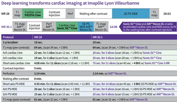
Figure 1.
Table demonstrating the total examination time difference for CMR before and after Sonic DL™ Cine implementation (MR 28 vs. MR 30.1) on SIGNA™ Hero at Imapôle Lyon Villeurbanne. BH=breath hold; BH instruction=10 sec.
A
Figure 2.
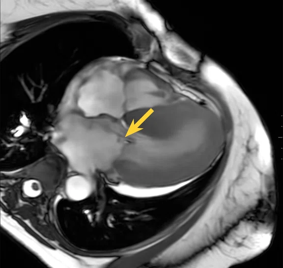
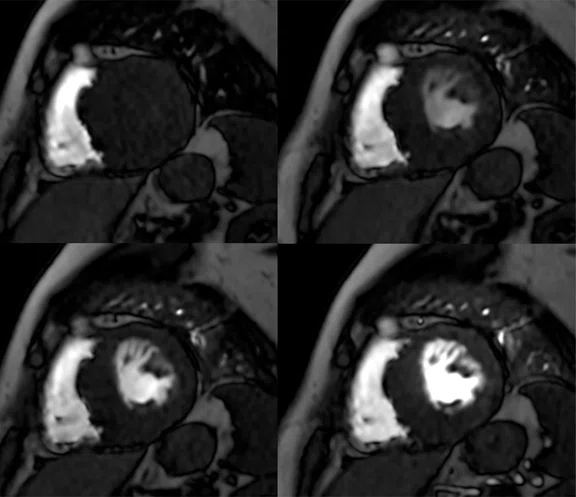
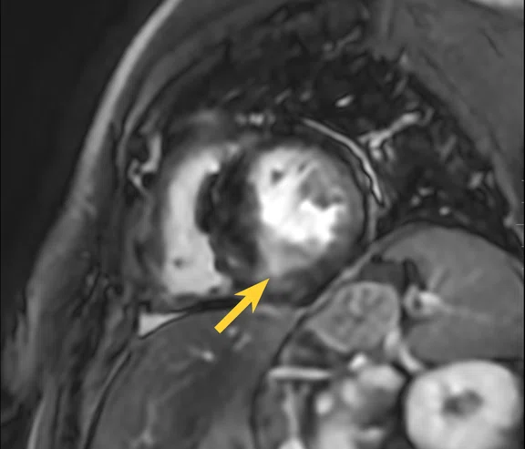
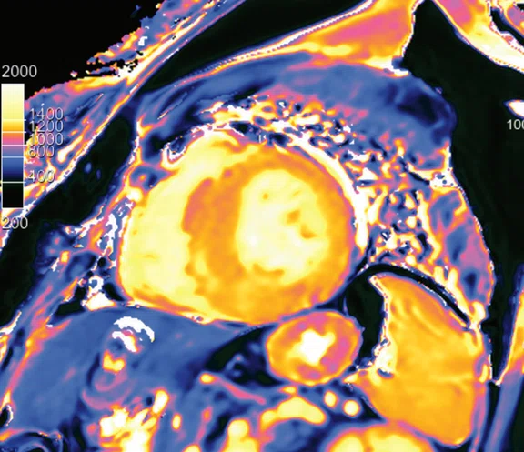
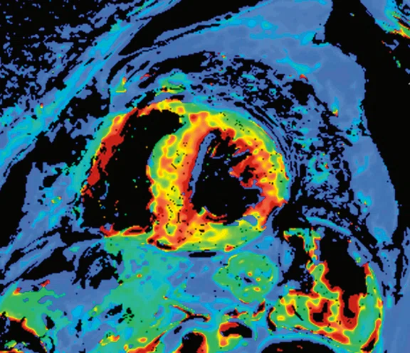
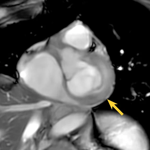
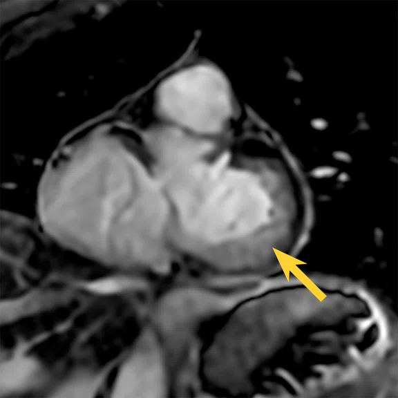
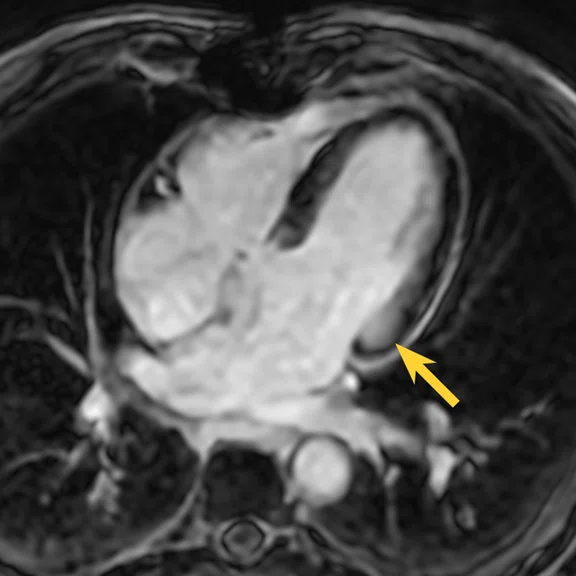
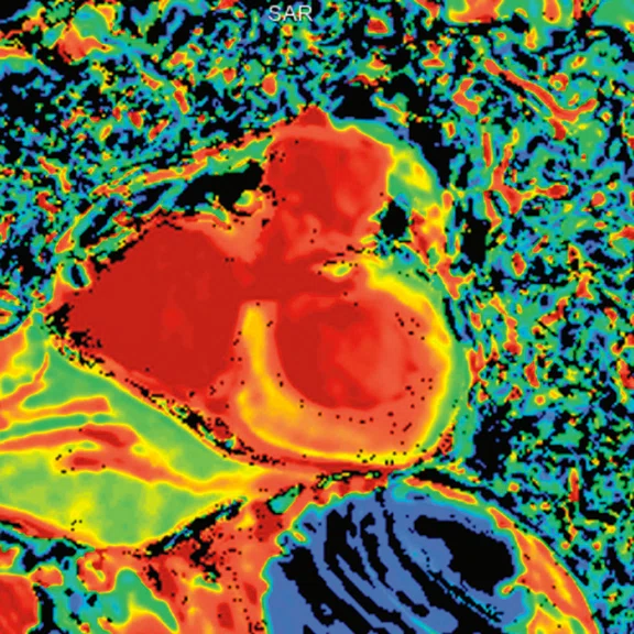
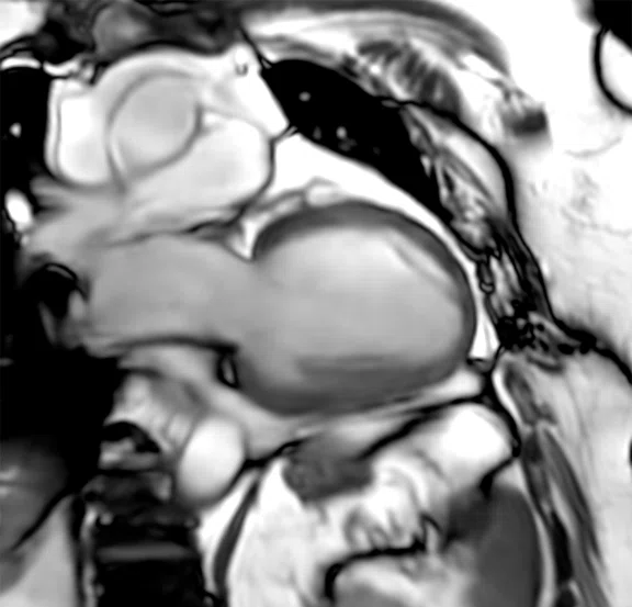
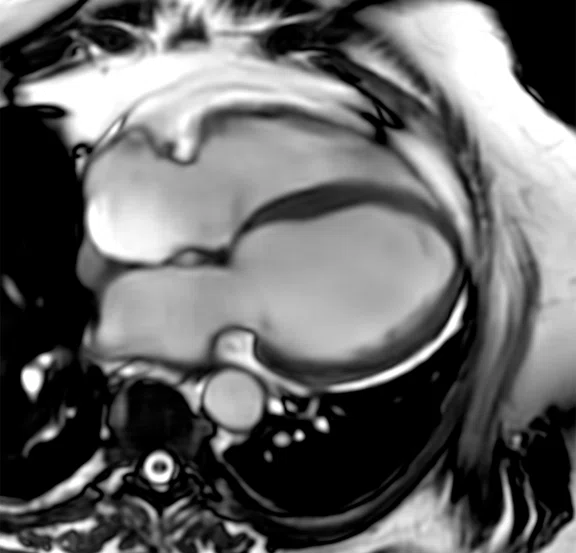
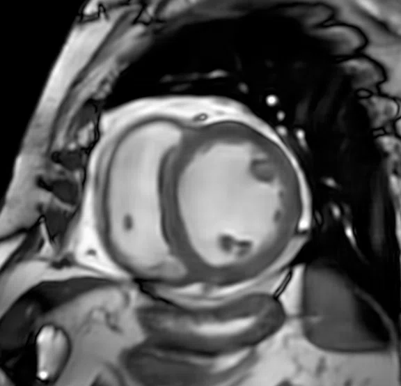
Case 1, patient with suspected hypertrophic cardiomyopathy. High-resolution imaging was performed using Sonic DL™ Cine, AIR™ Recon DL and AIR™ Multi-Purpose 21ch Coil (large). (A) Breath-hold 2-chamber Sonic DL™ Cine 1.6 x 1.6 x 8 mm, 3 locations with 30 phases per location, 23 sec. (B) Breath-hold short axis Sonic DL™ Cine, 1.6 x 1.6 x 8 mm, 14 locations with 30 phases per location, 1:05 min. (C, D) Breath-hold 4-chamber Sonic DL™ Cine, 1.6 x 1.6 x 8 mm, 3 locations with 30 phases per location, 23 sec. (E) Short axis perfusion with MoCo and AIR™ Recon DL, 3 x 2.4 x 8 mm, 4 locations with 80 phases per location, 1:24 min. (F) short axis PS MDE with AIR™ Recon DL, 2.9 x 2.9 x 8 mm, 18 slices, 45 sec. T1 mapping: (G)T1 Native and (H) ECV map with AIR™ Recon DL, 2.4 x 2.9 x 8 mm, 3 slices, 30 sec.
B
Figure 2.
Case 1, patient with suspected hypertrophic cardiomyopathy. High-resolution imaging was performed using Sonic DL™ Cine, AIR™ Recon DL and AIR™ Multi-Purpose 21ch Coil (large). (A) Breath-hold 2-chamber Sonic DL™ Cine 1.6 x 1.6 x 8 mm, 3 locations with 30 phases per location, 23 sec. (B) Breath-hold short axis Sonic DL™ Cine, 1.6 x 1.6 x 8 mm, 14 locations with 30 phases per location, 1:05 min. (C, D) Breath-hold 4-chamber Sonic DL™ Cine, 1.6 x 1.6 x 8 mm, 3 locations with 30 phases per location, 23 sec. (E) Short axis perfusion with MoCo and AIR™ Recon DL, 3 x 2.4 x 8 mm, 4 locations with 80 phases per location, 1:24 min. (F) short axis PS MDE with AIR™ Recon DL, 2.9 x 2.9 x 8 mm, 18 slices, 45 sec. T1 mapping: (G)T1 Native and (H) ECV map with AIR™ Recon DL, 2.4 x 2.9 x 8 mm, 3 slices, 30 sec.
C
Figure 2.
Case 1, patient with suspected hypertrophic cardiomyopathy. High-resolution imaging was performed using Sonic DL™ Cine, AIR™ Recon DL and AIR™ Multi-Purpose 21ch Coil (large). (A) Breath-hold 2-chamber Sonic DL™ Cine 1.6 x 1.6 x 8 mm, 3 locations with 30 phases per location, 23 sec. (B) Breath-hold short axis Sonic DL™ Cine, 1.6 x 1.6 x 8 mm, 14 locations with 30 phases per location, 1:05 min. (C, D) Breath-hold 4-chamber Sonic DL™ Cine, 1.6 x 1.6 x 8 mm, 3 locations with 30 phases per location, 23 sec. (E) Short axis perfusion with MoCo and AIR™ Recon DL, 3 x 2.4 x 8 mm, 4 locations with 80 phases per location, 1:24 min. (F) short axis PS MDE with AIR™ Recon DL, 2.9 x 2.9 x 8 mm, 18 slices, 45 sec. T1 mapping: (G)T1 Native and (H) ECV map with AIR™ Recon DL, 2.4 x 2.9 x 8 mm, 3 slices, 30 sec.
D
Figure 2.
Case 1, patient with suspected hypertrophic cardiomyopathy. High-resolution imaging was performed using Sonic DL™ Cine, AIR™ Recon DL and AIR™ Multi-Purpose 21ch Coil (large). (A) Breath-hold 2-chamber Sonic DL™ Cine 1.6 x 1.6 x 8 mm, 3 locations with 30 phases per location, 23 sec. (B) Breath-hold short axis Sonic DL™ Cine, 1.6 x 1.6 x 8 mm, 14 locations with 30 phases per location, 1:05 min. (C, D) Breath-hold 4-chamber Sonic DL™ Cine, 1.6 x 1.6 x 8 mm, 3 locations with 30 phases per location, 23 sec. (E) Short axis perfusion with MoCo and AIR™ Recon DL, 3 x 2.4 x 8 mm, 4 locations with 80 phases per location, 1:24 min. (F) short axis PS MDE with AIR™ Recon DL, 2.9 x 2.9 x 8 mm, 18 slices, 45 sec. T1 mapping: (G)T1 Native and (H) ECV map with AIR™ Recon DL, 2.4 x 2.9 x 8 mm, 3 slices, 30 sec.
E
Figure 2.
Case 1, patient with suspected hypertrophic cardiomyopathy. High-resolution imaging was performed using Sonic DL™ Cine, AIR™ Recon DL and AIR™ Multi-Purpose 21ch Coil (large). (A) Breath-hold 2-chamber Sonic DL™ Cine 1.6 x 1.6 x 8 mm, 3 locations with 30 phases per location, 23 sec. (B) Breath-hold short axis Sonic DL™ Cine, 1.6 x 1.6 x 8 mm, 14 locations with 30 phases per location, 1:05 min. (C, D) Breath-hold 4-chamber Sonic DL™ Cine, 1.6 x 1.6 x 8 mm, 3 locations with 30 phases per location, 23 sec. (E) Short axis perfusion with MoCo and AIR™ Recon DL, 3 x 2.4 x 8 mm, 4 locations with 80 phases per location, 1:24 min. (F) short axis PS MDE with AIR™ Recon DL, 2.9 x 2.9 x 8 mm, 18 slices, 45 sec. T1 mapping: (G)T1 Native and (H) ECV map with AIR™ Recon DL, 2.4 x 2.9 x 8 mm, 3 slices, 30 sec.
F
Figure 2.
Case 1, patient with suspected hypertrophic cardiomyopathy. High-resolution imaging was performed using Sonic DL™ Cine, AIR™ Recon DL and AIR™ Multi-Purpose 21ch Coil (large). (A) Breath-hold 2-chamber Sonic DL™ Cine 1.6 x 1.6 x 8 mm, 3 locations with 30 phases per location, 23 sec. (B) Breath-hold short axis Sonic DL™ Cine, 1.6 x 1.6 x 8 mm, 14 locations with 30 phases per location, 1:05 min. (C, D) Breath-hold 4-chamber Sonic DL™ Cine, 1.6 x 1.6 x 8 mm, 3 locations with 30 phases per location, 23 sec. (E) Short axis perfusion with MoCo and AIR™ Recon DL, 3 x 2.4 x 8 mm, 4 locations with 80 phases per location, 1:24 min. (F) short axis PS MDE with AIR™ Recon DL, 2.9 x 2.9 x 8 mm, 18 slices, 45 sec. T1 mapping: (G)T1 Native and (H) ECV map with AIR™ Recon DL, 2.4 x 2.9 x 8 mm, 3 slices, 30 sec.
G
Figure 2.
Case 1, patient with suspected hypertrophic cardiomyopathy. High-resolution imaging was performed using Sonic DL™ Cine, AIR™ Recon DL and AIR™ Multi-Purpose 21ch Coil (large). (A) Breath-hold 2-chamber Sonic DL™ Cine 1.6 x 1.6 x 8 mm, 3 locations with 30 phases per location, 23 sec. (B) Breath-hold short axis Sonic DL™ Cine, 1.6 x 1.6 x 8 mm, 14 locations with 30 phases per location, 1:05 min. (C, D) Breath-hold 4-chamber Sonic DL™ Cine, 1.6 x 1.6 x 8 mm, 3 locations with 30 phases per location, 23 sec. (E) Short axis perfusion with MoCo and AIR™ Recon DL, 3 x 2.4 x 8 mm, 4 locations with 80 phases per location, 1:24 min. (F) short axis PS MDE with AIR™ Recon DL, 2.9 x 2.9 x 8 mm, 18 slices, 45 sec. T1 mapping: (G)T1 Native and (H) ECV map with AIR™ Recon DL, 2.4 x 2.9 x 8 mm, 3 slices, 30 sec.
H
Figure 2.
Case 1, patient with suspected hypertrophic cardiomyopathy. High-resolution imaging was performed using Sonic DL™ Cine, AIR™ Recon DL and AIR™ Multi-Purpose 21ch Coil (large). (A) Breath-hold 2-chamber Sonic DL™ Cine 1.6 x 1.6 x 8 mm, 3 locations with 30 phases per location, 23 sec. (B) Breath-hold short axis Sonic DL™ Cine, 1.6 x 1.6 x 8 mm, 14 locations with 30 phases per location, 1:05 min. (C, D) Breath-hold 4-chamber Sonic DL™ Cine, 1.6 x 1.6 x 8 mm, 3 locations with 30 phases per location, 23 sec. (E) Short axis perfusion with MoCo and AIR™ Recon DL, 3 x 2.4 x 8 mm, 4 locations with 80 phases per location, 1:24 min. (F) short axis PS MDE with AIR™ Recon DL, 2.9 x 2.9 x 8 mm, 18 slices, 45 sec. T1 mapping: (G)T1 Native and (H) ECV map with AIR™ Recon DL, 2.4 x 2.9 x 8 mm, 3 slices, 30 sec.
A
Figure 3.
Case 2, patient referred to CMR to evaluate cardiac damage from AL amyloidosis. High-resolution imaging was performed using Sonic DL™ Cine, AIR™ Recon DL and AIR™ Multi-Purpose 21ch Coil (large). (A) Breath hold short axis Sonic DL™ Cine, 1.6 x 1.6 x 8 mm, 16 locations with 30 phases per location, 54 sec. (B) Short axis PS MDE with AIR™ Recon DL, 2.9 x 2.9 x 8 mm, 39 sec. (C) Long axis PS MDE with AIR™ Recon DL, 2.9 x 2.9 x 8 mm, 12 slices, 27 sec. (D) ECV map with AIR™ Recon DL, 2.4 x 2.9 x 8 mm, 3 slices, 26 sec.
B
Figure 3.
Case 2, patient referred to CMR to evaluate cardiac damage from AL amyloidosis. High-resolution imaging was performed using Sonic DL™ Cine, AIR™ Recon DL and AIR™ Multi-Purpose 21ch Coil (large). (A) Breath hold short axis Sonic DL™ Cine, 1.6 x 1.6 x 8 mm, 16 locations with 30 phases per location, 54 sec. (B) Short axis PS MDE with AIR™ Recon DL, 2.9 x 2.9 x 8 mm, 39 sec. (C) Long axis PS MDE with AIR™ Recon DL, 2.9 x 2.9 x 8 mm, 12 slices, 27 sec. (D) ECV map with AIR™ Recon DL, 2.4 x 2.9 x 8 mm, 3 slices, 26 sec.
C
Figure 3.
Case 2, patient referred to CMR to evaluate cardiac damage from AL amyloidosis. High-resolution imaging was performed using Sonic DL™ Cine, AIR™ Recon DL and AIR™ Multi-Purpose 21ch Coil (large). (A) Breath hold short axis Sonic DL™ Cine, 1.6 x 1.6 x 8 mm, 16 locations with 30 phases per location, 54 sec. (B) Short axis PS MDE with AIR™ Recon DL, 2.9 x 2.9 x 8 mm, 39 sec. (C) Long axis PS MDE with AIR™ Recon DL, 2.9 x 2.9 x 8 mm, 12 slices, 27 sec. (D) ECV map with AIR™ Recon DL, 2.4 x 2.9 x 8 mm, 3 slices, 26 sec.
D
Figure 3.
Case 2, patient referred to CMR to evaluate cardiac damage from AL amyloidosis. High-resolution imaging was performed using Sonic DL™ Cine, AIR™ Recon DL and AIR™ Multi-Purpose 21ch Coil (large). (A) Breath hold short axis Sonic DL™ Cine, 1.6 x 1.6 x 8 mm, 16 locations with 30 phases per location, 54 sec. (B) Short axis PS MDE with AIR™ Recon DL, 2.9 x 2.9 x 8 mm, 39 sec. (C) Long axis PS MDE with AIR™ Recon DL, 2.9 x 2.9 x 8 mm, 12 slices, 27 sec. (D) ECV map with AIR™ Recon DL, 2.4 x 2.9 x 8 mm, 3 slices, 26 sec.
A
Figure 4.
Case 3, patient referred for assessment of dilated heart disease. High-resolution imaging was performed using Sonic DL™ Cine, AIR™ Recon DL and AIR™ Multi-Purpose 21ch Coil (large). (A) Free-breathing, 2-chamber Sonic DL™ Cine, 2.1 x 2.5 x 8 mm, 3 locations with 30 phases per location, 23 sec. (B) Free-breathing 4-chamber Sonic DL™ Cine, 2.1 x 2.5 x 8 mm, 3 locations with 30 phases per location, 20 sec. (C) Free-breathing short axis Sonic DL™ Cine, 2.1 x 2.1 x 8 mm, 15 locations with 30 phases per location, 25 sec. (D) Short axis perfusion with MoCo and AIR™ Recon DL, 2.4 x 2.4 x 8 mm, 3 locations with 80 phases per location, 1:08 min.
B
Figure 4.
Case 3, patient referred for assessment of dilated heart disease. High-resolution imaging was performed using Sonic DL™ Cine, AIR™ Recon DL and AIR™ Multi-Purpose 21ch Coil (large). (A) Free-breathing, 2-chamber Sonic DL™ Cine, 2.1 x 2.5 x 8 mm, 3 locations with 30 phases per location, 23 sec. (B) Free-breathing 4-chamber Sonic DL™ Cine, 2.1 x 2.5 x 8 mm, 3 locations with 30 phases per location, 20 sec. (C) Free-breathing short axis Sonic DL™ Cine, 2.1 x 2.1 x 8 mm, 15 locations with 30 phases per location, 25 sec. (D) Short axis perfusion with MoCo and AIR™ Recon DL, 2.4 x 2.4 x 8 mm, 3 locations with 80 phases per location, 1:08 min.
C
Figure 4.
Case 3, patient referred for assessment of dilated heart disease. High-resolution imaging was performed using Sonic DL™ Cine, AIR™ Recon DL and AIR™ Multi-Purpose 21ch Coil (large). (A) Free-breathing, 2-chamber Sonic DL™ Cine, 2.1 x 2.5 x 8 mm, 3 locations with 30 phases per location, 23 sec. (B) Free-breathing 4-chamber Sonic DL™ Cine, 2.1 x 2.5 x 8 mm, 3 locations with 30 phases per location, 20 sec. (C) Free-breathing short axis Sonic DL™ Cine, 2.1 x 2.1 x 8 mm, 15 locations with 30 phases per location, 25 sec. (D) Short axis perfusion with MoCo and AIR™ Recon DL, 2.4 x 2.4 x 8 mm, 3 locations with 80 phases per location, 1:08 min.
D
Figure 4.
Case 3, patient referred for assessment of dilated heart disease. High-resolution imaging was performed using Sonic DL™ Cine, AIR™ Recon DL and AIR™ Multi-Purpose 21ch Coil (large). (A) Free-breathing, 2-chamber Sonic DL™ Cine, 2.1 x 2.5 x 8 mm, 3 locations with 30 phases per location, 23 sec. (B) Free-breathing 4-chamber Sonic DL™ Cine, 2.1 x 2.5 x 8 mm, 3 locations with 30 phases per location, 20 sec. (C) Free-breathing short axis Sonic DL™ Cine, 2.1 x 2.1 x 8 mm, 15 locations with 30 phases per location, 25 sec. (D) Short axis perfusion with MoCo and AIR™ Recon DL, 2.4 x 2.4 x 8 mm, 3 locations with 80 phases per location, 1:08 min.
result


PREVIOUS
${prev-page}
NEXT
${next-page}
Subscribe Now
Manage Subscription
FOLLOW US
Contact Us • Cookie Preferences • Privacy Policy • California Privacy PolicyDo Not Sell or Share My Personal Information • Terms & Conditions • Security
© 2024 GE HealthCare. GE is a trademark of General Electric Company. Used under trademark license.
In Practice
A 15-minute cardiac MR exam enabled by Sonic DL Cine and AIR Recon DL
A 15-minute cardiac MR exam enabled by Sonic DL Cine and AIR Recon DL
Cardiac MR (CMR) is one of the more complex and lengthy imaging examinations. Patients undergoing this examination are typically acutely ill with heart disease or injury, and often cannot endure the one-hour or longer examination, which can lead to suboptimal results. Scheduling patients can be difficult due to the scanner time needed, and facilities may opt to forgo CMR altogether due to the time, expense and clinical expertise required.
Imapôle Lyon Villeurbanne is a high-volume imaging clinic in Lyon, France, located within the Medipole Lyon-Villeurbanne, one of the largest private medical centers in the country. The imaging clinic is well equipped with two MR systems – a SIGNA™ Hero 3.0T and a SIGNA™ Voyager 1.5T – three CT systems, three radiography rooms, one cone beam CT system, four ultrasound rooms and one mammography machine.
According to Cédric Poullaouëc, lead technologist, Imapôle Lyon Villeurbanne performs 450 MR examinations each week on two MR systems across all anatomies. However, it did not offer CMR until after the installation of SIGNA™ Hero in 2022. With Medipole Lyon-Villeurbanne having a successful medical and surgical cardiology department, providing CMR on-site was an important service to add for patient care.
“It was difficult to integrate the cardiac patients into our MR imaging schedule,” he explains. “We had to free up an hour of scheduling time, as well as have an experienced CMR technologist present at that time to conduct the examination. Then, on average, 33% of the examinations were poor quality due to either the patient being unable to hold their breath, or they have arrhythmia.”
If the CMR examination was non-diagnostic or poor quality due to the patient’s condition, then repeating the sequence further lengthened the examination time, potentially leading to delays in the MR imaging schedule and impacting care for other patients.
Despite the increased interest to perform CMR, including referrals from the staff at Medipole, the imaging team could not fulfill these requests in an efficient, high-quality manner. To solve this conundrum, Imapôle Lyon Villeurbanne decided to acquire Sonic DL™ Cine once it became commercially available in mid-2023 for the center’s SIGNA™ Hero 3.0T system.
Sonic DL™ Cine is GE HealthCare’s cutting-edge, deep-learning based cardiac cine sequence that delivers up to 12x acceleration with up to 83% scan time reduction compared to a fully sampled cine acquisition – all while maintaining diagnostic quality. Sonic DL™ Cine enables rapid cardiac MR functional imaging in as fast as a single heartbeat (i.e., 1-RR), matching the speed of MR to the speed of physiology. This advancement minimizes or removes the need for repetitive patient breath holds, simplifying procedures and expanding the pool of patients eligible for cardiac MR to include arrhythmic patients and those with difficulty holding their breath.
Implementing a new CMR protocol
GE HealthCare’s clinical applications team in France worked with Imapôle Lyon Villeurbanne to optimize the center’s CMR protocol with Sonic DL™ Cine in order to obtain the best possible results for patients and referrers.
Typically, in a CMR protocol the morphological sequences are acquired first as these are longer in duration and require patient breath hold – sometimes multiple breath holds depending on the patient’s condition. Then, the contrast agent is administered and the perfusion sequence is acquired, followed by the late gadolinium enhancement (PS MDE) sequence.
However, with the speed of Sonic DL™ Cine in the cine acquisition, both acquisition and patient breath hold times have significantly decreased. This improvement has enabled Imapôle Lyon Villeurbanne to schedule the morphological cine acquisition post-contrast injection. After the localization scans, the short-axis T1 mapping is conducted, followed by short-axis perfusion using motion correction (MoCo) with AIR™ Recon DL, during which the contrast agent is administered to the patient. Late enhancement is achieved using the one-beat real-time PS MDE sequence (Figure 1).
This approach is frequently employed in challenging patient cases, such as those involving arrhythmia or irregular heartbeat. Moreover, Sonic DL™ Cine now allows the technologist to acquire multiple slices in a single breath hold, in contrast to acquiring just one slice per breath hold before the use of Sonic DL™ Cine. This contributes significantly to reducing the number of breath holds required to cover the entire heart and, consequently, the total examination time.
Imapôle Lyon Villeurbanne also utilizes AIR™ Recon DL to improve image quality and shorten scan times for the short axis T1 map (MOLLI), the short axis perfusion with motion correction and the late gadolinium enhancement in all three planes (SS PS MDE).
“The result is a 15- to 20-minute CMR exam, the exact time being dependent upon the patient’s condition, allowing for better patient care and significantly reduced MR scanning time,” says Poullaouëc. “I am impressed by the quality of the images thanks to Sonic DL™ Cine, which allowed us better spatial resolution and reduced breath-hold times, thus allowing better post-processing.”
Even in cases where the patient incorrectly held their breath or the cine acquisition is reacquired for better image quality, the technologist now has the time to repeat the sequence. This capability greatly diminished the stress level for technologists performing CMR.
AIR™ Coils are primarily used for CMR exams, providing increased comfort for patients and shorter patient positioning time. Due to the flexibility of the AIR™ Coils, they adapt to the patient body habitus and thus bring the coil elements closer to the anatomy for an increase in signal and image quality. As a result, Poullaouëc has also seen greater reproducibility of the CMR examinations.
Currently, Imapôle Lyon Villeurbanne conducts approximately five CMR exams each week on the SIGNA™ Hero, carried out by a single technologist specialized in cardiac imaging.
“Planning exams in the weekly schedule is simplified, allowing me to accommodate these referrals,” says Poullaouëc. With the short CMR examination times, he can also be present at the console with the technologist to help guide them in becoming more familiar with cardiac activity and anatomy.
“When a patient presents with a heart rhythm disorder, the technician does not necessarily need to modify the parameters due to the implementation of a 1-RR sequence using Sonic DL™ Cine,” he adds.
The significantly reduced CMR examination times also allows the center to accommodate emergency cardiac cases for suspected myocarditis.
“The combination of AIR™ Coils, AIR™ Recon DL and Sonic DL™ Cine has allowed us to more easily integrate a cardiac examination into our MR schedule without impacting the care of other patients,” Poullaouëc explains. “These three technologies take a complicated exam and immediately make it simpler and more reproducible.”
For centers like Imapôle Lyon Villeurbanne, where CMR is not part of the imaging service, Poullaouëc says Sonic DL™ Cine is the perfect solution because it simplifies the acquisition by significantly shortening scan time and allowing for 1-RR image acquisition. Sonic DL™ Cine also provides the image quality the radiologist needs for a confident diagnosis.
“Having performed CMR at the start of my professional career, I find that Sonic DL™ Cine is a real advance in the performance of these examinations, allowing them to be reproducible with reduced machine time for better care of patients with serious pathologies,” Poullaouëc says.
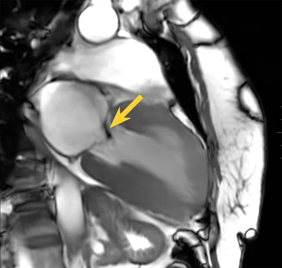
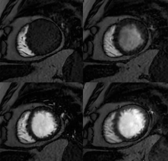
Figure 2.
Case 1, patient with suspected hypertrophic cardiomyopathy. High-resolution imaging was performed using Sonic DL™ Cine, AIR™ Recon DL and AIR™ Multi-Purpose 21ch Coil (large). (A) Breath-hold 2-chamber Sonic DL™ Cine 1.6 x 1.6 x 8 mm, 3 locations with 30 phases per location, 23 sec. (B) Breath-hold short axis Sonic DL™ Cine, 1.6 x 1.6 x 8 mm, 14 locations with 30 phases per location, 1:05 min. (C, D) Breath-hold 4-chamber Sonic DL™ Cine, 1.6 x 1.6 x 8 mm, 3 locations with 30 phases per location, 23 sec. (E) Short axis perfusion with MoCo and AIR™ Recon DL, 3 x 2.4 x 8 mm, 4 locations with 80 phases per location, 1:24 min. (F) short axis PS MDE with AIR™ Recon DL, 2.9 x 2.9 x 8 mm, 18 slices, 45 sec. T1 mapping: (G)T1 Native and (H) ECV map with AIR™ Recon DL, 2.4 x 2.9 x 8 mm, 3 slices, 30 sec.
Case 1
Patient with suspected hypertrophic cardiomyopathy was referred for CMR. Sonic DL™ Cine was utilized to examine the four cavities, short axis and long axis, and left ventricular outflow tract (LVOT). T1 mapping was performed before and after gadolinium injection.
Morphological study of the left ventricle and the right ventricle: Circumferential pericardial fluid effusion blade measuring a maximum of 8 mm thick next to the lateral basal wall of the left ventricle, without character constrictive. Absence of notable dilation of the right ear or tubular appearance of the right ventricle. Absence of intracavitary mass or detectable thrombus. The left ventricular mass is estimated at 198.6g or 128.9 g/m2. Marked thickening of the left ventricular wall at the height of:
- Anterobasal segment: 15.1 mm
- Anteroseptal basal segment: 22.3 mm
- Inferoseptobasal segment: 22.5 mm
- Lower basal segment: 19.5 mm
- Anteroseptomedial segment: 20.1 mm
- Lower septomedial segment: 21.4 mm
- Inferomedial segment: 21 mm
- Anteroapical segment: 19.5 mm
- Lateroapical segment: 18.1 mm
Absence of image suggestive of non-compaction of the left ventricle. The native T1 appears discreetly increased, evaluated at 1225.9 ms.
Dynamic study of the left ventricle: LVEF 84.2%
The end-diastolic volume of the left ventricle is equal to 170 mL or 110.4 mL/m2 of body surface.
The end-systolic volume of the left ventricle is equal to 26.9 mL or 17.4 mL/m2 of body surface.
The qualitative analysis of the segmental kinetics of the left ventricle does not highlight evidence of hypokinesia, akinesia or dyskinesia. Highlighting a systolic anterior motion effect with a jet of mitral regurgitation related to subaortic obstruction.
Dynamic study of the right ventricle: FEVD 71.4%
The end-diastolic volume of the right ventricle is equal to 122.6 mL or 79.6 mL/m2 of body surface. The end-systolic volume of the right ventricle is equal to 35.1 mL or 22.8 mL/m2 of body surface. The qualitative analysis of the segmental kinetics of the right ventricle does not highlight evidence of hypokinesia, akinesia or dyskinesia.
Study after gadolinium injection
Discreet subendocardial enhancement predominantly at the lateral wall of the ventricle left and the septo-basal and septo-medial wall of the left ventricle.
Absence of pathological contrast uptake of the pericardium.
Conclusion
Appearance of hypertrophic cardiomyopathy septal predominance with fibrosing changes and signs of subaortic obstruction. LVEF and FEVD appear preserved.
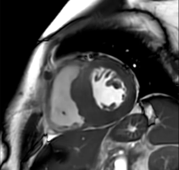
Figure 3.
Case 2, patient referred to CMR to evaluate cardiac damage from AL amyloidosis. High-resolution imaging was performed using Sonic DL™ Cine, AIR™ Recon DL and AIR™ Multi-Purpose 21ch Coil (large). (A) Breath hold short axis Sonic DL™ Cine, 1.6 x 1.6 x 8 mm, 16 locations with 30 phases per location, 54 sec. (B) Short axis PS MDE with AIR™ Recon DL, 2.9 x 2.9 x 8 mm, 39 sec. (C) Long axis PS MDE with AIR™ Recon DL, 2.9 x 2.9 x 8 mm, 12 slices, 27 sec. (D) ECV map with AIR™ Recon DL, 2.4 x 2.9 x 8 mm, 3 slices, 26 sec.
Case 2
Patient was referred to CMR to evaluate cardiac damage from primary or light chain (AL) amyloidosis, the most common type of system amyloidosis. It is a rare disease characterized by the abnormal accumulation of amyloid protein fibrils in various tissues and organs. Sonic DL™ Cine was utilized to examine the four cavities, short axis and long axis. T1 mapping was performed before and after gadolinium injection.
Morphological study of the left ventricle and the right ventricle
Absence of significant effusion of the pericardium.
Absence of notable dilation of the right ear or tubular appearance of the right ventricle.
Absence of intracavitary mass or detectable thrombus.
The left ventricular mass is estimated at 129.9 g or 74.6 g/m2.
Absence of image suggestive of non-compaction of the left ventricle.
Thickening of the inferoapical wall of the left ventricle with a thickness maximum rated at 15.3 mm.
Wall of the anteroapical, lateroapical, inferolateral, inferomedial, inferomedial segments septo medial, infero Iaterobasal and inferobasal of the left ventricle of thickness in the upper limits of normal, estimated between 13.6 and 14.8 mm.
Dynamic study of the left ventricle: LVEF 55.6%
The end-diastolic volume of the left ventricle is equal to 122.7 mL or 70.5 mL/m2 of body surface.
The end-systolic volume of the left ventricle is equal to 54.5 mL or 31.3 mL/m2 of body surface.The qualitative analysis of the segmental kinetics of the left ventricle does not highlight evidence of hypokinesia, akinesia or dyskinesia.
Dynamic study of the right ventricle: FEVD 42.8%
The end-diastolic volume of the right ventricle is equal to 138.4 mL or 795 mL/m2 of body surface.
The end-systolic volume of the right ventricle is equal to 79.1 mL or 45.4 mL/m2 of body surface.
The qualitative analysis of the segmental kinetics of the right ventricle does not highlight evidence of hypokinesia, akinesia or dyskinesia.
Study after gadolinium injection
Persistence of late subendocardial myocardial enhancement, predominant basal, which can be part of underlying cardiac amyloidosis associated with an elevation of native T1 to 1352 ms. Extracellular volume (ECV) rated at 35.5%. Absence of pathological contrast uptake of the pericardium.
Conclusion
The myocardial wall appears less thickened than in the previous CMR examination; however, the persistence of late subendocardial enhancement, predominant basal and an elongation of the native T1 can be indicative of underlying cardiac amyloidosis.
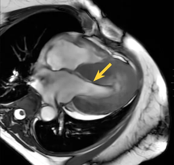
Figure 4.
Case 3, patient referred for assessment of dilated heart disease. High-resolution imaging was performed using Sonic DL™ Cine, AIR™ Recon DL and AIR™ Multi-Purpose 21ch Coil (large). (A) Free-breathing, 2-chamber Sonic DL™ Cine, 2.1 x 2.5 x 8 mm, 3 locations with 30 phases per location, 23 sec. (B) Free-breathing 4-chamber Sonic DL™ Cine, 2.1 x 2.5 x 8 mm, 3 locations with 30 phases per location, 20 sec. (C) Free-breathing short axis Sonic DL™ Cine, 2.1 x 2.1 x 8 mm, 15 locations with 30 phases per location, 25 sec. (D) Short axis perfusion with MoCo and AIR™ Recon DL, 2.4 x 2.4 x 8 mm, 3 locations with 80 phases per location, 1:08 min.
Case 3
Patient was referred for assessment of dilated heart disease. Sonic DL™ Cine was utilized to examine the four cavities, short axis and long axis. T1 mapping was performed before and after gadolinium injection.
Morphological study of the left ventricle and the right ventricle
Absence of significant effusion of the pericardium.
Absence of notable dilation of the right ear or tubular appearance of the right ventricle.
Absence of intracavitary mass or detectable thrombus. The left ventricular mass is estimated at 187.6 g or 77.7 g/m2. Absence of image suggestive of non-compaction of the left ventricle.
Dynamic study of the left ventricle: LVEF 30.5%
The end-diastolic volume of the left ventricle is equal to 301 mL or 124.7 mL/m2 of body surface.
The end-systolic volume of the left ventricle is equal to 209 mL or 86.6 mL/m2 of body surface.
The qualitative analysis of the segmental kinetics of the left ventricle highlights an appearance of diffuse hypokinesia.
Study after gadolinium injection
Absence of contrast enhancement suggestive of myocardial fibrosis or sequelae of myocardial necrosis.
Absence of pathological contrast uptake of the pericardium.
Conclusion
LVEF 30.5% with diffuse hypokinesia and enlargement of the cavities. However, MR analysis is limited. There is no image suggestive of sequelae of myocardial necrosis or fibrosis.
DOWNLOAD ARTICLE HERE


