A
Figure 1.
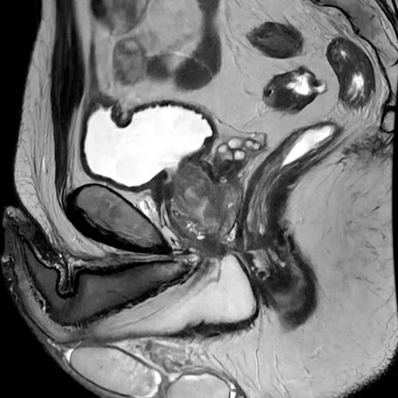
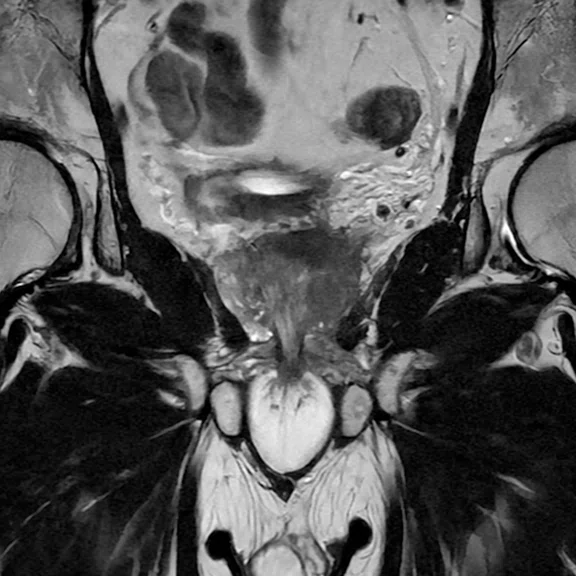
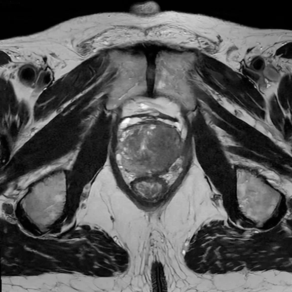
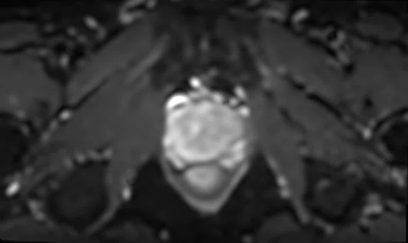
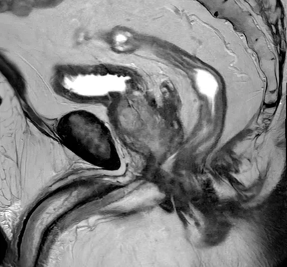
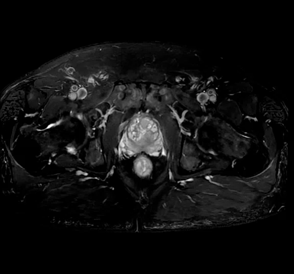
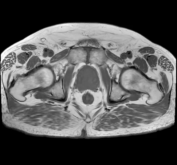
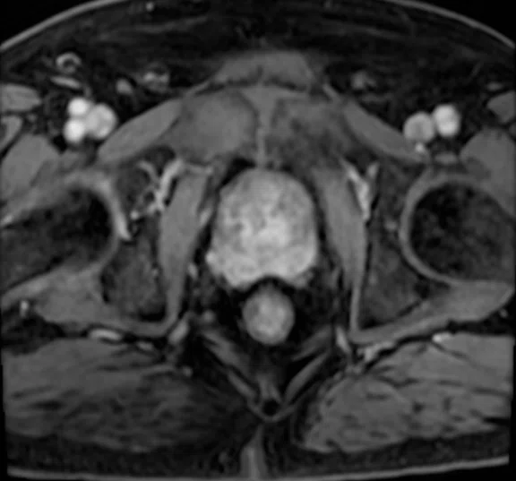
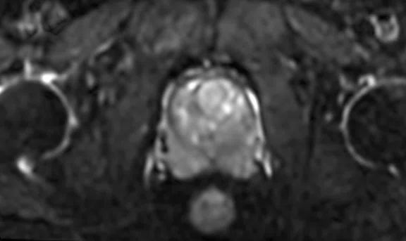
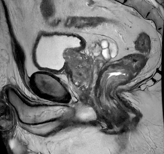
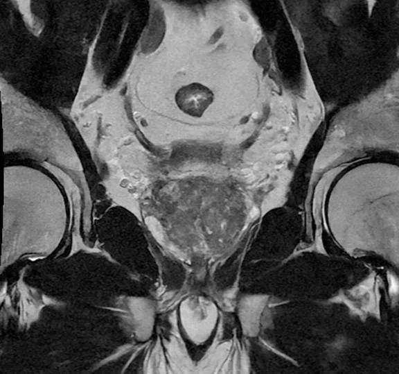
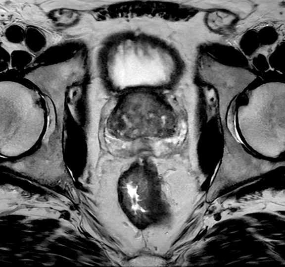
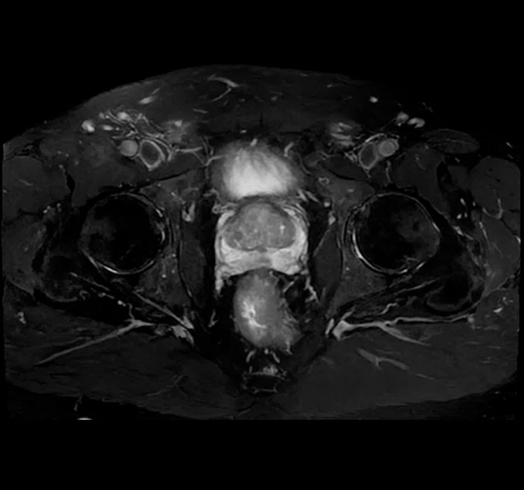
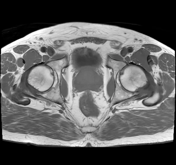
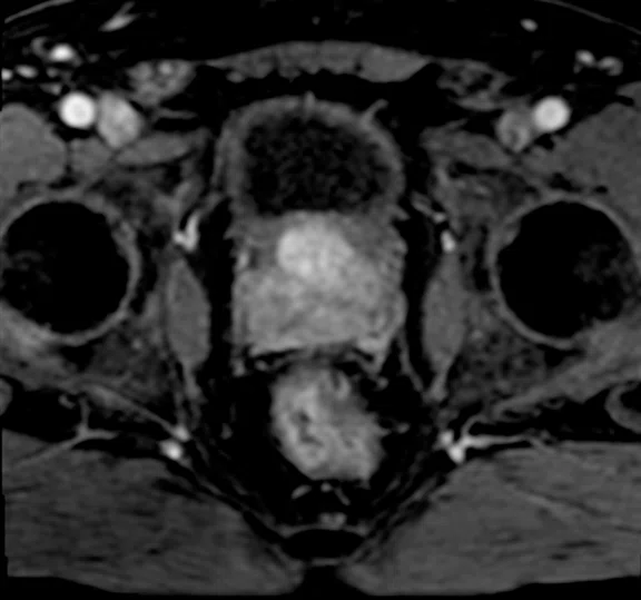
Prostate exam on SIGNA™ Voyager 1.5T with AIR™ Recon DL using a 16ch TDI coil. (A) Sagittal T2 FSE, 0.4 x 0.7 x 3 mm, 4:03 min.; (B) coronal T2 FSE, 0.4 x 0.7 x 3 mm, 4:05 min.; (C) axial T2 FSE, 0.4 x 0.7 x 3 mm, 4:07 min.; (D) axial T2 FSE FatSat, 0.9 x 0.9 x 5 mm, 1:56 min.; (E) axial T1 FSE, 1.1 x 1.2 x 5 mm, 2:42 min.; and (F) axial LAVA Flex (water), phase 6 of 11, 1.2 x 1.8 x 2 mm, 3:52 min.
B
Figure 1.
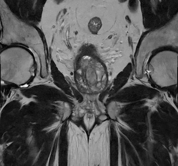
Prostate exam on SIGNA™ Voyager 1.5T with AIR™ Recon DL using a 16ch TDI coil. (A) Sagittal T2 FSE, 0.4 x 0.7 x 3 mm, 4:03 min.; (B) coronal T2 FSE, 0.4 x 0.7 x 3 mm, 4:05 min.; (C) axial T2 FSE, 0.4 x 0.7 x 3 mm, 4:07 min.; (D) axial T2 FSE FatSat, 0.9 x 0.9 x 5 mm, 1:56 min.; (E) axial T1 FSE, 1.1 x 1.2 x 5 mm, 2:42 min.; and (F) axial LAVA Flex (water), phase 6 of 11, 1.2 x 1.8 x 2 mm, 3:52 min.
C
Figure 1.
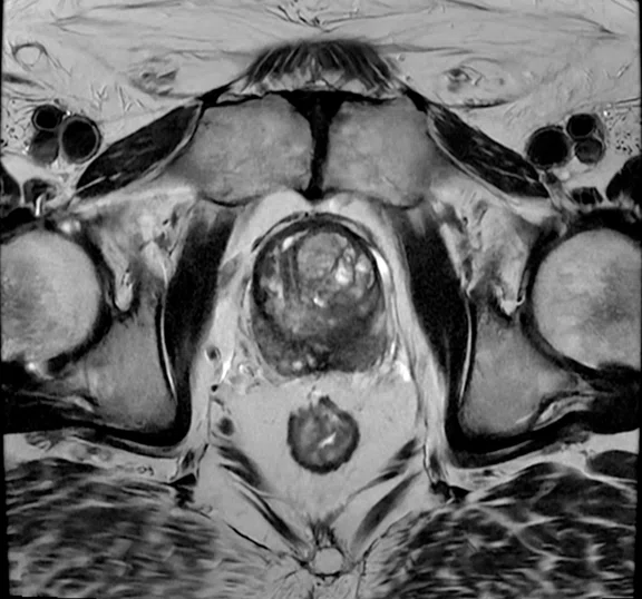
Prostate exam on SIGNA™ Voyager 1.5T with AIR™ Recon DL using a 16ch TDI coil. (A) Sagittal T2 FSE, 0.4 x 0.7 x 3 mm, 4:03 min.; (B) coronal T2 FSE, 0.4 x 0.7 x 3 mm, 4:05 min.; (C) axial T2 FSE, 0.4 x 0.7 x 3 mm, 4:07 min.; (D) axial T2 FSE FatSat, 0.9 x 0.9 x 5 mm, 1:56 min.; (E) axial T1 FSE, 1.1 x 1.2 x 5 mm, 2:42 min.; and (F) axial LAVA Flex (water), phase 6 of 11, 1.2 x 1.8 x 2 mm, 3:52 min.
D
Figure #.
Figure Copy.
E
Figure #.
Figure Copy.
F
Figure 1.
Prostate exam on SIGNA™ Voyager 1.5T with AIR™ Recon DL using a 16ch TDI coil. (A) Sagittal T2 FSE, 0.4 x 0.7 x 3 mm, 4:03 min.; (B) coronal T2 FSE, 0.4 x 0.7 x 3 mm, 4:05 min.; (C) axial T2 FSE, 0.4 x 0.7 x 3 mm, 4:07 min.; (D) axial T2 FSE FatSat, 0.9 x 0.9 x 5 mm, 1:56 min.; (E) axial T1 FSE, 1.1 x 1.2 x 5 mm, 2:42 min.; and (F) axial LAVA Flex (water), phase 6 of 11, 1.2 x 1.8 x 2 mm, 3:52 min.
A
Figure 2.
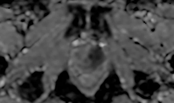
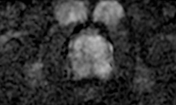
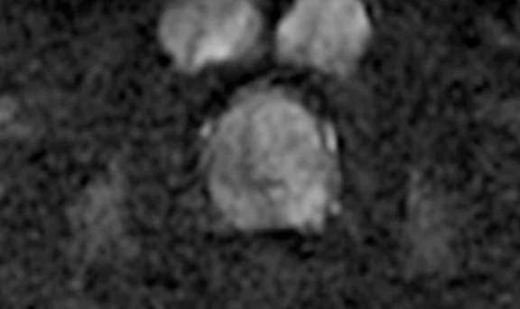
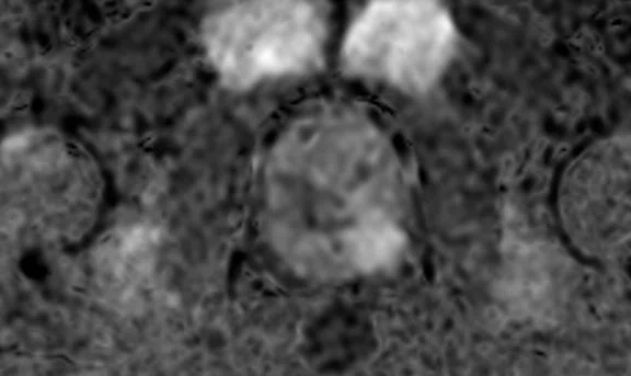
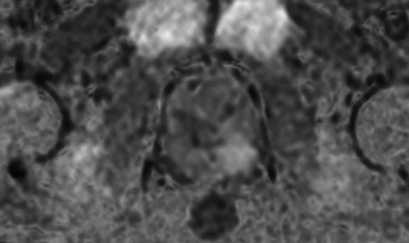
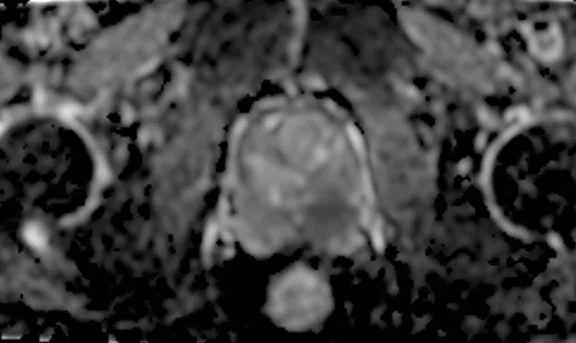
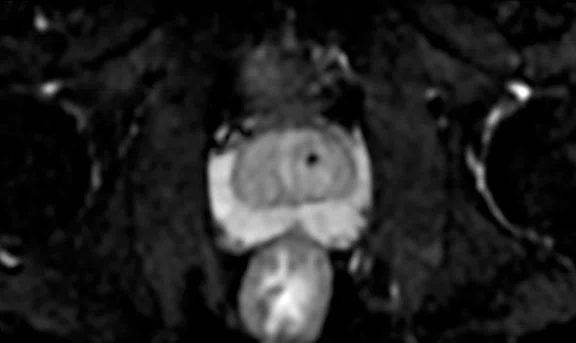
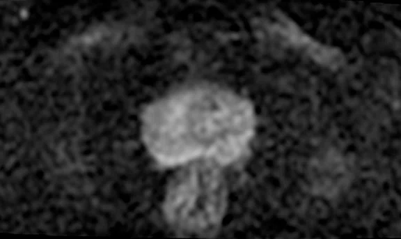
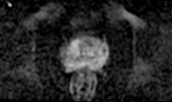
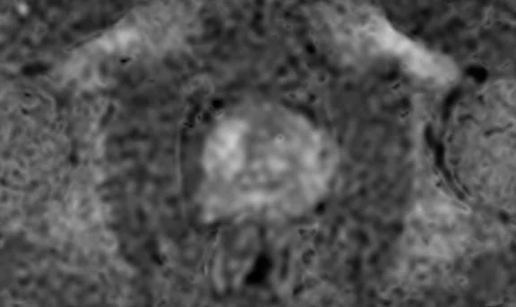
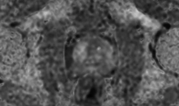
Same patient as Figure 1. Prostate exam on SIGNA™ Voyager 1.5T with AIR™ Recon DL using a 16ch TDI coil. Axial FOCUS DWI, 2 x 2 x 3.0 mm, 5:25 min. with (A) b50, (B) b500, (C) b1000, (D) synthetic b1500 with MAGiC DWI, (E) synthetic b2000 with MAGiC DWI and (F) ADC.
B
Figure 2.
Same patient as Figure 1. Prostate exam on SIGNA™ Voyager 1.5T with AIR™ Recon DL using a 16ch TDI coil. Axial FOCUS DWI, 2 x 2 x 3.0 mm, 5:25 min. with (A) b50, (B) b500, (C) b1000, (D) synthetic b1500 with MAGiC DWI, (E) synthetic b2000 with MAGiC DWI and (F) ADC.
C
Figure 2.
Same patient as Figure 1. Prostate exam on SIGNA™ Voyager 1.5T with AIR™ Recon DL using a 16ch TDI coil. Axial FOCUS DWI, 2 x 2 x 3.0 mm, 5:25 min. with (A) b50, (B) b500, (C) b1000, (D) synthetic b1500 with MAGiC DWI, (E) synthetic b2000 with MAGiC DWI and (F) ADC.
D
Figure 2.
Same patient as Figure 1. Prostate exam on SIGNA™ Voyager 1.5T with AIR™ Recon DL using a 16ch TDI coil. Axial FOCUS DWI, 2 x 2 x 3.0 mm, 5:25 min. with (A) b50, (B) b500, (C) b1000, (D) synthetic b1500 with MAGiC DWI, (E) synthetic b2000 with MAGiC DWI and (F) ADC.
E
Figure 2.
Same patient as Figure 1. Prostate exam on SIGNA™ Voyager 1.5T with AIR™ Recon DL using a 16ch TDI coil. Axial FOCUS DWI, 2 x 2 x 3.0 mm, 5:25 min. with (A) b50, (B) b500, (C) b1000, (D) synthetic b1500 with MAGiC DWI, (E) synthetic b2000 with MAGiC DWI and (F) ADC.
F
Figure 2.
Same patient as Figure 1. Prostate exam on SIGNA™ Voyager 1.5T with AIR™ Recon DL using a 16ch TDI coil. Axial FOCUS DWI, 2 x 2 x 3.0 mm, 5:25 min. with (A) b50, (B) b500, (C) b1000, (D) synthetic b1500 with MAGiC DWI, (E) synthetic b2000 with MAGiC DWI and (F) ADC.
A
Figure 3.
Prostate MR exam on SIGNA™ Voyager 1.5T with AIR™ Recon DL using the 21ch AIR™ Multi-Purpose Coil. (A) Sagittal T2 FSE, 0.4 x 0.7 x 3 mm, 4:03 min.; (B) coronal T2 FSE, 0.4 x 0.7 x 3 mm, 4:05 min.; (C) axial T2 FSE, 0.4 x 0.7 x 3 mm, 4:07 min.; (D) axial T2 FSE FatSat, 0.4 x 0.7 x 3 mm, 2:42 min.; (E) axial T1 FSE, 0.4 x 0.7 x 3 mm, 1:56 min.; and (F) axial LAVA Flex (water), phase 8 of 13, 1.2 x 1.8 x 2 mm, 3:52 min.
B
Figure 3.
Prostate MR exam on SIGNA™ Voyager 1.5T with AIR™ Recon DL using the 21ch AIR™ Multi-Purpose Coil. (A) Sagittal T2 FSE, 0.4 x 0.7 x 3 mm, 4:03 min.; (B) coronal T2 FSE, 0.4 x 0.7 x 3 mm, 4:05 min.; (C) axial T2 FSE, 0.4 x 0.7 x 3 mm, 4:07 min.; (D) axial T2 FSE FatSat, 0.4 x 0.7 x 3 mm, 2:42 min.; (E) axial T1 FSE, 0.4 x 0.7 x 3 mm, 1:56 min.; and (F) axial LAVA Flex (water), phase 8 of 13, 1.2 x 1.8 x 2 mm, 3:52 min.
C
Figure 3.
Prostate MR exam on SIGNA™ Voyager 1.5T with AIR™ Recon DL using the 21ch AIR™ Multi-Purpose Coil. (A) Sagittal T2 FSE, 0.4 x 0.7 x 3 mm, 4:03 min.; (B) coronal T2 FSE, 0.4 x 0.7 x 3 mm, 4:05 min.; (C) axial T2 FSE, 0.4 x 0.7 x 3 mm, 4:07 min.; (D) axial T2 FSE FatSat, 0.4 x 0.7 x 3 mm, 2:42 min.; (E) axial T1 FSE, 0.4 x 0.7 x 3 mm, 1:56 min.; and (F) axial LAVA Flex (water), phase 8 of 13, 1.2 x 1.8 x 2 mm, 3:52 min.
D
Figure 3.
Prostate MR exam on SIGNA™ Voyager 1.5T with AIR™ Recon DL using the 21ch AIR™ Multi-Purpose Coil. (A) Sagittal T2 FSE, 0.4 x 0.7 x 3 mm, 4:03 min.; (B) coronal T2 FSE, 0.4 x 0.7 x 3 mm, 4:05 min.; (C) axial T2 FSE, 0.4 x 0.7 x 3 mm, 4:07 min.; (D) axial T2 FSE FatSat, 0.4 x 0.7 x 3 mm, 2:42 min.; (E) axial T1 FSE, 0.4 x 0.7 x 3 mm, 1:56 min.; and (F) axial LAVA Flex (water), phase 8 of 13, 1.2 x 1.8 x 2 mm, 3:52 min.
E
Figure 3.
Prostate MR exam on SIGNA™ Voyager 1.5T with AIR™ Recon DL using the 21ch AIR™ Multi-Purpose Coil. (A) Sagittal T2 FSE, 0.4 x 0.7 x 3 mm, 4:03 min.; (B) coronal T2 FSE, 0.4 x 0.7 x 3 mm, 4:05 min.; (C) axial T2 FSE, 0.4 x 0.7 x 3 mm, 4:07 min.; (D) axial T2 FSE FatSat, 0.4 x 0.7 x 3 mm, 2:42 min.; (E) axial T1 FSE, 0.4 x 0.7 x 3 mm, 1:56 min.; and (F) axial LAVA Flex (water), phase 8 of 13, 1.2 x 1.8 x 2 mm, 3:52 min.
F
Figure 3.
Prostate MR exam on SIGNA™ Voyager 1.5T with AIR™ Recon DL using the 21ch AIR™ Multi-Purpose Coil. (A) Sagittal T2 FSE, 0.4 x 0.7 x 3 mm, 4:03 min.; (B) coronal T2 FSE, 0.4 x 0.7 x 3 mm, 4:05 min.; (C) axial T2 FSE, 0.4 x 0.7 x 3 mm, 4:07 min.; (D) axial T2 FSE FatSat, 0.4 x 0.7 x 3 mm, 2:42 min.; (E) axial T1 FSE, 0.4 x 0.7 x 3 mm, 1:56 min.; and (F) axial LAVA Flex (water), phase 8 of 13, 1.2 x 1.8 x 2 mm, 3:52 min.
A
Figure 4.
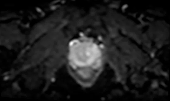
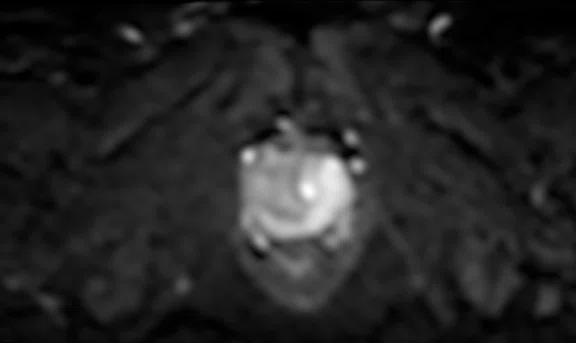
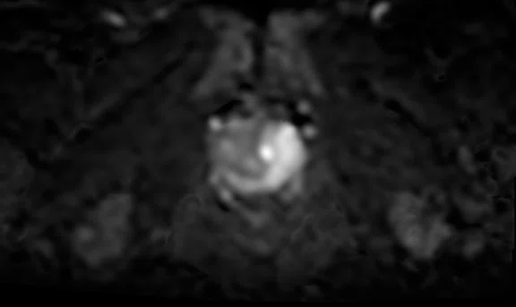
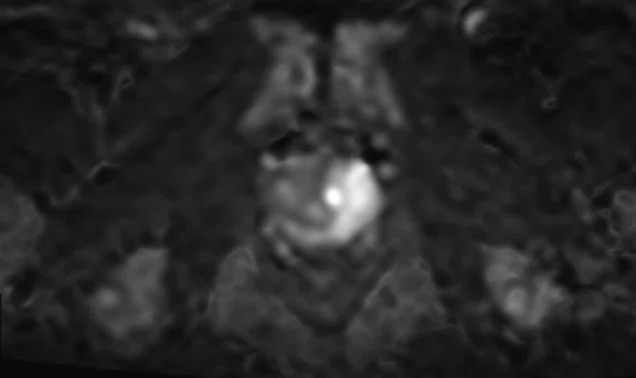
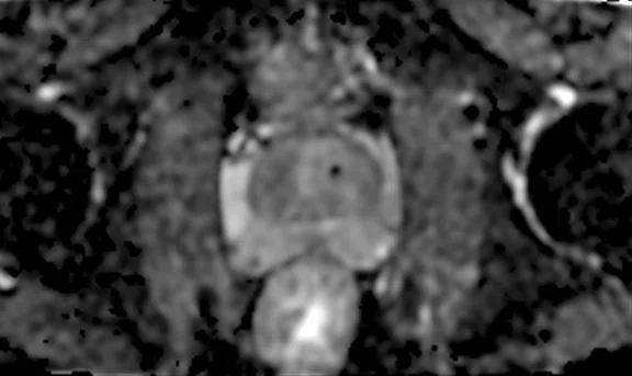
Same patient as Figure 3. Prostate exam on SIGNA™ Voyager 1.5T with AIR™ Recon DL using the 21ch AIR™ MP Coil. Axial FOCUS DWI, 2 x 2 x 3.0 mm, 5:25 min. with (A) b50, (B) b500, (C) b1000, (D) synthetic b1500 with MAGiC DWI, (E) synthetic b2000 with MAGiC DWI and (F) ADC.
B
Figure 4.
Same patient as Figure 3. Prostate exam on SIGNA™ Voyager 1.5T with AIR™ Recon DL using the 21ch AIR™ MP Coil. Axial FOCUS DWI, 2 x 2 x 3.0 mm, 5:25 min. with (A) b50, (B) b500, (C) b1000, (D) synthetic b1500 with MAGiC DWI, (E) synthetic b2000 with MAGiC DWI and (F) ADC.
C
Figure 4.
Same patient as Figure 3. Prostate exam on SIGNA™ Voyager 1.5T with AIR™ Recon DL using the 21ch AIR™ MP Coil. Axial FOCUS DWI, 2 x 2 x 3.0 mm, 5:25 min. with (A) b50, (B) b500, (C) b1000, (D) synthetic b1500 with MAGiC DWI, (E) synthetic b2000 with MAGiC DWI and (F) ADC.
D
Figure 4.
Same patient as Figure 3. Prostate exam on SIGNA™ Voyager 1.5T with AIR™ Recon DL using the 21ch AIR™ MP Coil. Axial FOCUS DWI, 2 x 2 x 3.0 mm, 5:25 min. with (A) b50, (B) b500, (C) b1000, (D) synthetic b1500 with MAGiC DWI, (E) synthetic b2000 with MAGiC DWI and (F) ADC.
E
Figure 4.
Same patient as Figure 3. Prostate exam on SIGNA™ Voyager 1.5T with AIR™ Recon DL using the 21ch AIR™ MP Coil. Axial FOCUS DWI, 2 x 2 x 3.0 mm, 5:25 min. with (A) b50, (B) b500, (C) b1000, (D) synthetic b1500 with MAGiC DWI, (E) synthetic b2000 with MAGiC DWI and (F) ADC.
F
Figure 4.
Same patient as Figure 3. Prostate exam on SIGNA™ Voyager 1.5T with AIR™ Recon DL using the 21ch AIR™ MP Coil. Axial FOCUS DWI, 2 x 2 x 3.0 mm, 5:25 min. with (A) b50, (B) b500, (C) b1000, (D) synthetic b1500 with MAGiC DWI, (E) synthetic b2000 with MAGiC DWI and (F) ADC.
A
Figure 5.
Same patient as Figure 5. Prostate exam on SIGNA™ Voyager 1.5T with AIR™ Recon DL using the 16ch AIR™ AA Coil. Axial FOCUS DWI, 2 x 2.4 x 3.0 mm, 6:34 min. with (A) b50, (B) b800, (C) b1000, (D) synthetic b1500 with MAGiC DWI, (E) synthetic b2000 with MAGiC DWI and (F) ADC.
B
Figure 6.
Same patient as Figure 5. Prostate exam on SIGNA™ Voyager 1.5T with AIR™ Recon DL using the 16ch AIR™ AA Coil. Axial FOCUS DWI, 2 x 2.4 x 3.0 mm, 6:34 min. with (A) b50, (B) b800, (C) b1000, (D) synthetic b1500 with MAGiC DWI, (E) synthetic b2000 with MAGiC DWI and (F) ADC.
C
Figure 6.
Same patient as Figure 5. Prostate exam on SIGNA™ Voyager 1.5T with AIR™ Recon DL using the 16ch AIR™ AA Coil. Axial FOCUS DWI, 2 x 2.4 x 3.0 mm, 6:34 min. with (A) b50, (B) b800, (C) b1000, (D) synthetic b1500 with MAGiC DWI, (E) synthetic b2000 with MAGiC DWI and (F) ADC.
D
Figure 6.
Same patient as Figure 5. Prostate exam on SIGNA™ Voyager 1.5T with AIR™ Recon DL using the 16ch AIR™ AA Coil. Axial FOCUS DWI, 2 x 2.4 x 3.0 mm, 6:34 min. with (A) b50, (B) b800, (C) b1000, (D) synthetic b1500 with MAGiC DWI, (E) synthetic b2000 with MAGiC DWI and (F) ADC.
E
Figure 6.
Same patient as Figure 5. Prostate exam on SIGNA™ Voyager 1.5T with AIR™ Recon DL using the 16ch AIR™ AA Coil. Axial FOCUS DWI, 2 x 2.4 x 3.0 mm, 6:34 min. with (A) b50, (B) b800, (C) b1000, (D) synthetic b1500 with MAGiC DWI, (E) synthetic b2000 with MAGiC DWI and (F) ADC.
F
Figure 6.
Same patient as Figure 5. Prostate exam on SIGNA™ Voyager 1.5T with AIR™ Recon DL using the 16ch AIR™ AA Coil. Axial FOCUS DWI, 2 x 2.4 x 3.0 mm, 6:34 min. with (A) b50, (B) b800, (C) b1000, (D) synthetic b1500 with MAGiC DWI, (E) synthetic b2000 with MAGiC DWI and (F) ADC.
A
Figure 5.
Prostate MR exam on SIGNA™ Voyager 1.5T with AIR™ Recon DL using the 16ch AIR™ AA Coil. (A) Sagittal T2 FSE, 0.4 x 0.7 x 3 mm, 4:03 min.; (B) coronal T2 FSE, 0.4 x 0.7 x 3 mm, 4:33 min.; (C) axial T2 FSE, 0.4 x 0.7 x 3 mm, 4:08 min.; (D) axial T2 FSE FatSat, 0.9 x 0.9 x 5 mm, 2:01 min.; (E) axial T1 FSE, 1.1 x 1.2 x 5 mm, 2:50 min.; and (F) axial LAVA Flex (water), phase 8 of 13, 1.8 x 1.8 x 2 mm, 3:52 min.
B
Figure 5.
Prostate MR exam on SIGNA™ Voyager 1.5T with AIR™ Recon DL using the 16ch AIR™ AA Coil. (A) Sagittal T2 FSE, 0.4 x 0.7 x 3 mm, 4:03 min.; (B) coronal T2 FSE, 0.4 x 0.7 x 3 mm, 4:33 min.; (C) axial T2 FSE, 0.4 x 0.7 x 3 mm, 4:08 min.; (D) axial T2 FSE FatSat, 0.9 x 0.9 x 5 mm, 2:01 min.; (E) axial T1 FSE, 1.1 x 1.2 x 5 mm, 2:50 min.; and (F) axial LAVA Flex (water), phase 8 of 13, 1.8 x 1.8 x 2 mm, 3:52 min.
C
Figure 5.
Prostate MR exam on SIGNA™ Voyager 1.5T with AIR™ Recon DL using the 16ch AIR™ AA Coil. (A) Sagittal T2 FSE, 0.4 x 0.7 x 3 mm, 4:03 min.; (B) coronal T2 FSE, 0.4 x 0.7 x 3 mm, 4:33 min.; (C) axial T2 FSE, 0.4 x 0.7 x 3 mm, 4:08 min.; (D) axial T2 FSE FatSat, 0.9 x 0.9 x 5 mm, 2:01 min.; (E) axial T1 FSE, 1.1 x 1.2 x 5 mm, 2:50 min.; and (F) axial LAVA Flex (water), phase 8 of 13, 1.8 x 1.8 x 2 mm, 3:52 min.
D
Figure 5.
Prostate MR exam on SIGNA™ Voyager 1.5T with AIR™ Recon DL using the 16ch AIR™ AA Coil. (A) Sagittal T2 FSE, 0.4 x 0.7 x 3 mm, 4:03 min.; (B) coronal T2 FSE, 0.4 x 0.7 x 3 mm, 4:33 min.; (C) axial T2 FSE, 0.4 x 0.7 x 3 mm, 4:08 min.; (D) axial T2 FSE FatSat, 0.9 x 0.9 x 5 mm, 2:01 min.; (E) axial T1 FSE, 1.1 x 1.2 x 5 mm, 2:50 min.; and (F) axial LAVA Flex (water), phase 8 of 13, 1.8 x 1.8 x 2 mm, 3:52 min.
E
Figure 5.
Prostate MR exam on SIGNA™ Voyager 1.5T with AIR™ Recon DL using the 16ch AIR™ AA Coil. (A) Sagittal T2 FSE, 0.4 x 0.7 x 3 mm, 4:03 min.; (B) coronal T2 FSE, 0.4 x 0.7 x 3 mm, 4:33 min.; (C) axial T2 FSE, 0.4 x 0.7 x 3 mm, 4:08 min.; (D) axial T2 FSE FatSat, 0.9 x 0.9 x 5 mm, 2:01 min.; (E) axial T1 FSE, 1.1 x 1.2 x 5 mm, 2:50 min.; and (F) axial LAVA Flex (water), phase 8 of 13, 1.8 x 1.8 x 2 mm, 3:52 min.
F
Figure 5.
Prostate MR exam on SIGNA™ Voyager 1.5T with AIR™ Recon DL using the 16ch AIR™ AA Coil. (A) Sagittal T2 FSE, 0.4 x 0.7 x 3 mm, 4:03 min.; (B) coronal T2 FSE, 0.4 x 0.7 x 3 mm, 4:33 min.; (C) axial T2 FSE, 0.4 x 0.7 x 3 mm, 4:08 min.; (D) axial T2 FSE FatSat, 0.9 x 0.9 x 5 mm, 2:01 min.; (E) axial T1 FSE, 1.1 x 1.2 x 5 mm, 2:50 min.; and (F) axial LAVA Flex (water), phase 8 of 13, 1.8 x 1.8 x 2 mm, 3:52 min.
1. Spektor M, Mathur M, Weinreb JC. Standards for MRI reporting-the evolution to PI-RADS v 2.0. Transl Androl Urol. 2017 Jun;6(3):355-367.
2. Hassanzadeh E, Glazer DI, Dunne RM, Fennessy FM, Harisinghani MG, Tempany CM. Prostate imaging reporting and data system version 2 (PI-RADS v2): a pictorial review. Abdom Radiol (NY). 2017 Jan;42(1):278-289.
3. Belue MJ, Yilmaz EC, Daryanani A, Turkbey B. Current Status of Biparametric MRI in Prostate Cancer Diagnosis: Literature Analysis. Life (Basel). 2022 May 28;12(6):804.
4. Greenberg JW, Koller CR, Casado C, Triche BL, Krane LS. A narrative review of biparametric MRI (bpMRI) implementation on screening, detection, and the overall accuracy for prostate cancer. Ther Adv Urol. 2022 May 4;14:17562872221096377.
5. Niu XK, Chen XH, Chen ZF, Chen L, Li J, Peng T. Diagnostic Performance of Biparametric MRI for Detection of Prostate Cancer: A Systematic Review and Meta-Analysis. AJR Am J Roentgenol. 2018 Aug;211(2):369-378.
6. Xu L, Zhang G, Shi B, Liu Y, Zou T, Yan W, Xiao Y, Xue H, Feng F, Lei J, Jin Z, Sun H. Comparison of biparametric and multiparametric MRI in the diagnosis of prostate cancer. Cancer Imaging. 2019 Dec 21;19(1):90.
result


PREVIOUS
${prev-page}
NEXT
${next-page}
Subscribe Now
Manage Subscription
FOLLOW US
Contact Us • Cookie Preferences • Privacy Policy • California Privacy PolicyDo Not Sell or Share My Personal Information • Terms & Conditions • Security
© 2024 GE HealthCare. GE is a trademark of General Electric Company. Used under trademark license.
In Practice
Developing a high-quality,
PI-RADS v2-compliant prostate MR service at 1.5T
Developing a high-quality,
PI-RADS v2-compliant prostate MR service at 1.5T
MR has become an integral tool in the diagnosis and management of prostate cancer. At Regional Medical Imaging (RMI) — an independent imaging center in Flint, Michigan with 10 locations throughout the east-central part of the state — prostate MR exams have grown exponentially in 2023, up an average of 250% compared to 2022 across three sites.
Much of this growth is tied to the acquisition of new fixed and mobile 1.5T MR systems with advanced imaging capabilities, as well as the addition of Michael J. Hicks, MD, who specialized in cross-sectional body imaging, including prostate MR, during his fellowship training at University of Michigan.
RMI’s main Flint location had an older MR system that was at the end of its life. While evaluating new scanners, leadership learned about GE HealthCare’s SIGNA™ MR Upgrade portfolio, which enables upgrades to an MR system without having to replace the magnet, extending the lifespan of an MR system by decades. They also learned about AIR™ Recon DL and believed the combination of the upgrade capability along with GE HealthCare’s advanced deep-learning technologies provided significant clinical and operational advantages for the practice, making GE HealthCare their vendor of choice for a new MR system purchase.
RMI decided SIGNA™ Voyager, a 1.5T wide bore MR system, was the right fit for the Flint site. SIGNA™ Voyager is also available as a mobile unit, providing a solution for its Novi and Southgate sites that are approximately 30 minutes apart. With these two new systems, RMI has the technology it needs to provide Prostate Imaging Reporting & Data System (PI-RADS®) v2.1-compliant prostate MR exams at 1.5T.
RMI is using the same high-resolution protocols, following the PI-RADS® standard, that Dr. Hicks used at his fellowship at the University of Michigan with one notable exception: the University
is using 3.0T MR for the vast majority of prostate exams while RMI uses a 1.5T MR.
“The imaging we are doing here at 1.5T is on par with what academic powerhouses are doing at 3.0T in terms of image quality and interpretability,” says Dr. Hicks. “With the technology on SIGNA™ Voyager, we are able to provide the quality exams urologists need to best manage their patients.”
Adds Mendy Caraway, MRI Department Manager at RMI, “We are performing prostate MR only on our SIGNA™ Voyager systems, and that is directly related to having AIR™ Recon DL providing the image quality our radiologists are comfortable with for making a diagnosis.”
“There is no denying the image quality has increased greatly with AIR™ Recon DL; we really can’t compare it to anything else,” says Caraway. “It also helps with the speed of the exam, specifically the diffusion imaging so patients aren’t on the table as long.”
D
E
F
Figure 1.
Prostate exam on SIGNA™ Voyager 1.5T with AIR™ Recon DL using a 16ch TDI coil. (A) Sagittal T2 FSE, 0.4 x 0.7 x 3 mm, 4:03 min.; (B) coronal T2 FSE, 0.4 x 0.7 x 3 mm, 4:05 min.; (C) axial T2 FSE, 0.4 x 0.7 x 3 mm, 4:07 min.; (D) axial T2 FSE FatSat, 0.9 x 0.9 x 5 mm, 1:56 min.; (E) axial T1 FSE, 1.1 x 1.2 x 5 mm, 2:42 min.; and (F) axial LAVA Flex (water), phase 6 of 11, 1.2 x 1.8 x 2 mm, 3:52 min.
Partnering with urology
The excellent image quality in prostate MR is a primary reason why RMI has been so successful attracting prostate MR referrals from urologists. RMI had an existing relationship with a local leading urology group based on decades of service providing quality CT imaging and urograms.
According to Dr. Hicks, urologists initially inquired whether the practice had a 3.0T system for prostate MR imaging. He demonstrated to the urology group that 3.0T-like image quality can be achieved on SIGNA™ Voyager with AIR™ Recon DL and explained how an advanced 1.5T MR system with the right technology can achieve the quality required for PI-RADS® v2.1-compliant prostate imaging.
“Prostate cancer is the number one cause of cancer in men, and there aren’t enough 3.0T MR systems across the country to provide the accessibility needed to image all these patients,” Dr. Hicks adds.
RMI is also using AIR™ Coils at the Flint location, further enhancing patient comfort and their overall imaging experience. When used with AIR™ Recon DL, these high-performance coils enable high SNR and spatial resolution, providing a viable alternative to endorectal coils. The referring urologists are also receiving positive feedback from patients regarding their prostate MR imaging experience at RMI, which can help strengthen the partnership between the two practices.
“The AIR Coils work with any body habitus,” adds Caraway. “Our technologists like that they are less bulky than the conventional coils, and our patients are happy because it feels like a weighted blanket being laid on them. So, it’s a great improvement for our patients and staff.”
Figure 3.
Prostate MR exam on SIGNA™ Voyager 1.5T with AIR™ Recon DL using the 21ch AIR™ Multi-Purpose Coil. (A) Sagittal T2 FSE, 0.4 x 0.7 x 3 mm, 4:03 min.; (B) coronal T2 FSE, 0.4 x 0.7 x 3 mm, 4:05 min.; (C) axial T2 FSE, 0.4 x 0.7 x 3 mm, 4:07 min.; (D) axial T2 FSE FatSat, 0.4 x 0.7 x 3 mm, 2:42 min.; (E) axial T1 FSE, 0.4 x 0.7 x 3 mm, 1:56 min.; and (F) axial LAVA Flex (water), phase 8 of 13, 1.2 x 1.8 x 2 mm, 3:52 min.
Growth in prostate MR
The partnership between RMI and the urology group has led to significant increases in prostate MR volume. Prostate MR volumes at two RMI locations doubled between August and November 2023 before tripling to over 60 cases in January 2024. RMI performed 25% of their 2023 prostate MR volume in just one month.
These significant increases in prostate MR volume were made possible by reducing scan times compared to conventional imaging techniques. With AIR™ Recon DL, RMI has balanced scan time reduction and image quality improvements to reduce acquisition times for T2-weighted imaging while simultaneously delivering higher resolution with a smaller field-of-view (FOV). In addition to AIR™ Recon DL, they also use MAGiC DWI, which generates multiple synthetic b-values from one DWI scanned series, allowing for higher b-values in shorter scan times without sacrificing contrast or anatomy coverage.
The combination of these technologies has allowed RMI to reduce overall prostate MR examination times by approximately 10 minutes, which allows these patients to fit into shorter scheduling slots of 30 minutes versus 45 minutes. Dr. Hicks and Caraway anticipate that once they implement MR 30 for SIGNA™, with AIR™ Recon DL for 3D and PROPELLER acquisitions, they can further reduce scan times to fit into a 20-minute scheduling slot.
With the shorter scan times, RMI has been able to add a wide FOV T1-weighted series without FatSat to evaluate pelvic lymph nodes that are outside the small FOV on DWI, as well as to evaluate for bone metastases.
Figure 5.
Prostate MR exam on SIGNA™ Voyager 1.5T with AIR™ Recon DL using the 16ch AIR™ AA Coil. (A) Sagittal T2 FSE, 0.4 x 0.7 x 3 mm, 4:03 min.; (B) coronal T2 FSE, 0.4 x 0.7 x 3 mm, 4:33 min.; (C) axial T2 FSE, 0.4 x 0.7 x 3 mm, 4:08 min.; (D) axial T2 FSE FatSat, 0.9 x 0.9 x 5 mm, 2:01 min.; (E) axial T1 FSE, 1.1 x 1.2 x 5 mm, 2:50 min.; and (F) axial LAVA Flex (water), phase 8 of 13, 1.8 x 1.8 x 2 mm, 3:52 min.
Improving the quality
Prostate MR imaging across the US has gained greater acceptance and adoption since the release of PI-RADS® by the American College of Radiology (ACR) in 2012. The update to PI-RADS® v2 in 2015 addressed several shortcomings, including unclear recommendations for scoring each of the multiparametric MR parameters by providing an overall assessment based on the appearance of a lesion on T2-weighted, diffusion and dynamic contrast-enhanced sequences.1,2
PI-RADS® v2.1 required improved SNR and improved soft-tissue contrast to further distinguish the different zones in the prostate gland to identify high-risk, more aggressive disease. It also required high-quality b-values in DWI.
RMI is currently involved with the ACR Prostate MR Image Quality Improvement Collaborative, which aims to improve the number of MR prostate exams that meet the Prostate Image Quality (PI-QUAL) criteria. The feedback Dr. Hicks and Caraway are receiving from this group regarding their prostate MR imaging studies has been overwhelmingly positive.
“We are getting confirmation from the ACR collaborative, which includes world leaders in prostate MR, that the images from the SIGNA™ Voyager are very high quality,” says Dr. Hicks. “In the PI-QUAL scoring system, any exam scored a 4 or 5 is considered great diagnostic quality, and our exams are right there nearly every time. That’s not only about the scanner and technology that we have, but also how good our technologists are at performing these studies.”
Clinicians, researchers and the industry continue to work toward improving prostate MR imaging, both in terms of quality and accessibility. In the past few years, several studies have suggested that bi-parametric prostate MR, which would exclude the use of contrast (DCE) imaging, could be used in initial diagnostic or screening examinations.3-6 While bi-parametric prostate MR needs further research and evaluation, Dr. Hicks sees it as an opportunity to provide greater accessibility to patients.
As more urologists are being trained on the clinical value of prostate MR and with a corresponding awareness among private practice physicians who may also refer patients with high PSA for prostate MR, it is increasingly important for radiology practices to find the right balance between shorter scanning times and higher quality examinations. RMI is helping to lead that charge by investing in leading MR imaging technology, participating with the ACR Collaborative to further ensure quality across the industry, and partnering with urology groups to provide them with clinical data needed to best manage their patients.
DOWNLOAD ARTICLE HERE


