A
Figure 3.
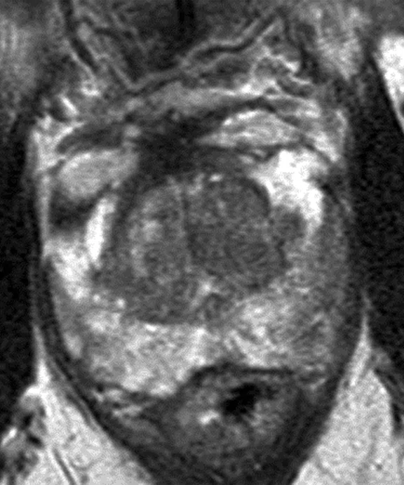
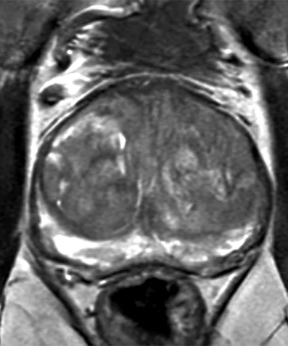
The image quality improvements as a result of the SIGNA™Works AIR™ IQ Edition upgrade now allow for high-quality prostate exams in nearly half the scan time. (A) Axial T2 FRFSE in 3:10 min. and (B) axial T2 FRFSE with AIR™ Recon DL in 1:36 min., representing a 49% reduction in scan time.
B
Figure 3.
The image quality improvements as a result of the SIGNA™Works AIR™ IQ Edition upgrade now allow for high-quality prostate exams in nearly half the scan time. (A) Axial T2 FRFSE in 3:10 min. and (B) axial T2 FRFSE with AIR™ Recon DL in 1:36 min., representing a 49% reduction in scan time.
A
Figure 1.
High SNR is achieved for excellent detail of the pituitary with SIGNA™ Architect Lift and AIR™ Recon DL.
B
Figure 1.
High SNR is achieved for excellent detail of the pituitary with SIGNA™ Architect Lift and AIR™ Recon DL.
A
Figure 2.
High-quality T2, T2 STIR and T2* imaging of the cervical spine using AIR™ Recon DL enables a more clear depiction of spinal cord lesions in a multiple sclerosis patient.
B
Figure 2.
High-quality T2, T2 STIR and T2* imaging of the cervical spine using AIR™ Recon DL enables a more clear depiction of spinal cord lesions in a multiple sclerosis patient.
D
Figure 2.
High-quality T2, T2 STIR and T2* imaging of the cervical spine using AIR™ Recon DL enables a more clear depiction of spinal cord lesions in a multiple sclerosis patient.
C
Figure 2.
High-quality T2, T2 STIR and T2* imaging of the cervical spine using AIR™ Recon DL enables a more clear depiction of spinal cord lesions in a multiple sclerosis patient.
A
Figure 4.
Pelvic imaging with AIR™ Recon DL in a patient with cervical carcinoma.
B
Figure 4.
Pelvic imaging with AIR™ Recon DL in a patient with cervical carcinoma.
C
Figure 4.
Pelvic imaging with AIR™ Recon DL in a patient with cervical carcinoma.
A
Figure 5.
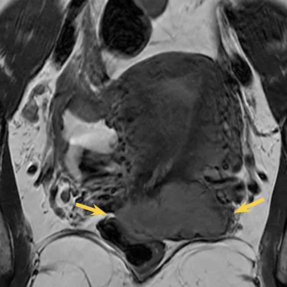
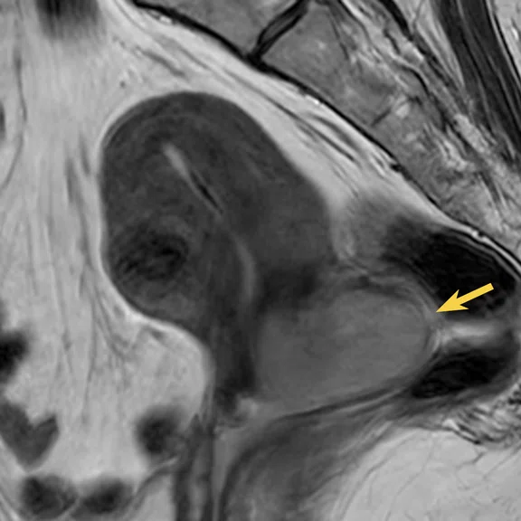
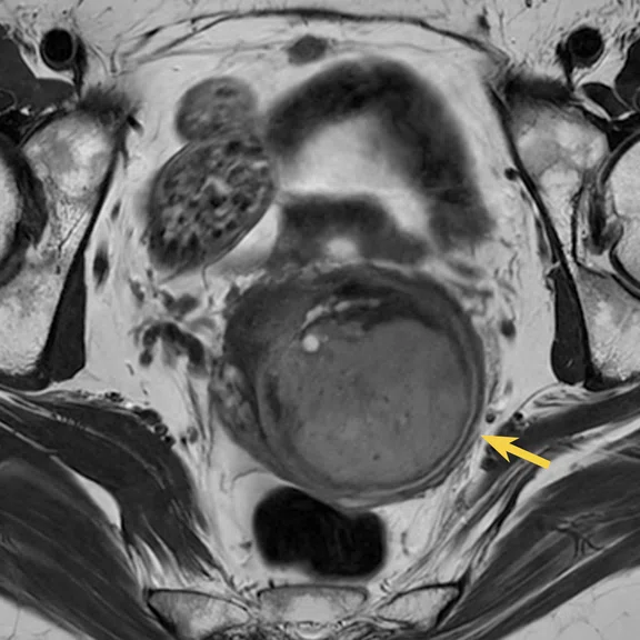
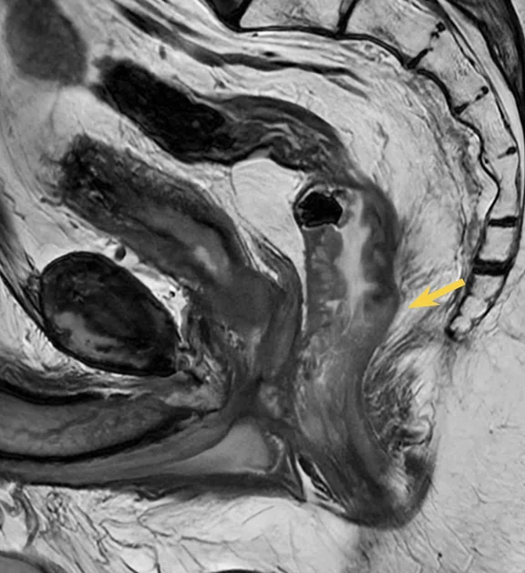
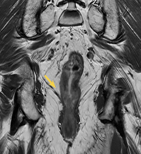
Pelvic imaging in a patient with rectal carcinoma using AIR™ Recon DL.
B
Figure 5.
Pelvic imaging in a patient with rectal carcinoma using AIR™ Recon DL.
‡ AIRTM Recon DL for 3D and PROPELLER is 510(k) pending at US FDA. Not yet CE Marked. Not available for sale in the US or the EU. Not commercially available in all markets.
result


PREVIOUS
${prev-page}
NEXT
${next-page}
Subscribe Now
Manage Subscription
FOLLOW US
Contact Us • Cookie Preferences • Privacy Policy • California Privacy PolicyDo Not Sell or Share My Personal Information • Terms & Conditions • Security
© 2024 GE HealthCare. GE is a trademark of General Electric Company. Used under trademark license.
SPOTLIGHT
A reinvestment that reaps rewards in image quality and scan time
A reinvestment that reaps rewards in image quality and scan time
Investing in a medical imaging system requires balancing cost with capabilities. For many institutions with GE Healthcare MR systems, there is also the option to upgrade an existing magnet with SIGNA™ Lift. SIGNA™ Lift works within a facility’s existing space to transform the whole system around a powerful magnet with all-new electronics, gradients and workstation, as well as the latest SIGNA™Works applications.
Investing in a medical imaging system requires balancing cost with capabilities. For many institutions with GE Healthcare MR systems, there is also the option to upgrade an existing magnet with SIGNA™ Lift. SIGNA™ Lift works within a facility’s existing space to transform the whole system around a powerful magnet with all-new electronics, gradients and workstation, as well as the latest SIGNA™Works applications.
As one of three hospitals in the ASZ healthcare network in Belgium, the 556-bed ASZ Aalst Hospital boasts a very high-volume radiology department, performing 240,000 imaging exams each year. The MR service accounts for 18,750 of these exams and is open 22 hours a day, seven days a week – closing only on Christmas and New Year’s Day.
The Belgian government regulates the number of MR systems installed at any hospital in the country – no MR systems can be installed outside of a hospital – and requires reinvestment in an MR system within seven years after it was first implemented. ASZ Aalst installed a Discovery™ MR750w in 2012.
“We have a lot of clinical activity in the hospital and, therefore, we would not be able to meet the demand with one MR system if we scanned for only 10 or 12 hours each day,” says William Simoens, MD, Chief of Radiology. In addition to inpatients requiring MR imaging, the hospital also accepts referrals for outpatients – those who are not admitted to the hospital.
Due to the large volumes, productivity is of the utmost importance.
“Optimization of the system is key,” he explains. “We also have many more oncologic and complex exams that are more time consuming than 10 years ago, so scan time per exam became increasingly important.”
The demand for oncologic imaging, including diffusion imaging of the breast, prostate, pancreas, rectum and liver, and cardiac MR exams has increased significantly. These are time-consuming exams that also require high spatial resolution and high signal-to-noise ratio (SNR) for accurate and confident diagnoses.
So, in 2019, when ASZ Aalst had to reinvest in MR technology by either buying a new system or upgrading it, Dr. Simoens chose the latter, deciding that the SIGNA™ Architect Lift was the best path for reinvestment.
“The upgrade cost more than the sum we had to reinvest, but we decided this choice provided the most benefit in terms of quality and productivity,” Dr. Simoens says. “The benefits outweighed the extra investment.”
Finding the right balance
One of the key factors in deciding to go with the SIGNA™ Lift upgrade was AIR™ Recon DL, GE’s deep-learning-based image reconstruction algorithm. Beyond just this technology, SIGNA™ Architect delivers a platform for the future that the radiology department at ASZ Aalst can continue to build upon.
Figure 3.
The image quality improvements as a result of the SIGNA™Works AIR™ IQ Edition upgrade now allow for high-quality prostate exams in nearly half the scan time. (A) Axial T2 FRFSE in 3:10 min. and (B) axial T2 FRFSE with AIR™ Recon DL in 1:36 min., representing a 49% reduction in scan time.
AIR™ Recon DL improves SNR and image sharpness without having to overcompensate in the scanning protocol, enabling shorter scan times. Dr. Simoens is utilizing AIR™ Recon DL to achieve a balance between increasing image quality and using technologies such as flexible NEX to reduce scan time.
With the help of GE application specialists, the department reduced scan times as low as possible. Then, Dr. Simoens and other radiologists worked with the applications team to increase scan time to achieve an image quality/ resolution that was satisfactory for each protocol.
“We are now scanning 55 patients each day, an increase of 10% prior to the upgrade. That’s pretty good considering we perform a lot of time-consuming exams such as oncology, cardiac and other special requests, such as limbs.”
Dr. William Simoens
It’s not just the shorter scan times that are having an impact on productivity. The better image quality also gives the radiologists more confidence when reading the exams.
“There is certainly an impact on better lesion detectability because the resolution of each slice is far better, including T2-weighting, and the diffusion is much better, especially with FOCUS,” he explains. “So, our confidence is far higher than it was several years ago.”
Technologists are also pleased with the use of the AIR™ Anterior Array Coil and the AIR™ Multi-Purpose Coil. Previously, they would have to change coils during the exam for some of the more complex studies. Now, with a coil that can be used for multiple body areas, the technologists’ productivity has also increased such that the schedule has been adjusted to reduce patient setup time.
Lumbar spine, cervical spine and pelvic MR exam slots have been reduced in the schedule from 20 to 15 minutes. Dr. Simoens excitedly awaits the regulatory clearance of AIR™ Recon DL for use with 3D and PROPELLER acquisitions‡ so he may further reduce scan times and schedule slots for musculoskeletal exams.
“We were never fully satisfied with our prostate imaging, so we used this technology to increase image quality. On the other hand, our lumbar spine exams were very good, so we invested in scan time reductions. It’s very dependent on the type of exam and the evaluation or choices made by the radiologist on what is most important for the patient,” Dr. Simoens says.
“Every hospital has to make its own decisions and balance costs versus advantages that come with a new system or new technology. For my department, the SIGNA™ Architect with AIR™ Recon DL was definitely a step forward and we cannot wait for this technology to become available on other types of sequences.”
Dr. William Simoens


