A
Figure 2.
Lumbar spine exam on a patient scanned in the lateral decubitus position performed with only the AIR™ AA Coil for high-quality imaging results. (A) T2 FSE; (B) T1 FSE; (C) T2 ASPIR; (D) T2 STIR PROPELLER; and (E-G) T2 FSE. Images courtesy of Emsey Hospital

B
Figure 2.
Lumbar spine exam on a patient scanned in the lateral decubitus position performed with only the AIR™ AA Coil for high-quality imaging results. (A) T2 FSE; (B) T1 FSE; (C) T2 ASPIR; (D) T2 STIR PROPELLER; and (E-G) T2 FSE. Images courtesy of Emsey Hospital

C
Figure 2.
Lumbar spine exam on a patient scanned in the lateral decubitus position performed with only the AIR™ AA Coil for high-quality imaging results. (A) T2 FSE; (B) T1 FSE; (C) T2 ASPIR; (D) T2 STIR PROPELLER; and (E-G) T2 FSE. Images courtesy of Emsey Hospital

D
Figure 2.
Lumbar spine exam on a patient scanned in the lateral decubitus position performed with only the AIR™ AA Coil for high-quality imaging results. (A) T2 FSE; (B) T1 FSE; (C) T2 ASPIR; (D) T2 STIR PROPELLER; and (E-G) T2 FSE. Images courtesy of Emsey Hospital
E
Figure 2.
Lumbar spine exam on a patient scanned in the lateral decubitus position performed with only the AIR™ AA Coil for high-quality imaging results. (A) T2 FSE; (B) T1 FSE; (C) T2 ASPIR; (D) T2 STIR PROPELLER; and (E-G) T2 FSE. Images courtesy of Emsey Hospital
F
Figure 2.
Lumbar spine exam on a patient scanned in the lateral decubitus position performed with only the AIR™ AA Coil for high-quality imaging results. (A) T2 FSE; (B) T1 FSE; (C) T2 ASPIR; (D) T2 STIR PROPELLER; and (E-G) T2 FSE. Images courtesy of Emsey Hospital
G
Figure 2.
Lumbar spine exam on a patient scanned in the lateral decubitus position performed with only the AIR™ AA Coil for high-quality imaging results. (A) T2 FSE; (B) T1 FSE; (C) T2 ASPIR; (D) T2 STIR PROPELLER; and (E-G) T2 FSE. Images courtesy of Emsey Hospital
A
Figure 3.
Knee imaging with the AIR™ AA Coil. (A) PD FatSat PROPELLER; (B) T1 PROPELLER; and (C) PD FatSat PROPELLER. All sequences acquired with a pixel size of 0.7 x 0.7 x 3.5 mm. Images courtesy of Emsey Hospital
B
Figure 3.
Knee imaging with the AIR™ AA Coil. (A) PD FatSat PROPELLER; (B) T1 PROPELLER; and (C) PD FatSat PROPELLER. All sequences acquired with a pixel size of 0.7 x 0.7 x 3.5 mm. Images courtesy of Emsey Hospital
C
Figure 3.
Knee imaging with the AIR™ AA Coil. (A) PD FatSat PROPELLER; (B) T1 PROPELLER; and (C) PD FatSat PROPELLER. All sequences acquired with a pixel size of 0.7 x 0.7 x 3.5 mm. Images courtesy of Emsey Hospital
A
Figure 1.
AIR x™ provides excellent accuracy in slice prescription and reproducible slice planning even in extreme conditions. (A-C) 3-plane localizer helps determine (D) image slice/orientation in the first position so that it matches (E-G) 3-plane localizer and (H) image slice/orientation in the second position. Images courtesy of Emsey Hospital
D
Figure 1.
AIR x™ provides excellent accuracy in slice prescription and reproducible slice planning even in extreme conditions. (A-C) 3-plane localizer helps determine (D) image slice/orientation in the first position so that it matches (E-G) 3-plane localizer and (H) image slice/orientation in the second position. Images courtesy of Emsey Hospital
B
Figure 1.
AIR x™ provides excellent accuracy in slice prescription and reproducible slice planning even in extreme conditions. (A-C) 3-plane localizer helps determine (D) image slice/orientation in the first position so that it matches (E-G) 3-plane localizer and (H) image slice/orientation in the second position. Images courtesy of Emsey Hospital
C
Figure 1.
AIR x™ provides excellent accuracy in slice prescription and reproducible slice planning even in extreme conditions. (A-C) 3-plane localizer helps determine (D) image slice/orientation in the first position so that it matches (E-G) 3-plane localizer and (H) image slice/orientation in the second position. Images courtesy of Emsey Hospital
E
Figure 1.
AIR x™ provides excellent accuracy in slice prescription and reproducible slice planning even in extreme conditions. (A-C) 3-plane localizer helps determine (D) image slice/orientation in the first position so that it matches (E-G) 3-plane localizer and (H) image slice/orientation in the second position. Images courtesy of Emsey Hospital
H
Figure 1.
AIR x™ provides excellent accuracy in slice prescription and reproducible slice planning even in extreme conditions. (A-C) 3-plane localizer helps determine (D) image slice/orientation in the first position so that it matches (E-G) 3-plane localizer and (H) image slice/orientation in the second position. Images courtesy of Emsey Hospital
F
Figure 1.
AIR x™ provides excellent accuracy in slice prescription and reproducible slice planning even in extreme conditions. (A-C) 3-plane localizer helps determine (D) image slice/orientation in the first position so that it matches (E-G) 3-plane localizer and (H) image slice/orientation in the second position. Images courtesy of Emsey Hospital
G
Figure 1.
AIR x™ provides excellent accuracy in slice prescription and reproducible slice planning even in extreme conditions. (A-C) 3-plane localizer helps determine (D) image slice/orientation in the first position so that it matches (E-G) 3-plane localizer and (H) image slice/orientation in the second position. Images courtesy of Emsey Hospital
A
Figure 4.
Patient underwent MR imaging of the pituitary on two different days. With AIR x™, the technologist was able to start and finish the acquisition at the same anatomic position to obtain identical coverage and slice positioning data for comparison of the two studies. (A-C) First day exam with (D) 3-plane localizer and (E-G) the second day exam with (H) 3-plane localizer. Notice the slightly different translation and rotation in the 3-plane localizer images between the first and second day. Images courtesy of Kremlin-Bicetre Hospital
C
Figure 4.
Patient underwent MR imaging of the pituitary on two different days. With AIR x™, the technologist was able to start and finish the acquisition at the same anatomic position to obtain identical coverage and slice positioning data for comparison of the two studies. (A-C) First day exam with (D) 3-plane localizer and (E-G) the second day exam with (H) 3-plane localizer. Notice the slightly different translation and rotation in the 3-plane localizer images between the first and second day. Images courtesy of Kremlin-Bicetre Hospital
D
Figure #.
Figure Copy.
D
Figure 4.
Patient underwent MR imaging of the pituitary on two different days. With AIR x™, the technologist was able to start and finish the acquisition at the same anatomic position to obtain identical coverage and slice positioning data for comparison of the two studies. (A-C) First day exam with (D) 3-plane localizer and (E-G) the second day exam with (H) 3-plane localizer. Notice the slightly different translation and rotation in the 3-plane localizer images between the first and second day. Images courtesy of Kremlin-Bicetre Hospital
B
Figure 4.
Patient underwent MR imaging of the pituitary on two different days. With AIR x™, the technologist was able to start and finish the acquisition at the same anatomic position to obtain identical coverage and slice positioning data for comparison of the two studies. (A-C) First day exam with (D) 3-plane localizer and (E-G) the second day exam with (H) 3-plane localizer. Notice the slightly different translation and rotation in the 3-plane localizer images between the first and second day. Images courtesy of Kremlin-Bicetre Hospital
D
Figure 4.
Patient underwent MR imaging of the pituitary on two different days. With AIR x™, the technologist was able to start and finish the acquisition at the same anatomic position to obtain identical coverage and slice positioning data for comparison of the two studies. (A-C) First day exam with (D) 3-plane localizer and (E-G) the second day exam with (H) 3-plane localizer. Notice the slightly different translation and rotation in the 3-plane localizer images between the first and second day. Images courtesy of Kremlin-Bicetre Hospital
E
Figure 4.
Patient underwent MR imaging of the pituitary on two different days. With AIR x™, the technologist was able to start and finish the acquisition at the same anatomic position to obtain identical coverage and slice positioning data for comparison of the two studies. (A-C) First day exam with (D) 3-plane localizer and (E-G) the second day exam with (H) 3-plane localizer. Notice the slightly different translation and rotation in the 3-plane localizer images between the first and second day. Images courtesy of Kremlin-Bicetre Hospital
F
Figure 4.
Patient underwent MR imaging of the pituitary on two different days. With AIR x™, the technologist was able to start and finish the acquisition at the same anatomic position to obtain identical coverage and slice positioning data for comparison of the two studies. (A-C) First day exam with (D) 3-plane localizer and (E-G) the second day exam with (H) 3-plane localizer. Notice the slightly different translation and rotation in the 3-plane localizer images between the first and second day. Images courtesy of Kremlin-Bicetre Hospital
G
Figure 4.
Patient underwent MR imaging of the pituitary on two different days. With AIR x™, the technologist was able to start and finish the acquisition at the same anatomic position to obtain identical coverage and slice positioning data for comparison of the two studies. (A-C) First day exam with (D) 3-plane localizer and (E-G) the second day exam with (H) 3-plane localizer. Notice the slightly different translation and rotation in the 3-plane localizer images between the first and second day. Images courtesy of Kremlin-Bicetre Hospital
H
Figure 4.
Patient underwent MR imaging of the pituitary on two different days. With AIR x™, the technologist was able to start and finish the acquisition at the same anatomic position to obtain identical coverage and slice positioning data for comparison of the two studies. (A-C) First day exam with (D) 3-plane localizer and (E-G) the second day exam with (H) 3-plane localizer. Notice the slightly different translation and rotation in the 3-plane localizer images between the first and second day. Images courtesy of Kremlin-Bicetre Hospital
D
Figure #.
Figure Copy.
D
Figure 4.
Patient underwent MR imaging of the pituitary on two different days. With AIR x™, the technologist was able to start and finish the acquisition at the same anatomic position to obtain identical coverage and slice positioning data for comparison of the two studies. (A-C) First day exam with (D) 3-plane localizer and (E-G) the second day exam with (H) 3-plane localizer. Notice the slightly different translation and rotation in the 3-plane localizer images between the first and second day. Images courtesy of Kremlin-Bicetre Hospital
D
Figure #.
Figure Copy.


result
PREVIOUS
${prev-page}
NEXT
${next-page}
Subscribe Now
Manage Subscription
FOLLOW US
Contact Us • Cookie Preferences • Privacy Policy • California Privacy PolicyDo Not Sell or Share My Personal Information • Terms & Conditions • Security
© 2024 GE HealthCare. GE is a trademark of General Electric Company. Used under trademark license.
Ahmet Kemal Firat, MD
Emsey Hospital
Istanbul, Turkey

Huseyin Cagil, MD
Emsey Hospital
Istanbul, Turkey
Farida Benoudiba-
Bataille, MD
Kremlin-Bicêtre Hospital
Kremlin-Bicêtre, France
SPOTLIGHT
A simply better MR experience
A simply better MR experience

Ahmet Kemal Firat, MD
Emsey Hospital
Istanbul, Turkey
Huseyin Cagil, MD
Emsey Hospital
Istanbul, Turkey
Farida Benoudiba-
Bataille, MD
Kremlin-Bicêtre Hospital
Kremlin-Bicêtre, France
In patient neuro MR follow-up studies, consistency of image slices is crucial for determining response to therapy or progression of disease. The quality of the exam is also often dependent upon the technologist’s experience. AIR x™ is an AI based automated workflow tool for MR neuro exams that increases consistency and productivity in slice prescriptions for less variability between technologists and between exams.
In patient neuro MR follow-up studies, consistency of image slices is crucial for determining response to therapy or progression of disease. The quality of the exam is also often dependent upon the technologist’s experience. AIR x™ is an AI based automated workflow tool for MR neuro exams that increases consistency and productivity in slice prescriptions for less variability between technologists and between exams.
As one of Turkey’s leading private hospitals, Emsey Hospital provides high-quality healthcare to residents of Istanbul, as well as patients from throughout the region. The radiology department provides a full complement of imaging and interventional services and relies on the advanced technology found across the breadth of GE Healthcare systems. As the hospital embarked on its vision to expand services to international patients, it became apparent that a new 3.0T MR system was needed to complement its existing Brivo™ MR355 1.5T.
To fill this need, the department installed a 3.0T SIGNA™ Pioneer. According to Huseyin Cagil, MD, radiologist, a key factor in selecting the system was the foundation of innovative technologies — from Total Digital Imaging to the AIR™ Coils and AIR x™ to the SIGNA™Works productivity platform of advanced applications. Today, SIGNA™ Pioneer is the fi rst choice for MR imaging at Emsey Hospital, especially for prostate, liver, MSK and neuro.
“AIR x™ has become one of the favorite applications for the technologists and neuroradiologists,” Dr. Cagil explains. “This deep-learning automatic slice prescription tool has brought standardized improvement for almost all our head exams today.”
In addition to automatically prescribing slices for routine brain exams, AIR x™ is also being used for MR exams of the temporal lobe, internal auditory canal, orbits, optic nerves, pituitary gland and the Circle of Willis.
AIR x™ also enables the department to improve the patient experience. Dr. Cagil and Mahmut Erol, MD, radiologist, explain that if the patient needs to move their head or take a break during the exam, they can. That’s because AIR x™ automatically detects anatomy and prescribes the slices to produce images with less variability between scans and technologists. After the technologist performs a scan localizer, AIR x™ handles the rest for reproducible slice planning.
“We see consistently correct slices regardless of the technologist’s experience, which contributes to an easier and more reliable evaluation of head exams by the radiologists,” Dr. Cagil adds.
Neuroimaging is often dependent upon the technologist’s experience in slice prescription. Dr. Erol explains, “This is also important for patients who we follow longitudinally to evaluate the course of their neurological disease. Patients have unique morphologies and we need robust algorithms for these challenging clinical needs. We believe that our decision to implement AIR x™ and SIGNA™ Pioneer now will help with patient follow-up well into the future.”
Dr. Erol adds that patients with neurological or neurodegenerative diseases are often assessed for signal changes in brain structures bilaterally with symmetrical evaluations. He believes the consistency in slices provided by AIR x™ will further improve patient management.
Figure 1.
AIR x™ provides excellent accuracy in slice prescription and reproducible slice planning even in extreme conditions. (A-C) 3-plane localizer helps determine (D) image slice/orientation in the first position so that it matches (E-G) 3-plane localizer and (H) image slice/orientation in the second position. Images courtesy of Emsey Hospital
A
B
C
D
E
F
G
Figure 2.
Lumbar spine exam on a patient scanned in the lateral decubitus position performed with only the AIR™ AA Coil for high-quality imaging results. (A) T2 FSE; (B) T1 FSE; (C) T2 ASPIR; (D) T2 STIR PROPELLER; and (E-G) T2 FSE. Images courtesy of Emsey Hospital
It’s not just AIR x™ that is making a difference in patient care. Emsey Hospital also acquired the AIR™ Anterior Array (AA) Coil with the SIGNA™ Pioneer. Although the AIR™ AA Coil was not yet commercially available when the new system was installed, the radiologists believed the concept of MR coils that conform to the body like a blanket would transform patient comfort and improve image quality.
“The AIR™ AA Coil has significantly improved our SNR and we can use it for many imaging exams, from large to small fields of view and it never disappoints us,” Dr. Cagil says. “In patients who are difficult to position or where traditional coils cannot sufficiently cover their anatomy, the AIR™ AA Coil resolves these situations.”
For example, patients with severe back pain could only be positioned in the decubitus position in lumbar spine exams. Now, the AIR™ AA Coil is placed on the back of these patients. In long bone scans where no dedicated coil was available, the large coverage and proximity to the anatomy with the AIR™ AA Coil provides better images for patient management. In MSK imaging, combining the Posterior Array (PA) Coil with the AIR™ AA Coil enables the technologist to scan both knees simultaneously with excellent resolution from the approximately 30 active channels (between both coils). This approach also helps save exam time by eliminating patient re-positioning.
A
B
C
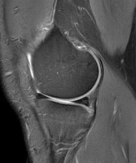
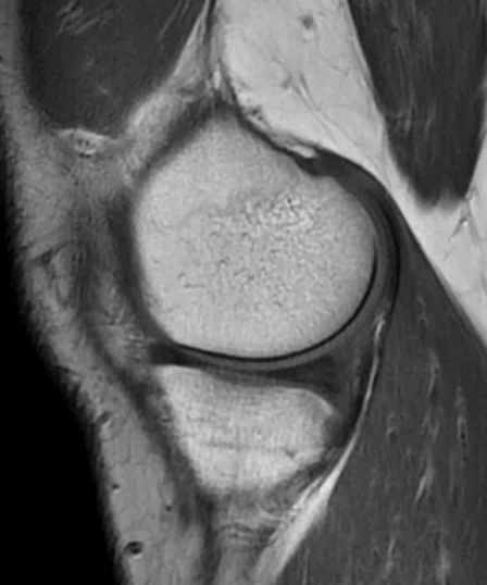
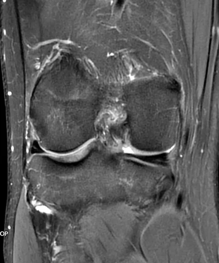
Figure 3.
Knee imaging with the AIR™ AA Coil. (A) PD FatSat PROPELLER; (B) T1 PROPELLER; and (C) PD FatSat PROPELLER. All sequences acquired with a pixel size of 0.7 x 0.7 x 3.5 mm. Images courtesy of Emsey Hospital
“Signal uniformity has significantly improved across all anatomies and coil combinations,” Dr. Cagil adds. “We’ve seen impressive results in dedicated organ studies requiring a small FOV, such as the prostate, female pelvis, pancreas, etc.”
AIR™ AA Coils are also impressive in large FOV exams, particularly in abdominal-pelvic oncology cases. According to Dr. Erol, two traditional hard shell AA coils were previously needed for the large coverage on some patients.
“Now, patients are much more comfortable with the AIR™ AA Coil because it helps them tolerate the exam — and that impacts image quality,” Dr. Erol says. “Also, having acceleration techniques available with the sequences, especially SSFSE, provides clearer imaging results due to less motion captured during the acquisition.”
The feedback from the technologists further supports the radiologists’ view on improved patient comfort. Dr. Erol says they are very positive about the easier patient positioning and flexibility of coverage. They can even slide the AIR™ AA Coil up and down on the patient while they are still inside the bore.
Beyond the benefits of AIR™, Emsey Hospital is also exploring the use of MR Touch, IDEAL IQ and StarMap for a comprehensive liver health program.
According to Ahmet Kemal Firat, MD, Associate Professor of Radiology and an interventional radiologist, these sequences enable the department to tackle the growing incidence of fatty liver diseases.
“We are currently treating approximately 200 patients each year with interventional radio/chemo-embolization,” Professor Firat explains. “We are planning to use these imaging techniques to address liver disease in a larger cohort of patients.”
Professor Firat also anticipates participating in research exploring the use of 3D MR elastography techniques in collaboration with other institutions worldwide.
Reproducibility in slice prescription
Kremlin-Bicêtre Hospital is one of three hospitals in the Paris-Sud University Hospitals that provides comprehensive healthcare within the framework of a teaching and research environment. The hospital is renowned for specializing in neurosurgery, interventional neuroradiology, neurooncology and polytrauma cases.
In 2017, the hospital implemented SIGNA™ Architect and the AIR™ 48-channel Head Coil to further support neuroimaging for surgery, interventions, radiation therapy and head trauma cases. Recently, AIR x™ was added as part of a SIGNA™Works AIR™ Edition productivity platform upgrade at the hospital.
Many patients receiving a neuro MR exam at Kremlin-Bicêtre Hospital are undergoing some type of therapy or intervention for a neuro disease or injury. According to Farida Benoudiba- Bataille, MD, neuroradiologist, AIR x™ saves time for the patient, technologist and the radiologist.
“We have found that AIR x™ can help save time in the patient set up and that means less time that they are immobilized and in the scanner,” Dr. Benoudiba-Bataille explains.
Adds Laure Cacheux, RT(R), technologist, “AIR x™ is very fast in the slice positioning regardless of the required plan. That saves us time because we just need to check the slice prescription from AIR x™.” As a result, the technologists can spend more time addressing any patient concerns, anxiety or stress.
More importantly, since the slice prescription is no longer technologist-dependent, there is greater reproducibility and similarity of the MR slices (images) across studies. This is very important for patient follow-up in oncology, including primary and secondary tumors, neurodegenerative diseases, such as multiple sclerosis, and arteriovenous malformation or other neurosurgery cases.
“In the case of a patient follow-up for tumor assessment, the slice positioning must be as identical as possible to the prior exams in both the axis and the coverage,” says Antony Morel, RT(R), technologist. “While AIR x™ is clearly useful for a newer technologist, even the most experienced technologists may not be able to achieve this precision with every follow-up exam.”
“In the case of a patient follow-up for tumor assessment, the slice positioning must be as identical as possible to the prior exams in both the axis and the coverage,” says Antony Morel, RT(R), technologist. “While AIR x™ is clearly useful for a newer technologist, even the most experienced technologists may not be able to achieve this precision with every follow-up exam.”
Figure 4.
Patient underwent MR imaging of the pituitary on two different days. With AIR x™, the technologist was able to start and finish the acquisition at the same anatomic position to obtain identical coverage and slice positioning data for comparison of the two studies. (A-C) First day exam with (D) 3-plane localizer and (E-G) the second day exam with (H) 3-plane localizer. Notice the slightly different translation and rotation in the 3-plane localizer images between the first and second day. Images courtesy of Kremlin-Bicetre Hospital
Dr. Benoudiba-Bataille explains, “With the same slice prescription and patient position, we are more confident that we can detect a residual tumor in our follow-up MR study, for example. It also assists with precise contouring and reliable measurements needed for therapy planning independent of the radiology reviewer.”
The reliable and reproducible measurement of tumors is a requirement of the Response Evaluation Criteria in Solid Tumors (RECIST), a set of published rules that define when patients respond, stay the same or worsen during treatment. RECIST is used in many cancer trials to evaluate the efficacy of treatments.
D
“The reproducibility in imaging that we can obtain with AIR x™ enhances my confidence in patient follow-up cases, and that allows me to better support my colleagues in oncology and surgery.”
Dr. Farida Benoudiba-Bataille