result
A
Figure 1.
AIR™ Recon DL for consistent image quality. Shown are (A-E) conventional and (F-J) AIR™ Recon DL reconstructions of a shoulder with varied protocol parameters. The standard protocol is depicted in A and F. B and G were obtained with a higher receive bandwidth, C and H with a reduced NEX, D and I had a thinner slice thickness and E and J were obtained with a higher echo train length. Note how the conventional reconstructed images (A-E) show a lower SNR and blurring as a response to parameter changes; however, the AIR™ Recon DL images (F-J) all show excellent SNR and sharp image quality.
B
Figure 1.
AIR™ Recon DL for consistent image quality. Shown are (A-E) conventional and (F-J) AIR™ Recon DL reconstructions of a shoulder with varied protocol parameters. The standard protocol is depicted in A and F. B and G were obtained with a higher receive bandwidth, C and H with a reduced NEX, D and I had a thinner slice thickness and E and J were obtained with a higher echo train length. Note how the conventional reconstructed images (A-E) show a lower SNR and blurring as a response to parameter changes; however, the AIR™ Recon DL images (F-J) all show excellent SNR and sharp image quality.
C
Figure 1.
AIR™ Recon DL for consistent image quality. Shown are (A-E) conventional and (F-J) AIR™ Recon DL reconstructions of a shoulder with varied protocol parameters. The standard protocol is depicted in A and F. B and G were obtained with a higher receive bandwidth, C and H with a reduced NEX, D and I had a thinner slice thickness and E and J were obtained with a higher echo train length. Note how the conventional reconstructed images (A-E) show a lower SNR and blurring as a response to parameter changes; however, the AIR™ Recon DL images (F-J) all show excellent SNR and sharp image quality.
D
Figure 1.
AIR™ Recon DL for consistent image quality. Shown are (A-E) conventional and (F-J) AIR™ Recon DL reconstructions of a shoulder with varied protocol parameters. The standard protocol is depicted in A and F. B and G were obtained with a higher receive bandwidth, C and H with a reduced NEX, D and I had a thinner slice thickness and E and J were obtained with a higher echo train length. Note how the conventional reconstructed images (A-E) show a lower SNR and blurring as a response to parameter changes; however, the AIR™ Recon DL images (F-J) all show excellent SNR and sharp image quality.
E
Figure 1.
AIR™ Recon DL for consistent image quality. Shown are (A-E) conventional and (F-J) AIR™ Recon DL reconstructions of a shoulder with varied protocol parameters. The standard protocol is depicted in A and F. B and G were obtained with a higher receive bandwidth, C and H with a reduced NEX, D and I had a thinner slice thickness and E and J were obtained with a higher echo train length. Note how the conventional reconstructed images (A-E) show a lower SNR and blurring as a response to parameter changes; however, the AIR™ Recon DL images (F-J) all show excellent SNR and sharp image quality.
F
Figure 1.
AIR™ Recon DL for consistent image quality. Shown are (A-E) conventional and (F-J) AIR™ Recon DL reconstructions of a shoulder with varied protocol parameters. The standard protocol is depicted in A and F. B and G were obtained with a higher receive bandwidth, C and H with a reduced NEX, D and I had a thinner slice thickness and E and J were obtained with a higher echo train length. Note how the conventional reconstructed images (A-E) show a lower SNR and blurring as a response to parameter changes; however, the AIR™ Recon DL images (F-J) all show excellent SNR and sharp image quality.
G
Figure 1.
AIR™ Recon DL for consistent image quality. Shown are (A-E) conventional and (F-J) AIR™ Recon DL reconstructions of a shoulder with varied protocol parameters. The standard protocol is depicted in A and F. B and G were obtained with a higher receive bandwidth, C and H with a reduced NEX, D and I had a thinner slice thickness and E and J were obtained with a higher echo train length. Note how the conventional reconstructed images (A-E) show a lower SNR and blurring as a response to parameter changes; however, the AIR™ Recon DL images (F-J) all show excellent SNR and sharp image quality.
H
Figure 1.
AIR™ Recon DL for consistent image quality. Shown are (A-E) conventional and (F-J) AIR™ Recon DL reconstructions of a shoulder with varied protocol parameters. The standard protocol is depicted in A and F. B and G were obtained with a higher receive bandwidth, C and H with a reduced NEX, D and I had a thinner slice thickness and E and J were obtained with a higher echo train length. Note how the conventional reconstructed images (A-E) show a lower SNR and blurring as a response to parameter changes; however, the AIR™ Recon DL images (F-J) all show excellent SNR and sharp image quality.
I
Figure 1.
AIR™ Recon DL for consistent image quality. Shown are (A-E) conventional and (F-J) AIR™ Recon DL reconstructions of a shoulder with varied protocol parameters. The standard protocol is depicted in A and F. B and G were obtained with a higher receive bandwidth, C and H with a reduced NEX, D and I had a thinner slice thickness and E and J were obtained with a higher echo train length. Note how the conventional reconstructed images (A-E) show a lower SNR and blurring as a response to parameter changes; however, the AIR™ Recon DL images (F-J) all show excellent SNR and sharp image quality.
J
Figure 1.
AIR™ Recon DL for consistent image quality. Shown are (A-E) conventional and (F-J) AIR™ Recon DL reconstructions of a shoulder with varied protocol parameters. The standard protocol is depicted in A and F. B and G were obtained with a higher receive bandwidth, C and H with a reduced NEX, D and I had a thinner slice thickness and E and J were obtained with a higher echo train length. Note how the conventional reconstructed images (A-E) show a lower SNR and blurring as a response to parameter changes; however, the AIR™ Recon DL images (F-J) all show excellent SNR and sharp image quality.
1. Peters RD, Harris H, Lawson S. The clinical benefits of AIR™ Recon DL for MR image reconstruction. GE SIGNA Pulse of MR, Autumn 2020, pages 77-80. Longer version available http://tinyurl.com/AIR-Recon-DL-whitepaper
2. Peters RD, Blahnik H, Harris H, Lawson S. Practical protocol conversion and optimization with AIR Recon DL. GE SIGNA Pulse of MR, Autumn 2020, pages 81-83.
2. Peters RD, Blahnik H, Harris H, Lawson S. Practical protocol conversion and optimization with AIR Recon DL. GE SIGNA Pulse of MR, Autumn 2020, pages 81-83.
A
Figure 1.
AIR™ Recon DL for consistent image quality. Shown are (A-E) conventional and (F-J) AIR™ Recon DL reconstructions of a shoulder with varied protocol parameters. The standard protocol is depicted in A and F. B and G were obtained with a higher receive bandwidth, C and H with a reduced NEX, D and I had a thinner slice thickness and E and J were obtained with a higher echo train length. Note how the conventional reconstructed images (A-E) show a lower SNR and blurring as a response to parameter changes; however, the AIR™ Recon DL images (F-J) all show excellent SNR and sharp image quality.
B
Figure 1.
AIR™ Recon DL for consistent image quality. Shown are (A-E) conventional and (F-J) AIR™ Recon DL reconstructions of a shoulder with varied protocol parameters. The standard protocol is depicted in A and F. B and G were obtained with a higher receive bandwidth, C and H with a reduced NEX, D and I had a thinner slice thickness and E and J were obtained with a higher echo train length. Note how the conventional reconstructed images (A-E) show a lower SNR and blurring as a response to parameter changes; however, the AIR™ Recon DL images (F-J) all show excellent SNR and sharp image quality.
C
Figure 1.
AIR™ Recon DL for consistent image quality. Shown are (A-E) conventional and (F-J) AIR™ Recon DL reconstructions of a shoulder with varied protocol parameters. The standard protocol is depicted in A and F. B and G were obtained with a higher receive bandwidth, C and H with a reduced NEX, D and I had a thinner slice thickness and E and J were obtained with a higher echo train length. Note how the conventional reconstructed images (A-E) show a lower SNR and blurring as a response to parameter changes; however, the AIR™ Recon DL images (F-J) all show excellent SNR and sharp image quality.
D
Figure 1.
AIR™ Recon DL for consistent image quality. Shown are (A-E) conventional and (F-J) AIR™ Recon DL reconstructions of a shoulder with varied protocol parameters. The standard protocol is depicted in A and F. B and G were obtained with a higher receive bandwidth, C and H with a reduced NEX, D and I had a thinner slice thickness and E and J were obtained with a higher echo train length. Note how the conventional reconstructed images (A-E) show a lower SNR and blurring as a response to parameter changes; however, the AIR™ Recon DL images (F-J) all show excellent SNR and sharp image quality.
E
Figure 1.
AIR™ Recon DL for consistent image quality. Shown are (A-E) conventional and (F-J) AIR™ Recon DL reconstructions of a shoulder with varied protocol parameters. The standard protocol is depicted in A and F. B and G were obtained with a higher receive bandwidth, C and H with a reduced NEX, D and I had a thinner slice thickness and E and J were obtained with a higher echo train length. Note how the conventional reconstructed images (A-E) show a lower SNR and blurring as a response to parameter changes; however, the AIR™ Recon DL images (F-J) all show excellent SNR and sharp image quality.
F
Figure 1.
AIR™ Recon DL for consistent image quality. Shown are (A-E) conventional and (F-J) AIR™ Recon DL reconstructions of a shoulder with varied protocol parameters. The standard protocol is depicted in A and F. B and G were obtained with a higher receive bandwidth, C and H with a reduced NEX, D and I had a thinner slice thickness and E and J were obtained with a higher echo train length. Note how the conventional reconstructed images (A-E) show a lower SNR and blurring as a response to parameter changes; however, the AIR™ Recon DL images (F-J) all show excellent SNR and sharp image quality.
G
Figure 1.
AIR™ Recon DL for consistent image quality. Shown are (A-E) conventional and (F-J) AIR™ Recon DL reconstructions of a shoulder with varied protocol parameters. The standard protocol is depicted in A and F. B and G were obtained with a higher receive bandwidth, C and H with a reduced NEX, D and I had a thinner slice thickness and E and J were obtained with a higher echo train length. Note how the conventional reconstructed images (A-E) show a lower SNR and blurring as a response to parameter changes; however, the AIR™ Recon DL images (F-J) all show excellent SNR and sharp image quality.
H
Figure 1.
AIR™ Recon DL for consistent image quality. Shown are (A-E) conventional and (F-J) AIR™ Recon DL reconstructions of a shoulder with varied protocol parameters. The standard protocol is depicted in A and F. B and G were obtained with a higher receive bandwidth, C and H with a reduced NEX, D and I had a thinner slice thickness and E and J were obtained with a higher echo train length. Note how the conventional reconstructed images (A-E) show a lower SNR and blurring as a response to parameter changes; however, the AIR™ Recon DL images (F-J) all show excellent SNR and sharp image quality.
I
Figure 1.
AIR™ Recon DL for consistent image quality. Shown are (A-E) conventional and (F-J) AIR™ Recon DL reconstructions of a shoulder with varied protocol parameters. The standard protocol is depicted in A and F. B and G were obtained with a higher receive bandwidth, C and H with a reduced NEX, D and I had a thinner slice thickness and E and J were obtained with a higher echo train length. Note how the conventional reconstructed images (A-E) show a lower SNR and blurring as a response to parameter changes; however, the AIR™ Recon DL images (F-J) all show excellent SNR and sharp image quality.
J
Figure 1.
AIR™ Recon DL for consistent image quality. Shown are (A-E) conventional and (F-J) AIR™ Recon DL reconstructions of a shoulder with varied protocol parameters. The standard protocol is depicted in A and F. B and G were obtained with a higher receive bandwidth, C and H with a reduced NEX, D and I had a thinner slice thickness and E and J were obtained with a higher echo train length. Note how the conventional reconstructed images (A-E) show a lower SNR and blurring as a response to parameter changes; however, the AIR™ Recon DL images (F-J) all show excellent SNR and sharp image quality.


PREVIOUS
${prev-page}
NEXT
${next-page}
Subscribe Now
Manage Subscription
FOLLOW US
Contact Us • Cookie Preferences • Privacy Policy • California Privacy PolicyDo Not Sell or Share My Personal Information • Terms & Conditions • Security
© 2024 GE HealthCare. GE is a trademark of General Electric Company. Used under trademark license.
SPOTLIGHT
AIR Recon DL for consistent image quality and exam time reduction
AIR Recon DL for consistent image quality and exam time reduction
by Robert D. Peters, PhD, Global Product Marketing Director, Nishant Gupta, Business Analytics Leader and Yuko Ueda, Global Product Marketing Director, GE Healthcare
The benefits of AIR™ Recon DL
The clinical, operational and financial benefits of AIR™ Recon DL have been described in several recent articles1,2. In addition to the well-known capability of increasing SNR and delivering sharper images, AIR™ Recon DL provides for improved contrast-to-noise ratio (CNR), reconstructs images directly at the MR console and is compatible across all anatomies. When used with optimized protocols, AIR™ Recon DL can lead to considerably reduced scan and exam times2. Moreover, early-adopter feedback suggests that AIR™ Recon DL images are faster and easier to read, resulting in less eye fatigue.
Figure 1.
AIR™ Recon DL for consistent image quality. Shown are (A-E) conventional and (F-J) AIR™ Recon DL reconstructions of a shoulder with varied protocol parameters. The standard protocol is depicted in A and F. B and G were obtained with a higher receive bandwidth, C and H with a reduced NEX, D and I had a thinner slice thickness and E and J were obtained with a higher echo train length. Note how the conventional reconstructed images (A-E) show a lower SNR and blurring as a response to parameter changes; however, the AIR™ Recon DL images (F-J) all show excellent SNR and sharp image quality.
Tolerant of protocol variation, leading to greater consistency
AIR™ Recon DL is a tool that can expand the clinical protocol space, which is typically bound by clinically acceptable thresholds for SNR, voxel volume and scan time. Essentially, by increasing this usable clinical protocol space, AIR™ Recon DL allows scan operators to modify a multitude of sequence parameters without the consequences of poor SNR and image quality.
For example, repeating a conventional scan acquired with acceptable SNR and image quality could result in an unpredictable mix of poor SNR and blurriness if some of the scan operators were to change protocol parameters. However, the same scans obtained with AIR™ Recon DL will be considerably more tolerant of protocol parameter changes, due to the larger clinical protocol space in which SNR and image quality is preserved.
Consider the shoulder image example in Figure 1. Due to respiratory motion artifacts, shoulder scans are frequently repeated. Figure 1A-E images represent repeat conventional scans with variations in bandwidth, number of excitations (NEX), slice thickness and echo train length (ETL), all resulting in a mix of poor SNR and blurriness. The same scan data reconstructed with AIR™ Recon DL (Figure 1F-J) all present excellent SNR and sharpness. Simply put, AIR™ Recon DL is tolerant of protocol variation and delivers greater image quality consistency.
More predictable scan times
In September 2020, AIR™ Recon DL was installed at several early adopter sites globally to provide feedback in clinical practice. During this period, GE Healthcare received usage statistics both before and after the installation.
Of particular importance was how AIR™ Recon DL led to shorter individual scan times and, ultimately, total exam times.
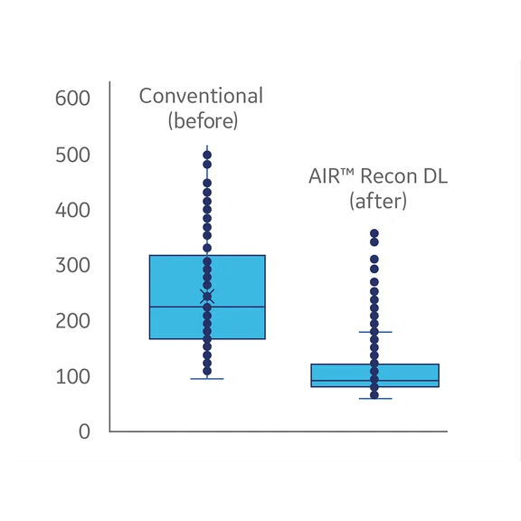
Figure 2 contains a representative summary from one such early adopter site showing the mean and standard deviation of individual anatomical scans with conventional and AIR™ Recon DL reconstruction. In this example of 2D FSE scans in the pelvis, the mean scan time is reduced from 242 seconds to 108 seconds. The standard deviation also reduced from 100 seconds to 44 seconds, which suggests that AIR™ Recon DL delivers shorter and more predictable scan times.
Shorter exam and slot times
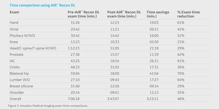
Capable of delivering consistently shorter individual scan times while improving image quality, AIR™ Recon DL has generated enthusiasm among the early adopters for reducing the total exam times, allowing for reduced slot times and greater patient throughput. As presented in a recent GE webinar, Randall Stenoien, MD, owner and CEO of Houston Medical Imaging, summarized his center’s exam time reductions for various anatomies with AIR™ Recon DL (Figure 3). Through consistent and predictable individual scan time reductions, this site realized an average 48% reduction in exam times and was able to adjust its schedule with considerably shorter slot times.
Summary
AIR™ Recon DL offers many clinical, operational and financial benefits – yet one benefit that can be overlooked is its tolerance to protocol variation and, thus, its capability to deliver consistent image quality. Consistent image quality, in turn, leads to shorter and more predictable scan times, which can now be quantified at AIR™ Recon DL early adopter sites. In conclusion, AIR™ Recon DL can lead to shorter net exam times, more patients scanned per day and a faster return on investment.
Figure 1.
AIR™ Recon DL for consistent image quality. Shown are (A-E) conventional and (F-J) AIR™ Recon DL reconstructions of a shoulder with varied protocol parameters. The standard protocol is depicted in A and F. B and G were obtained with a higher receive bandwidth, C and H with a reduced NEX, D and I had a thinner slice thickness and E and J were obtained with a higher echo train length. Note how the conventional reconstructed images (A-E) show a lower SNR and blurring as a response to parameter changes; however, the AIR™ Recon DL images (F-J) all show excellent SNR and sharp image quality.

Figure 2.
AIR™ Recon DL for consistent scan times. Shown is a box plot of the distribution of individual 2D FSE pelvis scans from the same early-adopter site, before and after installation of AIR™ Recon DL.


