‡Not licensed in accordance with Canadian law. Not available for sale in Canada. Not CE marked. Not available for sale in all regions.
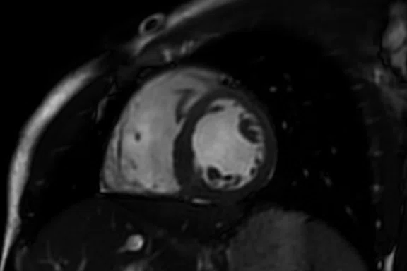
Figure 1.
Short axis 2D FIESTA. The combination of SIGNA™ Premier and AIR Technology™ delivers high SNR and high image quality for excellent cardiac MR imaging results at 3.0T.
A
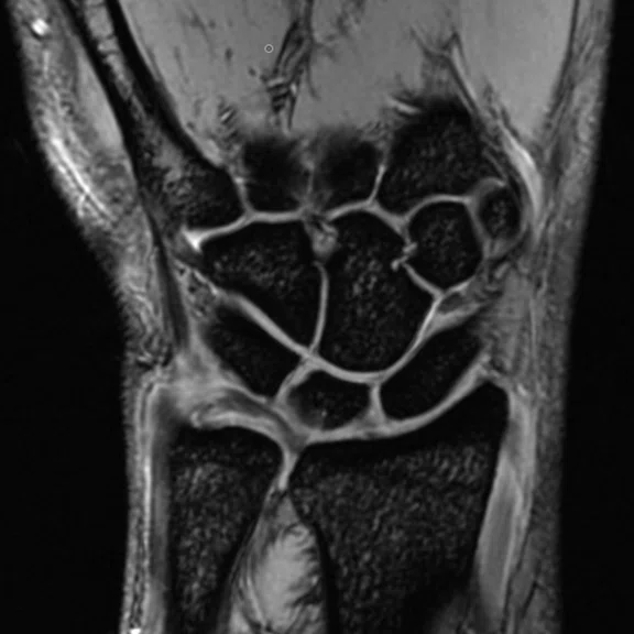
Figure 2.
AIR Technology™ Suite is flexible and assists with patient positioning in areas where coil selection may be more difficult, such as the
wrist. (A) Coronal 3D MERGE; (B) Coronal PD FatSat; and (C) Coronal T2 Flex.
B
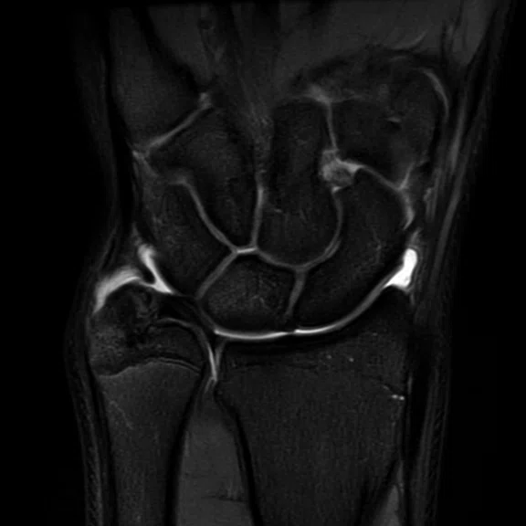
Figure 2.
AIR Technology™ Suite is flexible and assists with patient positioning in areas where coil selection may be more difficult, such as the
wrist. (A) Coronal 3D MERGE; (B) Coronal PD FatSat; and (C) Coronal T2 Flex.
C
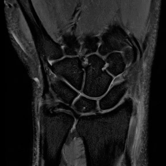
Figure 2.
AIR Technology™ Suite is flexible and assists with patient positioning in areas where coil selection may be more difficult, such as the
wrist. (A) Coronal 3D MERGE; (B) Coronal PD FatSat; and (C) Coronal T2 Flex.
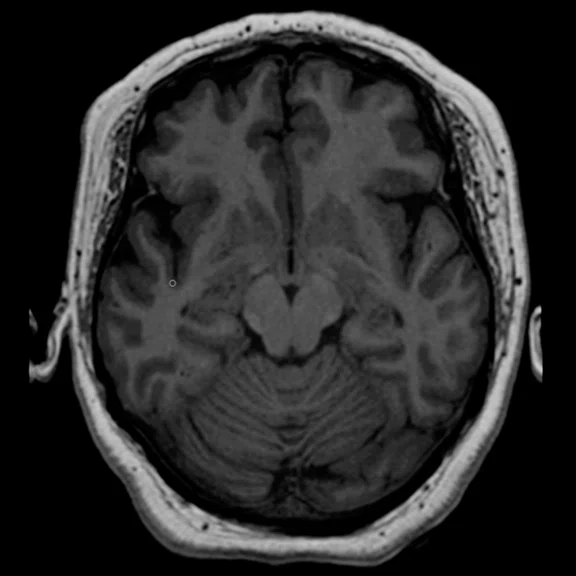
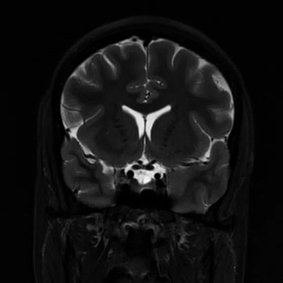
A
Figure 3.
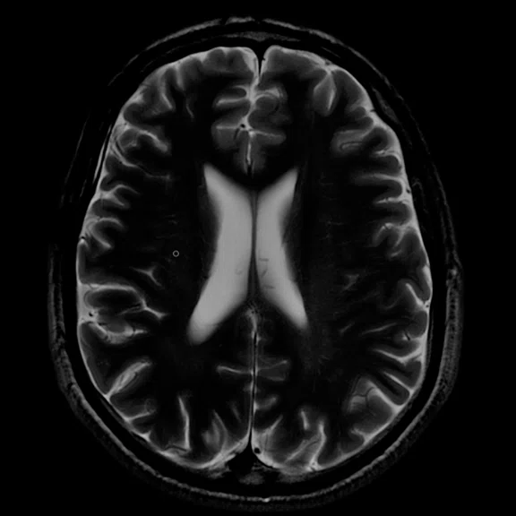
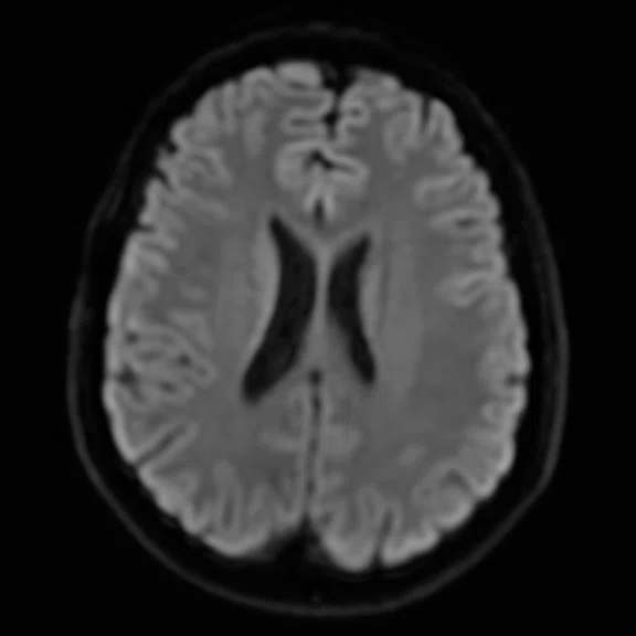
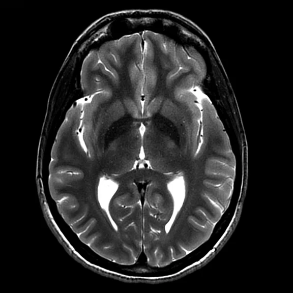
The 48-channel Head Coil delivers more signal from the cortical, basal ganglia and deep brain. (A) Axial 3D T1 FSPGR; (B) Axial T2; (C) Axial DWI b1000; (D) Axial T2 PROPELLER; and (E) Coronal T2 PROPELLER FatSat.
B
Figure 3.
The 48-channel Head Coil delivers more signal from the cortical, basal ganglia and deep brain. (A) Axial 3D T1 FSPGR; (B) Axial T2; (C) Axial DWI b1000; (D) Axial T2 PROPELLER; and (E) Coronal T2 PROPELLER FatSat.
C
Figure 3.
The 48-channel Head Coil delivers more signal from the cortical, basal ganglia and deep brain. (A) Axial 3D T1 FSPGR; (B) Axial T2; (C) Axial DWI b1000; (D) Axial T2 PROPELLER; and (E) Coronal T2 PROPELLER FatSat.
D
Figure 3.
The 48-channel Head Coil delivers more signal from the cortical, basal ganglia and deep brain. (A) Axial 3D T1 FSPGR; (B) Axial T2; (C) Axial DWI b1000; (D) Axial T2 PROPELLER; and (E) Coronal T2 PROPELLER FatSat.
E
Figure 3.
The 48-channel Head Coil delivers more signal from the cortical, basal ganglia and deep brain. (A) Axial 3D T1 FSPGR; (B) Axial T2; (C) Axial DWI b1000; (D) Axial T2 PROPELLER; and (E) Coronal T2 PROPELLER FatSat.
‡‡ Drug products should be used in accordance with their approved labeling. Gadolinium-based contrast agents have not been approved for cardiac use in all regions.
result


PREVIOUS
${prev-page}
NEXT
${next-page}
Subscribe Now
Manage Subscription
FOLLOW US
Contact Us • Cookie Preferences • Privacy Policy • California Privacy PolicyDo Not Sell or Share My Personal Information • Terms & Conditions • Security
© 2024 GE HealthCare. GE is a trademark of General Electric Company. Used under trademark license.
IN PRACTICE
AIR Technology: a brilliant improvement in high-quality imaging and patient comfort
AIR Technology: a brilliant improvement in high-quality imaging and patient comfort
As one of the first sites in the world to install SIGNA™ Premier and AIR Technology™‡, Erasmus Medical Center is a leader in adopting cutting-edge technologies. These new solutions are providing a better patient experience while delivering high-quality imaging and advanced applications, further enhancing the excellent care provided by clinicians at Erasmus.
As one of the first sites in the world to install SIGNA™ Premier and AIR Technology™‡, Erasmus Medical Center is a leader in adopting cutting-edge technologies. These new solutions are providing a better patient experience while delivering high-quality imaging and advanced applications, further enhancing the excellent care provided by clinicians at Erasmus.
Erasmus Medical Center in Rotterdam, Netherlands, is a leading university medical center in Europe and has long been recognized for its adoption of cutting-edge technologies and advanced medical solutions. For the last few years, Erasmus has collaborated with GE Healthcare to evaluate the introduction of new technologies into the clinical environment. One of these is AIR Technology™.
AIR Technology™ Coils are designed to fit all patients, allow flexibility in any direction and closely wrap around the patient’s anatomy for greater visibility of hard-to-scan areas with excellent image quality. By conforming to the patient habitus and bringing the coil elements closer to the patient, AIR Technology™ improves signal quality and signal-to-noise ratio (SNR) and reduces imaging artifacts when compared to previous generations of conventional coil technology.
Recently, several clinicians from Erasmus shared their initial impression of AIR Technology™ on the SIGNA™ Premier 3.0T MR system, including the AIR Technology™ Anterior Array (AA), the 48-channel Head Coil and AIR Touch™.
Cardiac imaging
Alexander Hirsch, MD, cardiologist, specializes in non-invasive cardiac imaging. In cardiac patients, Dr. Hirsch scans cardiomyopathy and ischemic heart disease patients on SIGNA™ Premier. Typically, the 2D FIESTA, first-pass perfusion and MDE images are the most common sequences for these patients.‡‡ Image quality is important, particularly in the late enhancement (MDE) sequence where Dr. Hirsch evaluates myocardial viability. With the 2D FIESTA sequence, he is looking at cardiac function. However, 2D FIESTA sequences have historically been problematic at 3.0T.
"The new SIGNA™ Premier system is especially good for late enhancement images and also for perfusion," Dr. Hirsch says. "I was able to see the anatomy and the function, as well as differentiate the contrast between the blood and the myocardium. Previously in a 3.0T system, that was a problem, however, with the SIGNA™ Premier this has improved a lot."
A key factor in the improved image quality is AIR Technology™. Dr. Hirsch says he gets a more homogeneous signal and better contrast between the blood and the myocardium.
IN PRACTICE
AIR Technology: a brilliant improvement in high-quality imaging and patient comfort
AIR Technology: a brilliant improvement in high-quality imaging and patient comfort
As one of the first sites in the world to install SIGNA™ Premier and AIR Technology™‡, Erasmus Medical Center is a leader in adopting cutting-edge technologies. These new solutions are providing a better patient experience while delivering high-quality imaging and advanced applications, further enhancing the excellent care provided by clinicians at Erasmus
As one of the first sites in the world to install SIGNA™ Premier and AIR Technology™‡, Erasmus Medical Center is a leader in adopting cutting-edge technologies. These new solutions are providing a better patient experience while delivering high-quality imaging and advanced applications, further enhancing the excellent care provided by clinicians at Erasmus
Erasmus Medical Center in Rotterdam, Netherlands, is a leading university medical center in Europe and has long been recognized for its adoption of cutting-edge technologies and advanced medical solutions. For the last few years, Erasmus has collaborated with GE Healthcare to evaluate the introduction of new technologies into the clinical environment. One of these is AIR Technology™.
AIR Technology™ Coils are designed to fit all patients, allow flexibility in any direction and closely wrap around the patient’s anatomy for greater visibility of hard-to-scan areas with excellent image quality. By conforming to the patient habitus and bringing the coil elements closer to the patient, AIR Technology™ improves signal quality and signal-to-noise ratio (SNR) and reduces imaging artifacts when compared to previous generations of conventional coil technology.
Recently, several clinicians from Erasmus shared their initial impression of AIR Technology™ on the SIGNA™ Premier 3.0T MR system, including the AIR Technology™ Anterior Array (AA), the 48-channel Head Coil and AIR Touch™.
Cardiac imaging
Alexander Hirsch, MD, cardiologist, specializes in non-invasive cardiac imaging. In cardiac patients, Dr. Hirsch scans cardiomyopathy and ischemic heart disease patients on SIGNA™ Premier. Typically, the 2D FIESTA, first-pass perfusion and MDE images are the most common sequences for these patients.‡‡ Image quality is important, particularly in the late enhancement (MDE) sequence where Dr. Hirsch evaluates myocardial viability. With the 2D FIESTA sequence, he is looking at cardiac function. However, 2D FIESTA sequences have historically been problematic at 3.0T.
"The new SIGNA™ Premier system is especially good for late enhancement images and also for perfusion," Dr. Hirsch says. "I was able to see the anatomy and the function, as well as differentiate the contrast between the blood and the myocardium. Previously in a 3.0T system, that was a problem, however, with the SIGNA™ Premier this has improved a lot."
A key factor in the improved image quality is AIR Technology™. Dr. Hirsch says he gets a more homogeneous signal and better contrast between the blood and the myocardium.
"Because of the specialized nature of our facility, with referrals from all over the Netherlands, it is important to have the latest technology," he says. "With the new GE SIGNA™ Premier and AIR Technology™, we can provide high-quality care for our patients."
"The new AIR Technology™ AA has a major advantage in that it helps provide high image quality," Dr. Hirsch adds. Plus, with SIGNA™ Premier he has been able to achieve high SNR, which is very important for the sequences he is using. Dr. Hirsch also expects to see improvements in 4D Flow (ViosWorks), as well as the new 3D MDE sequence.
"When we started working with SIGNA™ Premier, I was pleasantly surprised to see the image quality, especially for the 2D FIESTA sequence," he says.
Brendan Bakker, MR radiographer, has developed cardiac MR (CMR) protocols at Erasmus with Dr. Hirsch. While 1.5T was typically preferred for CMR, he worked with Dr. Hirsch to evaluate CMR exams on the SIGNA™ Premier 3.0T MR system with AIR Technology™.
"The AIR Technology™ AA is brilliant and it’s an improvement for the patient. It is very easy to handle, very lightweight and the quality is very good for cardiac imaging, especially on the SIGNA™ Premier system," Bakker says.
"AIR Technology™ is very flexible, you can put it around the chest or stomach but also use it around the knee or shoulders," Bakker says. "With other coils that are more rigid, this is not possible."
In pediatric imaging, the AIR Technology™ Coils fit almost like a blanket on the child, he adds.
MSK imaging
Edwin Oei, MD, PhD, is an Associate Professor of Musculoskeletal Imaging and Section Chief of Musculoskeletal Radiology at Erasmus Medical Center. He dedicates half his time to research and working with MR physicists and PhD students to improve technologies and apply MR imaging in population health studies.
"SIGNA™ Premier offers advantages in musculoskeletal imaging because of its higher gradient performance, especially when it is used with the AIR Technology™ Coil," Professor Oei says.
According to Professor Oei, musculoskeletal (MSK) MR imaging tends to suffer from artifacts and movement more than in other body parts. Often, there are difficulties with positioning patients due to their injury or ailment, as well as using the right coil. While coil selection is not as problematic in the knee or ankle, it can be more difficult when imaging the shoulder, wrists or ribs.
"With AIR Technology™, we are more flexible in choosing the coil, which allows for imaging specific body parts with greater accuracy. For patients with chronic diseases such as arthritis, it may not be easy for them to lie still in the scanner for a long time with a rigid coil. AIR Technology™ is lighter and more comfortable for the patient so, indirectly, I think it will also reduce movement artifacts."
Professor Edwin Oei
AIR Technology™ also assists with patient positioning. When using traditional rigid coils, the body part being imaged had to be positioned precisely in the coil. With AIR Technology™, this is less of an issue.
"We mainly now use the blanket-type AA Coil and have achieved great imaging results in the chest wall and in joints," adds Professor Oei. "I think AIR Technology™ is beneficial for diverse patient groups, including pediatric and elderly patients."
Professor Oei believes there is a movement in MR imaging toward whole-body imaging, particularly for oncology. He anticipates that AIR Technology™ will provide excellent results over existing coil technology due to its wide coverage.
"Since the introduction of SIGNA™ Premier and AIR Technology™ at Erasmus, I’ve seen image quality improve over previous scans and I believe that AIR Technology™ can greatly improve patient throughput," Professor Oei says.
The AIR Technology™ Suite also includes a 48-channel Head Coil. Jean Paul Laarhoven, MR radiographer, has scanned patients with both the 48-channel Head Coil and the AIR Technology™ AA on SIGNA™ Premier. With the ability to adjust the coil for larger-sized heads and necks, he can accommodate more patients. He has found that patients with anxiety or claustrophobia can better tolerate the 48-channel Head Coil because the front part of the coil is slightly smaller and doesn’t cover the patient’s entire face.
"You can immediately see the high-quality images that the AIR Technology™ Coil captures," Laarhoven explains. "Of course, we also have the AIR Technology™ Posterior Array (PA) in the table so we only have to position the AIR Technology™ AA on top of the patient."
Improving the patient experience
Sita Ramman has been an MR radiographer for nearly 28 years at Erasmus. Often, she has had to comfort and reassure patients who are nervous about their MR exams. She will explain that they have to remain very still and may have to hold their breath while the system acquires the images.
Since the introduction of AIR Technology™, she has seen a noticeable difference.
"The patients like the AIR Technology™ Coils because they are very lightweight and flexible, and mold to the patient’s anatomy," Ramman says. "For us, it is very easy to position. You just put it on the patient and that’s it. That’s all you have to do."
She has also used AIR Touch™, an intelligent coil localization and selection tool that enables automatic coil element selection and uniquely optimizes uniformity and SNR. AIR Touch™ informs the system when the coil is connected, allows the technologist to landmark the patient with a single touch and even optimizes the element configuration. Coil coverage, uniformity and parallel imaging acceleration are generated dynamically to optimize image quality. A simplified user interface allows the technologist to focus on the patient and also maximizes examination efficiency.
"We just put the AIR Technology™ Coil on the patient, localize using the AIR Touch™ button on the table and move the patient inside the SIGNA Premier," Ramman explains. "With AIR Technology™ and AIR Touch™, we don’t need to do any calibration as it is done automatically. This makes a difference in our daily routine because it takes less time to position a patient."
A remarkable advance
Juan Hernandez Tamames, PhD, Associate Professor (MR) and Head of the MR Physics group in the radiology department at Erasmus, facilitates the introduction of new technology in MR imaging for both clinical and research purposes.
"SIGNA™ Premier incorporates several new approaches and breakthroughs in technology," Professor Tamames says. "For example, the AIR Technology™ Coils are one of the most remarkable innovations I’ve seen because they increase SNR."
He also discovered that the HyperBand capability on SIGNA™ Premier enables the possibility to simultaneously scan several slices, accelerating acquisition with the potential to shorten scan times when using DWI. With the parallel transmission, he can tailor the RF for specific tissues in a more appropriate way.
"Compressed sensing is another remarkable advance on SIGNA™ Premier," Professor Tamames adds. "When used with AIR Technology™, which improves signal due to the closer proximity to the patient anatomy and tissue, we can increase the acceleration with compressed sensing and parallel imaging to reduce scan times."
For example, since the lungs are filled with air, it is often difficult to obtain good SNR. Because the AIR Technology™ AA lays on the patient’s chest, it is as close to the body as possible. This enables a high SNR.
Another advantage is in pediatric imaging. Professor Tamames says a baby can be wrapped in the coil, which makes them more comfortable and enables the coil to get closer to the anatomy.
"In general, AIR Technology™ is more convenient and it can fit almost any sized anatomy," adds Professor Tamames.
Professor Tamames is interested in testing the AIR Technology™ AA with a conventional head coil and also with the 48-channel Head Coil.
"With 48-channels we can accelerate more because we have a really good, high-quality signal," Professor Tamames explains. "By accelerating, we reduce the echo time, which means less distortion in an EPI sequence. And that is important for exploring the basal ganglia and frontal or temporal areas. Not only is the signal better, but the anatomy and morphology of the tissue is more realistic."
And, because the headset for the 48-channel Head Coil is compatible with EEG systems, clinicians at Erasmus can simultaneously record EEG and capture MR images. Professor Tamames also sees the potential for continued innovation in technology and sequences to shorten MR scan times, in some cases to as quick as five or 10 minutes.
"I think SIGNA™ Premier and AIR Technology™ are paving the way to achieve this goal," Professor Tamames adds.


