A
Figure 2.
Same patient as Figure 1. (A) Sagittal Inhance 3D Velocity NCE MRA; (B) Sagittal 3D MERGE; and (C) Volume rendered 3D MERGE fused with NCE MRA and FSE Flex.
B
Figure 2.
Same patient as Figure 1. (A) Sagittal Inhance 3D Velocity NCE MRA; (B) Sagittal 3D MERGE; and (C) Volume rendered 3D MERGE fused with NCE MRA and FSE Flex.
C
Figure 2.
Same patient as Figure 1. (A) Sagittal Inhance 3D Velocity NCE MRA; (B) Sagittal 3D MERGE; and (C) Volume rendered 3D MERGE fused with NCE MRA and FSE Flex.
G
Figure #.
Figure Copy.
D
Figure #.
Figure Copy.
A
Figure 1.
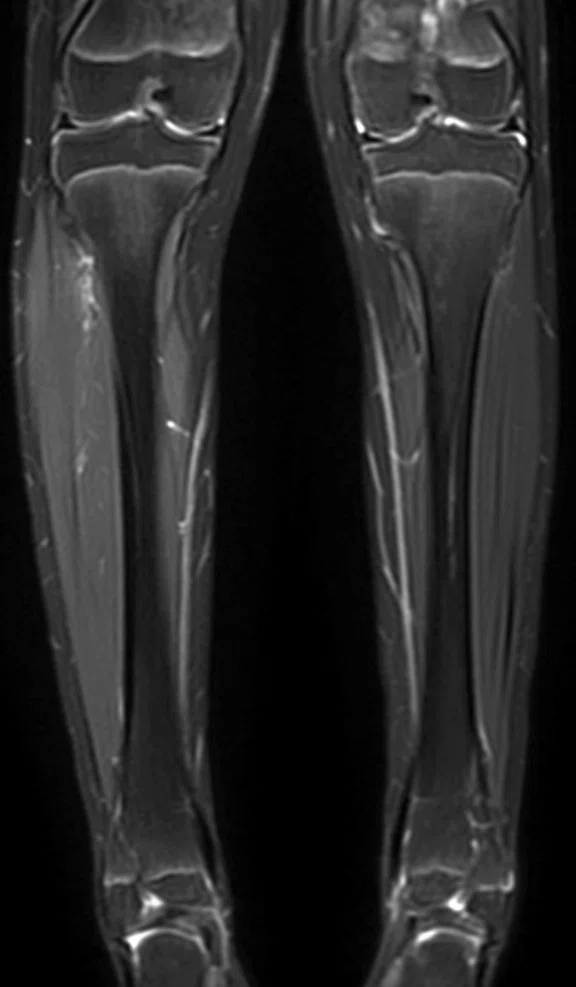
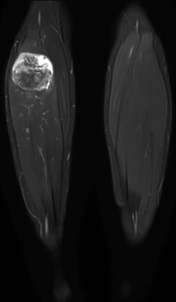
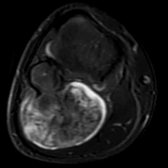
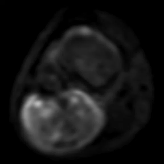
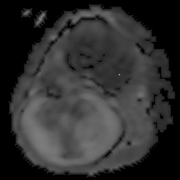
Long bone/soft tissue assessment in 10-year-old patient, both legs imaged simultaneously with AIR Technology™ AA. (A, B) Coronal T2 FSE Flex, 0.9 x 1.2 x 5 mm, 2:54 min.; (B) Water image; (C) Axial T2 FSE Flex (Water), 1.2 x 0.8 x 5 mm, 2:31 min.; and (D, E) Axial DWI with ADC Map.
B
Figure 1.
Long bone/soft tissue assessment in 10-year-old patient, both legs imaged simultaneously with AIR Technology™ AA. (A, B) Coronal T2 FSE Flex, 0.9 x 1.2 x 5 mm, 2:54 min.; (B) Water image; (C) Axial T2 FSE Flex (Water), 1.2 x 0.8 x 5 mm, 2:31 min.; and (D, E) Axial DWI with ADC Map.
C
Figure 1.
Long bone/soft tissue assessment in 10-year-old patient, both legs imaged simultaneously with AIR Technology™ AA. (A, B) Coronal T2 FSE Flex, 0.9 x 1.2 x 5 mm, 2:54 min.; (B) Water image; (C) Axial T2 FSE Flex (Water), 1.2 x 0.8 x 5 mm, 2:31 min.; and (D, E) Axial DWI with ADC Map.
D
Figure 1.
Long bone/soft tissue assessment in 10-year-old patient, both legs imaged simultaneously with AIR Technology™ AA. (A, B) Coronal T2 FSE Flex, 0.9 x 1.2 x 5 mm, 2:54 min.; (B) Water image; (C) Axial T2 FSE Flex (Water), 1.2 x 0.8 x 5 mm, 2:31 min.; and (D, E) Axial DWI with ADC Map.
E
Figure 1.
Long bone/soft tissue assessment in 10-year-old patient, both legs imaged simultaneously with AIR Technology™ AA. (A, B) Coronal T2 FSE Flex, 0.9 x 1.2 x 5 mm, 2:54 min.; (B) Water image; (C) Axial T2 FSE Flex (Water), 1.2 x 0.8 x 5 mm, 2:31 min.; and (D, E) Axial DWI with ADC Map.
‡Not licensed in accordance with Canadian law. Not available for sale in Canada. Not CE marked. Not available for sale in all regions.
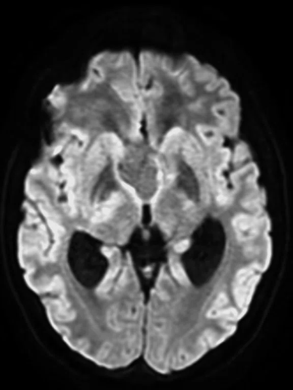
A
Figure 6.
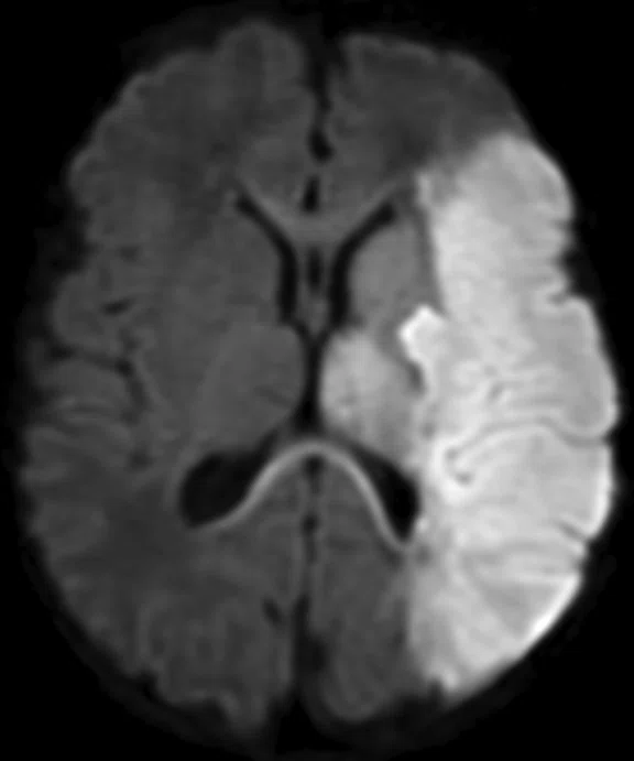
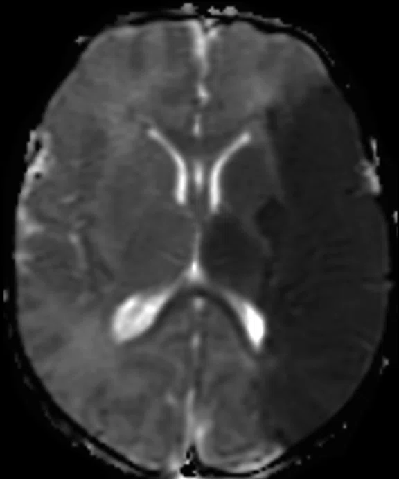
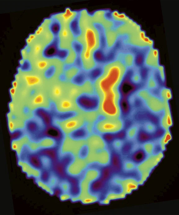
Neuro imaging with the 48-channel Head Coil in a three-day-old patient. (A-B) MUSE DWI with ADC Map, 1.2 x 1 x 3 mm, 3:09 min.; and (C) Axial ASL CBF Map.
B
Figure 6.
Neuro imaging with the 48-channel Head Coil in a three-day-old patient. (A-B) MUSE DWI with ADC Map, 1.2 x 1 x 3 mm, 3:09 min.; and (C) Axial ASL CBF Map.
C
Figure 6.
Neuro imaging with the 48-channel Head Coil in a three-day-old patient. (A-B) MUSE DWI with ADC Map, 1.2 x 1 x 3 mm, 3:09 min.; and (C) Axial ASL CBF Map.
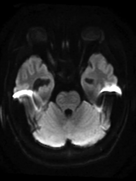
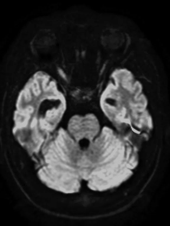
A
Figure 5.
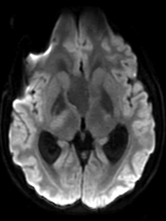
Neuro imaging with the 48-channel Head Coil in a three-year-old patient. Note the same resolution with less blurring in the MUSE DWI sequence. (A-B) Traditional single-shot DWI, 1.8 x 1.4 x 3.6 mm, 2 shots, acceleration factor of 2, 1:58 min.; and (C, D) MUSE DWI, 1.8 x 1.4 x 3.6 mm, 2 shots, acceleration factor of 2, 2:16 min.
B
Figure 5.
Neuro imaging with the 48-channel Head Coil in a three-year-old patient. Note the same resolution with less blurring in the MUSE DWI sequence. (A-B) Traditional single-shot DWI, 1.8 x 1.4 x 3.6 mm, 2 shots, acceleration factor of 2, 1:58 min.; and (C, D) MUSE DWI, 1.8 x 1.4 x 3.6 mm, 2 shots, acceleration factor of 2, 2:16 min.
C
Figure 5.
Neuro imaging with the 48-channel Head Coil in a three-year-old patient. Note the same resolution with less blurring in the MUSE DWI sequence. (A-B) Traditional single-shot DWI, 1.8 x 1.4 x 3.6 mm, 2 shots, acceleration factor of 2, 1:58 min.; and (C, D) MUSE DWI, 1.8 x 1.4 x 3.6 mm, 2 shots, acceleration factor of 2, 2:16 min.
D
Figure 5.
Neuro imaging with the 48-channel Head Coil in a three-year-old patient. Note the same resolution with less blurring in the MUSE DWI sequence. (A-B) Traditional single-shot DWI, 1.8 x 1.4 x 3.6 mm, 2 shots, acceleration factor of 2, 1:58 min.; and (C, D) MUSE DWI, 1.8 x 1.4 x 3.6 mm, 2 shots, acceleration factor of 2, 2:16 min.
A
Figure 4.
Free-breathing kidney exam with Auto Navigator in a six-year-old patient using a combination of the AIR Technology™ AA and the PA embedded in the SIGNA™ Architect table. (A) SSFSE, 0.9 x 1.3 x 6 mm, 17 sec.; (B) T2 PROPELLER with respiratory trigger, 0.9 x 0.9 x 4 mm, 4 min.; (C) SSFSE, 0.9 x 1.0 x 5 mm, 49 sec.; (D) LAVA Flex with Auto Navigator, 1 x 0.9 x 3 mm, 3:56 min.; and (E) T2 frFSE FatSat with Auto Navigator, 0.8 x 0.9 x 3 mm, 4 min.
B
Figure 4.
Free-breathing kidney exam with Auto Navigator in a six-year-old patient using a combination of the AIR Technology™ AA and the PA embedded in the SIGNA™ Architect table. (A) SSFSE, 0.9 x 1.3 x 6 mm, 17 sec.; (B) T2 PROPELLER with respiratory trigger, 0.9 x 0.9 x 4 mm, 4 min.; (C) SSFSE, 0.9 x 1.0 x 5 mm, 49 sec.; (D) LAVA Flex with Auto Navigator, 1 x 0.9 x 3 mm, 3:56 min.; and (E) T2 frFSE FatSat with Auto Navigator, 0.8 x 0.9 x 3 mm, 4 min.
C
Figure 4.
Free-breathing kidney exam with Auto Navigator in a six-year-old patient using a combination of the AIR Technology™ AA and the PA embedded in the SIGNA™ Architect table. (A) SSFSE, 0.9 x 1.3 x 6 mm, 17 sec.; (B) T2 PROPELLER with respiratory trigger, 0.9 x 0.9 x 4 mm, 4 min.; (C) SSFSE, 0.9 x 1.0 x 5 mm, 49 sec.; (D) LAVA Flex with Auto Navigator, 1 x 0.9 x 3 mm, 3:56 min.; and (E) T2 frFSE FatSat with Auto Navigator, 0.8 x 0.9 x 3 mm, 4 min.
D
Figure 4.
Free-breathing kidney exam with Auto Navigator in a six-year-old patient using a combination of the AIR Technology™ AA and the PA embedded in the SIGNA™ Architect table. (A) SSFSE, 0.9 x 1.3 x 6 mm, 17 sec.; (B) T2 PROPELLER with respiratory trigger, 0.9 x 0.9 x 4 mm, 4 min.; (C) SSFSE, 0.9 x 1.0 x 5 mm, 49 sec.; (D) LAVA Flex with Auto Navigator, 1 x 0.9 x 3 mm, 3:56 min.; and (E) T2 frFSE FatSat with Auto Navigator, 0.8 x 0.9 x 3 mm, 4 min.
E
Figure 4.
Free-breathing kidney exam with Auto Navigator in a six-year-old patient using a combination of the AIR Technology™ AA and the PA embedded in the SIGNA™ Architect table. (A) SSFSE, 0.9 x 1.3 x 6 mm, 17 sec.; (B) T2 PROPELLER with respiratory trigger, 0.9 x 0.9 x 4 mm, 4 min.; (C) SSFSE, 0.9 x 1.0 x 5 mm, 49 sec.; (D) LAVA Flex with Auto Navigator, 1 x 0.9 x 3 mm, 3:56 min.; and (E) T2 frFSE FatSat with Auto Navigator, 0.8 x 0.9 x 3 mm, 4 min.
A
Figure 3.
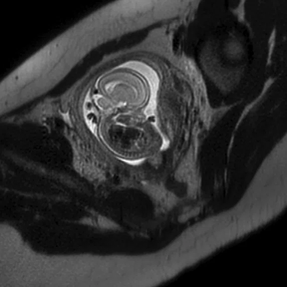
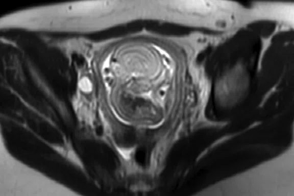
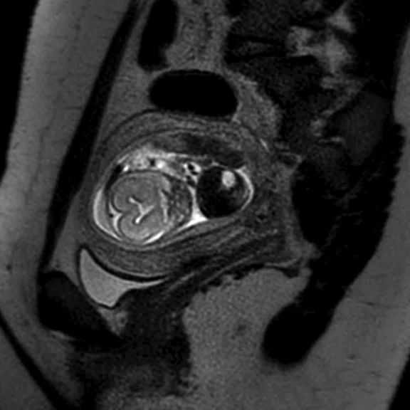
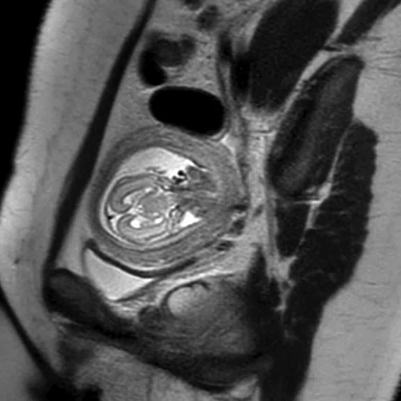
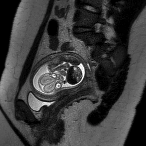
Fetal imaging with AIR Technology™ AA improved comfort for the woman by imaging her in the decubitus position. (A-B) Axial T2 SSFSE; and (C-E) Oblique T2 SSFSE
B
Figure 3.
Fetal imaging with AIR Technology™ AA improved comfort for the woman by imaging her in the decubitus position. (A-B) Axial T2 SSFSE; and (C-E) Oblique T2 SSFSE
C
Figure 3.
Fetal imaging with AIR Technology™ AA improved comfort for the woman by imaging her in the decubitus position. (A-B) Axial T2 SSFSE; and (C-E) Oblique T2 SSFSE
D
Figure 3.
Fetal imaging with AIR Technology™ AA improved comfort for the woman by imaging her in the decubitus position. (A-B) Axial T2 SSFSE; and (C-E) Oblique T2 SSFSE
E
Figure 3.
Fetal imaging with AIR Technology™ AA improved comfort for the woman by imaging her in the decubitus position. (A-B) Axial T2 SSFSE; and (C-E) Oblique T2 SSFSE
A
Figure 2.
Same patient as Figure 1. (A) Sagittal Inhance 3D Velocity NCE MRA; (B) Sagittal 3D MERGE; and (C) Volume rendered 3D MERGE fused with NCE MRA and FSE Flex.
B
Figure 2.
Same patient as Figure 1. (A) Sagittal Inhance 3D Velocity NCE MRA; (B) Sagittal 3D MERGE; and (C) Volume rendered 3D MERGE fused with NCE MRA and FSE Flex.
C
Figure 2.
Same patient as Figure 1. (A) Sagittal Inhance 3D Velocity NCE MRA; (B) Sagittal 3D MERGE; and (C) Volume rendered 3D MERGE fused with NCE MRA and FSE Flex.
A
Figure 4.
Free-breathing kidney exam with Auto Navigator in a six-year-old patient using a combination of the AIR Technology™ AA and the PA embedded in the SIGNA™ Architect table. (A) SSFSE, 0.9 x 1.3 x 6 mm, 17 sec.; (B) T2 PROPELLER with respiratory trigger, 0.9 x 0.9 x 4 mm, 4 min.; (C) SSFSE, 0.9 x 1.0 x 5 mm, 49 sec.; (D) LAVA Flex with Auto Navigator, 1 x 0.9 x 3 mm, 3:56 min.; and (E) T2 frFSE FatSat with Auto Navigator, 0.8 x 0.9 x 3 mm, 4 min.
B
Figure 4.
Free-breathing kidney exam with Auto Navigator in a six-year-old patient using a combination of the AIR Technology™ AA and the PA embedded in the SIGNA™ Architect table. (A) SSFSE, 0.9 x 1.3 x 6 mm, 17 sec.; (B) T2 PROPELLER with respiratory trigger, 0.9 x 0.9 x 4 mm, 4 min.; (C) SSFSE, 0.9 x 1.0 x 5 mm, 49 sec.; (D) LAVA Flex with Auto Navigator, 1 x 0.9 x 3 mm, 3:56 min.; and (E) T2 frFSE FatSat with Auto Navigator, 0.8 x 0.9 x 3 mm, 4 min.
C
Figure 4.
Free-breathing kidney exam with Auto Navigator in a six-year-old patient using a combination of the AIR Technology™ AA and the PA embedded in the SIGNA™ Architect table. (A) SSFSE, 0.9 x 1.3 x 6 mm, 17 sec.; (B) T2 PROPELLER with respiratory trigger, 0.9 x 0.9 x 4 mm, 4 min.; (C) SSFSE, 0.9 x 1.0 x 5 mm, 49 sec.; (D) LAVA Flex with Auto Navigator, 1 x 0.9 x 3 mm, 3:56 min.; and (E) T2 frFSE FatSat with Auto Navigator, 0.8 x 0.9 x 3 mm, 4 min.
D
Figure 4.
Free-breathing kidney exam with Auto Navigator in a six-year-old patient using a combination of the AIR Technology™ AA and the PA embedded in the SIGNA™ Architect table. (A) SSFSE, 0.9 x 1.3 x 6 mm, 17 sec.; (B) T2 PROPELLER with respiratory trigger, 0.9 x 0.9 x 4 mm, 4 min.; (C) SSFSE, 0.9 x 1.0 x 5 mm, 49 sec.; (D) LAVA Flex with Auto Navigator, 1 x 0.9 x 3 mm, 3:56 min.; and (E) T2 frFSE FatSat with Auto Navigator, 0.8 x 0.9 x 3 mm, 4 min.
E
Figure 4.
Free-breathing kidney exam with Auto Navigator in a six-year-old patient using a combination of the AIR Technology™ AA and the PA embedded in the SIGNA™ Architect table. (A) SSFSE, 0.9 x 1.3 x 6 mm, 17 sec.; (B) T2 PROPELLER with respiratory trigger, 0.9 x 0.9 x 4 mm, 4 min.; (C) SSFSE, 0.9 x 1.0 x 5 mm, 49 sec.; (D) LAVA Flex with Auto Navigator, 1 x 0.9 x 3 mm, 3:56 min.; and (E) T2 frFSE FatSat with Auto Navigator, 0.8 x 0.9 x 3 mm, 4 min.
A
Figure 5.
Neuro imaging with the 48-channel Head Coil in a three-year-old patient. Note the same resolution with less blurring in the MUSE DWI sequence. (A-B) Traditional single-shot DWI, 1.8 x 1.4 x 3.6 mm, 2 shots, acceleration factor of 2, 1:58 min.; and (C, D) MUSE DWI, 1.8 x 1.4 x 3.6 mm, 2 shots, acceleration factor of 2, 2:16 min.
B
Figure 5.
Neuro imaging with the 48-channel Head Coil in a three-year-old patient. Note the same resolution with less blurring in the MUSE DWI sequence. (A-B) Traditional single-shot DWI, 1.8 x 1.4 x 3.6 mm, 2 shots, acceleration factor of 2, 1:58 min.; and (C, D) MUSE DWI, 1.8 x 1.4 x 3.6 mm, 2 shots, acceleration factor of 2, 2:16 min.
C
Figure 5.
Neuro imaging with the 48-channel Head Coil in a three-year-old patient. Note the same resolution with less blurring in the MUSE DWI sequence. (A-B) Traditional single-shot DWI, 1.8 x 1.4 x 3.6 mm, 2 shots, acceleration factor of 2, 1:58 min.; and (C, D) MUSE DWI, 1.8 x 1.4 x 3.6 mm, 2 shots, acceleration factor of 2, 2:16 min.
D
Figure 5.
Neuro imaging with the 48-channel Head Coil in a three-year-old patient. Note the same resolution with less blurring in the MUSE DWI sequence. (A-B) Traditional single-shot DWI, 1.8 x 1.4 x 3.6 mm, 2 shots, acceleration factor of 2, 1:58 min.; and (C, D) MUSE DWI, 1.8 x 1.4 x 3.6 mm, 2 shots, acceleration factor of 2, 2:16 min.
A
Figure 6.
Neuro imaging with the 48-channel Head Coil in a three-day-old patient. (A-B) MUSE DWI with ADC Map, 1.2 x 1 x 3 mm, 3:09 min.; and (C) Axial ASL CBF Map.
B
Figure 6.
Neuro imaging with the 48-channel Head Coil in a three-day-old patient. (A-B) MUSE DWI with ADC Map, 1.2 x 1 x 3 mm, 3:09 min.; and (C) Axial ASL CBF Map.
C
Figure 6.
Neuro imaging with the 48-channel Head Coil in a three-day-old patient. (A-B) MUSE DWI with ADC Map, 1.2 x 1 x 3 mm, 3:09 min.; and (C) Axial ASL CBF Map.
‡Not licensed in accordance with Canadian law. Not available for sale in Canada. Not CE marked. Not available for sale in all regions.
A
Figure 1.
Long bone/soft tissue assessment in 10-year-old patient, both legs imaged simultaneously with AIR Technology™ AA. (A, B) Coronal T2 FSE Flex, 0.9 x 1.2 x 5 mm, 2:54 min.; (B) Water image; (C) Axial T2 FSE Flex (Water), 1.2 x 0.8 x 5 mm, 2:31 min.; and (D, E) Axial DWI with ADC Map.
B
Figure 1.
Long bone/soft tissue assessment in 10-year-old patient, both legs imaged simultaneously with AIR Technology™ AA. (A, B) Coronal T2 FSE Flex, 0.9 x 1.2 x 5 mm, 2:54 min.; (B) Water image; (C) Axial T2 FSE Flex (Water), 1.2 x 0.8 x 5 mm, 2:31 min.; and (D, E) Axial DWI with ADC Map.
C
Figure 1.
Long bone/soft tissue assessment in 10-year-old patient, both legs imaged simultaneously with AIR Technology™ AA. (A, B) Coronal T2 FSE Flex, 0.9 x 1.2 x 5 mm, 2:54 min.; (B) Water image; (C) Axial T2 FSE Flex (Water), 1.2 x 0.8 x 5 mm, 2:31 min.; and (D, E) Axial DWI with ADC Map.
D
Figure 1.
Long bone/soft tissue assessment in 10-year-old patient, both legs imaged simultaneously with AIR Technology™ AA. (A, B) Coronal T2 FSE Flex, 0.9 x 1.2 x 5 mm, 2:54 min.; (B) Water image; (C) Axial T2 FSE Flex (Water), 1.2 x 0.8 x 5 mm, 2:31 min.; and (D, E) Axial DWI with ADC Map.
E
Figure 1.
Long bone/soft tissue assessment in 10-year-old patient, both legs imaged simultaneously with AIR Technology™ AA. (A, B) Coronal T2 FSE Flex, 0.9 x 1.2 x 5 mm, 2:54 min.; (B) Water image; (C) Axial T2 FSE Flex (Water), 1.2 x 0.8 x 5 mm, 2:31 min.; and (D, E) Axial DWI with ADC Map.
result


PREVIOUS
${prev-page}
NEXT
${next-page}
Subscribe Now
Manage Subscription
FOLLOW US
Contact Us • Cookie Preferences • Privacy Policy • California Privacy PolicyDo Not Sell or Share My Personal Information • Terms & Conditions • Security
© 2024 GE HealthCare. GE is a trademark of General Electric Company. Used under trademark license.
IN PRACTICE
Transforming the MR imaging experience for one of Sweden’s largest pediatric hospitals
Transforming the MR imaging experience for one of Sweden’s largest pediatric hospitals
As part of a modernization project, The Queen Silvia Children’s Hospital upgraded to the SIGNA™ Architect with AIR Technology™‡. An initial evaluation by the Department of Pediatric Radiology found the new AIR Technology™Anterior Array (AA) delivers good SNR and homogeneity for high-resolution imaging. The coil also enables flexibility and ease of positioning patients, and when used with AIR Touch™ makes coil selection easier and helps with workflow.
As part of a modernization project, The Queen Silvia Children’s Hospital upgraded to the SIGNA™ Architect with AIR Technology™‡. An initial evaluation by the Department of Pediatric Radiology found the new AIR Technology™Anterior Array (AA) delivers good SNR and homogeneity for high-resolution imaging. The coil also enables flexibility and ease of positioning patients, and when used with AIR Touch™ makes coil selection easier and helps with workflow.
As one of the largest pediatric hospitals in Sweden, The Queen Silvia Children’s Hospital at Sahlgrenska University Hospital in Gothenburg provides care to children from newborns up to age 18. The Department of Pediatric Radiology performs around 45,000 exams each year. Named for the country’s current Queen, the hospital has undergone a modernization project to improve workflow and enhance clinical services as well as create a safe and secure healing environment for its young patients.
The pediatric radiology department recently upgraded its Discovery™ MR750w 3.0T wide-bore system to SIGNA™ Architect. With new gradients and Total Digital Imaging (TDI), the cutting-edge platform is designed to help facilities like The Queen Silvia Children’s Hospital adapt to existing and future advancements in MR imaging technologies, such as AIR Technology™ and the SIGNA™Works productivity platform.
"We want to be on the front line of new technology and prepare for the future," says Håkan Boström, MD, pediatric radiologist at The Queen Silvia Children’s Hospital.
In addition to the recently upgraded SIGNA™ Architect, the hospital also has an Optima™ MR450w 1.5T. Combined, these two MR systems enable the hospital to perform more than 2,000 MR exams each year.
According to Pär-Arne Svensson, MR research radiographer, a key motivation behind the SIGNA™ Architect upgrade was the ability to acquire the new AIR Technology™ Suite, including the 48-channel Head Coil, along with new MR sequences available in SIGNA™Works, specifically MUSE, PROPELLER MB (multi-blade), MPRAGE and distortion correction with diffusion-weighted imaging (DWI) like PROGRES.
The radiology department has been evaluating MUSE as a replacement sequence for single-shot diffusion imaging in neonates. With MUSE, Dr. Boström can obtain higher resolution and the distortion correction is better with less distortion artifacts. In addition to Dr. Boström finding that MUSE delivers high resolution and excellent image quality, PROPELLER MB is also helpful in avoiding metal artifacts. PROPELLER MB is particularly beneficial when imaging the cervical spine and temporal bone with diffusion sequences. Svensson and Dr. Boström are still evaluating MPRAGE, however, they are obtaining better contrast between white and gray matter in the brain compared to other conventional 3D FSPGR sequences. This sequence would be particularly helpful when imaging epilepsy patients before and after surgery.
In addition to the new sequences, the department received the AIR Technology™ AA and 48-channel Head Coil in early 2019. The AIR Technology™ AA has the highest channel count and coverage in the industry.
"Our initial experience is very good," says Dr. Boström. "We’ve used the AA for several exams, such as the abdomen, pelvic, lower extremities, shoulder and fetal imaging. The main advantages with the AIR Technology™ AA are the flexibility and ease of positioning on the patient."
It is lightweight—60 percent lighter than conventional, hard-shell coils—and children in pain may not tolerate a heavy coil on their body, Dr. Boström explains. This includes children who had open heart surgery at The Queen Silvia Children’s Hospital, one of only two pediatric cardiac surgery centers in Sweden.
"Fetal imaging is another area where we see an advantage with the AIR Technology™ AA," adds Svensson. "It can be difficult to put a conventional, hard-shell coil around a pregnant woman’s abdomen and get a good, homogeneous signal."
For women in the late stage of pregnancy, lying on their back can be uncomfortable. Svensson wants to try imaging them on their side with the AIR Technology™ AA wrapped around them and see what the impact is on patient comfort and image quality.
In musculoskeletal imaging of the shoulder and arm, lower extremities or imaging both legs, Svensson can wrap the coil around larger field-of-views (FOVs) and obtain a homogenous signal for good image quality. For example, patients with multifocal chronic osteomyelitis or muscular dystrophy/myositis will often require imaging of both legs simultaneously.
"With the AIR Technology™ AA, we can cover large areas but we also get good SNR, so we can provide detailed images of specific joints with high resolution," he says.
Positioning these precious patients is also easier now with AIR Technology™. There are many factors that can impact the overall time a child is in the MR scanner and any time saved in positioning means the sooner the patient can get back to his or her parents.
Figure 4.
Free-breathing kidney exam with Auto Navigator in a six-year-old patient using a combination of the AIR Technology™ AA and the PA embedded in the SIGNA™ Architect table. (A) SSFSE, 0.9 x 1.3 x 6 mm, 17 sec.; (B) T2 PROPELLER with respiratory trigger, 0.9 x 0.9 x 4 mm, 4 min.; (C) SSFSE, 0.9 x 1.0 x 5 mm, 49 sec.; (D) LAVA Flex with Auto Navigator, 1 x 0.9 x 3 mm, 3:56 min.; and (E) T2 frFSE FatSat with Auto Navigator, 0.8 x 0.9 x 3 mm, 4 min.
Figure 5.
Neuro imaging with the 48-channel Head Coil in a three-year-old patient. Note the same resolution with less blurring in the MUSE DWI sequence. (A-B) Traditional single-shot DWI, 1.8 x 1.4 x 3.6 mm, 2 shots, acceleration factor of 2, 1:58 min.; and (C, D) MUSE DWI, 1.8 x 1.4 x 3.6 mm, 2 shots, acceleration factor of 2, 2:16 min.
Another patient-centric feature of AIR Touch™ is that it assists with patient positioning. It automatically selects the best elements to use and uniquely optimizes uniformity, SNR, artifacts and parallel imaging.
"AIR Touch™ makes coil selection much easier and I don’t have to check what elements are activated because the system does it. It helps with workflow, but the most important factor is that it helps me focus more on the child."
Pär-Arne Svensson
AIR Touch™ even helps when using more than one coil. Embedded in the SIGNA Architect™ table is the Posterior Array (PA). With small children, Dr. Boström and Svensson are using GE’s Flex Coil in combination with the PA. They have seen excellent results using the combination of both coils in cardiac and abdominal exams.
After using the impressive AIR Technology™ AA for just two months Svensson and Dr. Boström no longer use the conventional AA. They look forward to receiving the new AIR Technology™ Multi-Purpose (MP) Coil, a smaller version of the AIR Technology™ AA.
There have been a few patients who had MR exams with both the conventional coil and the AIR Technology™ AA. Svensson says when asked, these patients preferred the new coil, especially because it was not so heavy and confining on their bodies.
"The most important benefit of AIR Technology™ is the patient comfort," says Dr. Boström. "It is lightweight and can lay on the patient like a blanket. We believe this also impacts patient compliance."
Overall, Dr. Boström and Svensson are impressed with SIGNA™ Architect, SIGNA™Works and especially AIR Technology™.
"This is a stable MR system with very good image quality," says Svensson. "We are satisfied with the upgrade and our initial experience with AIR Technology™."


