A
Figure 1.
HyperCube, HyperSense, MAGiC DWI (calculated b-values) and flexible phase oversampling are all excellent tools for reducing scan times. (A) Sagittal T2 3D Cube with HyperSense and (B, C) MPR reformats, 0.9 x 0.9 x 0.9 mm, 6:40 min.; (D) sagittal T2 3D HyperCube with (E) MPR reformats, 1.2 x 1.2 x 1.4 mm, 3:13 min.; (F) axial DWI FOCUS b800; and (G, H) MAGiC DWI b1400 and b2000.
B
Figure 1.
HyperCube, HyperSense, MAGiC DWI (calculated b-values) and flexible phase oversampling are all excellent tools for reducing scan times. (A) Sagittal T2 3D Cube with HyperSense and (B, C) MPR reformats, 0.9 x 0.9 x 0.9 mm, 6:40 min.; (D) sagittal T2 3D HyperCube with (E) MPR reformats, 1.2 x 1.2 x 1.4 mm, 3:13 min.; (F) axial DWI FOCUS b800; and (G, H) MAGiC DWI b1400 and b2000.
C
Figure 1.
HyperCube, HyperSense, MAGiC DWI (calculated b-values) and flexible phase oversampling are all excellent tools for reducing scan times. (A) Sagittal T2 3D Cube with HyperSense and (B, C) MPR reformats, 0.9 x 0.9 x 0.9 mm, 6:40 min.; (D) sagittal T2 3D HyperCube with (E) MPR reformats, 1.2 x 1.2 x 1.4 mm, 3:13 min.; (F) axial DWI FOCUS b800; and (G, H) MAGiC DWI b1400 and b2000.
A
Figure 2.
With HyperSense, 3D TOF angiography exam times have been reduced by nearly 50 percent. (A) Axial 3D TOF with HyperSense, 0.7 x 0.9 x 1 mm, 3:04 min. and (B) coronal 3D MRCP with HyperSense, 1.2 x 1.2 x 1.4 mm, 3:01 min.
B
Figure 2.
With HyperSense, 3D TOF angiography exam times have been reduced by nearly 50 percent. (A) Axial 3D TOF with HyperSense, 0.7 x 0.9 x 1 mm, 3:04 min. and (B) coronal 3D MRCP with HyperSense, 1.2 x 1.2 x 1.4 mm, 3:01 min.
A
Figure 3.
PROPELLER and FOCUS improve image quality in prostate exams. (A) Axial T2 PROPELLER, 0.8 x 0.8 x 3 mm, 3:57 min.; (B) axial diffusion FOCUS b800, 2 x 2 x 5 mm, 5:53 min.; and (C) axial T2 PROPELLER fused with axial DWI FOCUS b800.
B
Figure 3.
PROPELLER and FOCUS improve image quality in prostate exams. (A) Axial T2 PROPELLER, 0.8 x 0.8 x 3 mm, 3:57 min.; (B) axial diffusion FOCUS b800, 2 x 2 x 5 mm, 5:53 min.; and (C) axial T2 PROPELLER fused with axial DWI FOCUS b800.
C
Figure 3.
PROPELLER and FOCUS improve image quality in prostate exams. (A) Axial T2 PROPELLER, 0.8 x 0.8 x 3 mm, 3:57 min.; (B) axial diffusion FOCUS b800, 2 x 2 x 5 mm, 5:53 min.; and (C) axial T2 PROPELLER fused with axial DWI FOCUS b800.
A
Figure 4.
Every liver exam at Shields now includes IDEAL IQ for quantitative fat fraction and iron assessment of the liver. (A) Axial 3D IDEAL IQ, 20 sec. breath-hold fat fraction map; (B) axial 3D IDEAL IQ, 20 sec. breath-hold R2* map (Hz); and (C) axial 3D IDEAL IQ, 20 sec. breath-hold R2* map (Hz — colored).
B
Figure 4.
Every liver exam at Shields now includes IDEAL IQ for quantitative fat fraction and iron assessment of the liver. (A) Axial 3D IDEAL IQ, 20 sec. breath-hold fat fraction map; (B) axial 3D IDEAL IQ, 20 sec. breath-hold R2* map (Hz); and (C) axial 3D IDEAL IQ, 20 sec. breath-hold R2* map (Hz — colored).
C
Figure 4.
Every liver exam at Shields now includes IDEAL IQ for quantitative fat fraction and iron assessment of the liver. (A) Axial 3D IDEAL IQ, 20 sec. breath-hold fat fraction map; (B) axial 3D IDEAL IQ, 20 sec. breath-hold R2* map (Hz); and (C) axial 3D IDEAL IQ, 20 sec. breath-hold R2* map (Hz — colored).
A
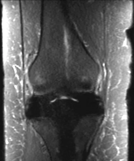
Figure 5.
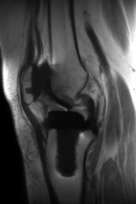
MAVRIC SL has opened the door to more orthopedic referrals for joint implant imaging. Knee exam with (A) sagittal PD MAVRIC SL, 0.6 x 0.9 x 4 mm; (B) coronal STIR MAVRIC SL, 0.8 x 1 x 4 mm; and (C) a standard coronal FSE T2 STIR that is showing considerable distortion.
B
Figure 5.
MAVRIC SL has opened the door to more orthopedic referrals for joint implant imaging. Knee exam with (A) sagittal PD MAVRIC SL, 0.6 x 0.9 x 4 mm; (B) coronal STIR MAVRIC SL, 0.8 x 1 x 4 mm; and (C) a standard coronal FSE T2 STIR that is showing considerable distortion.
C
Figure 5.
MAVRIC SL has opened the door to more orthopedic referrals for joint implant imaging. Knee exam with (A) sagittal PD MAVRIC SL, 0.6 x 0.9 x 4 mm; (B) coronal STIR MAVRIC SL, 0.8 x 1 x 4 mm; and (C) a standard coronal FSE T2 STIR that is showing considerable distortion.
‡MAVRIC SL should only be used with MR-Conditional implants and within the MR conditions specified for those implants.
D
Figure 1.
HyperCube, HyperSense, MAGiC DWI (calculated b-values) and flexible phase oversampling are all excellent tools for reducing scan times. (A) Sagittal T2 3D Cube with HyperSense and (B, C) MPR reformats, 0.9 x 0.9 x 0.9 mm, 6:40 min.; (D) sagittal T2 3D HyperCube with (E) MPR reformats, 1.2 x 1.2 x 1.4 mm, 3:13 min.; (F) axial DWI FOCUS b800; and (G, H) MAGiC DWI b1400 and b2000.
E
Figure 1.
HyperCube, HyperSense, MAGiC DWI (calculated b-values) and flexible phase oversampling are all excellent tools for reducing scan times. (A) Sagittal T2 3D Cube with HyperSense and (B, C) MPR reformats, 0.9 x 0.9 x 0.9 mm, 6:40 min.; (D) sagittal T2 3D HyperCube with (E) MPR reformats, 1.2 x 1.2 x 1.4 mm, 3:13 min.; (F) axial DWI FOCUS b800; and (G, H) MAGiC DWI b1400 and b2000.
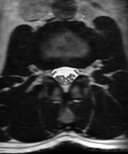
F
Figure 1.
HyperCube, HyperSense, MAGiC DWI (calculated b-values) and flexible phase oversampling are all excellent tools for reducing scan times. (A) Sagittal T2 3D Cube with HyperSense and (B, C) MPR reformats, 0.9 x 0.9 x 0.9 mm, 6:40 min.; (D) sagittal T2 3D HyperCube with (E) MPR reformats, 1.2 x 1.2 x 1.4 mm, 3:13 min.; (F) axial DWI FOCUS b800; and (G, H) MAGiC DWI b1400 and b2000.
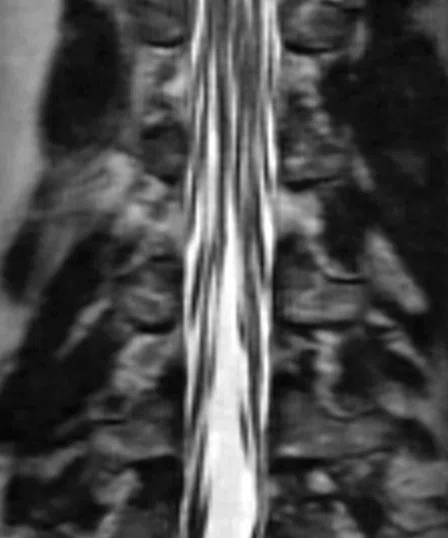
G
Figure 1.
HyperCube, HyperSense, MAGiC DWI (calculated b-values) and flexible phase oversampling are all excellent tools for reducing scan times. (A) Sagittal T2 3D Cube with HyperSense and (B, C) MPR reformats, 0.9 x 0.9 x 0.9 mm, 6:40 min.; (D) sagittal T2 3D HyperCube with (E) MPR reformats, 1.2 x 1.2 x 1.4 mm, 3:13 min.; (F) axial DWI FOCUS b800; and (G, H) MAGiC DWI b1400 and b2000.
H
Figure 1.
HyperCube, HyperSense, MAGiC DWI (calculated b-values) and flexible phase oversampling are all excellent tools for reducing scan times. (A) Sagittal T2 3D Cube with HyperSense and (B, C) MPR reformats, 0.9 x 0.9 x 0.9 mm, 6:40 min.; (D) sagittal T2 3D HyperCube with (E) MPR reformats, 1.2 x 1.2 x 1.4 mm, 3:13 min.; (F) axial DWI FOCUS b800; and (G, H) MAGiC DWI b1400 and b2000.
A
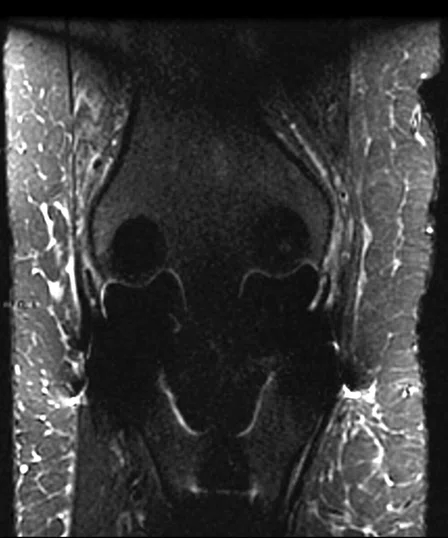
Figure 6.
PROPELLER MB is the go-to sequence at Shields for all shoulders, hips, wrists, elbows and even in the brain. All lumbar spine and shoulder exams are now completed in 20-minute slots. Wrist exam with (A) axial PD PROPELLER FatSat, 0.4 x 0.4 x 2 mm, 3:20 min. and (B) axial PD PROPELLER, 0.4 x 0.4 x 2 mm, 2:34 min. Elbow exam using the 16-channel small GEM Flex Coil with (C, D) sagittal and (E) axial 3D MENSA, 0.7 x 0.7 x 0.8 mm, 3:08 min.
B
Figure 6.
PROPELLER MB is the go-to sequence at Shields for all shoulders, hips, wrists, elbows and even in the brain. All lumbar spine and shoulder exams are now completed in 20-minute slots. Wrist exam with (A) axial PD PROPELLER FatSat, 0.4 x 0.4 x 2 mm, 3:20 min. and (B) axial PD PROPELLER, 0.4 x 0.4 x 2 mm, 2:34 min. Elbow exam using the 16-channel small GEM Flex Coil with (C, D) sagittal and (E) axial 3D MENSA, 0.7 x 0.7 x 0.8 mm, 3:08 min.
C
Figure 6.
PROPELLER MB is the go-to sequence at Shields for all shoulders, hips, wrists, elbows and even in the brain. All lumbar spine and shoulder exams are now completed in 20-minute slots. Wrist exam with (A) axial PD PROPELLER FatSat, 0.4 x 0.4 x 2 mm, 3:20 min. and (B) axial PD PROPELLER, 0.4 x 0.4 x 2 mm, 2:34 min. Elbow exam using the 16-channel small GEM Flex Coil with (C, D) sagittal and (E) axial 3D MENSA, 0.7 x 0.7 x 0.8 mm, 3:08 min.
D
Figure 6.
PROPELLER MB is the go-to sequence at Shields for all shoulders, hips, wrists, elbows and even in the brain. All lumbar spine and shoulder exams are now completed in 20-minute slots. Wrist exam with (A) axial PD PROPELLER FatSat, 0.4 x 0.4 x 2 mm, 3:20 min. and (B) axial PD PROPELLER, 0.4 x 0.4 x 2 mm, 2:34 min. Elbow exam using the 16-channel small GEM Flex Coil with (C, D) sagittal and (E) axial 3D MENSA, 0.7 x 0.7 x 0.8 mm, 3:08 min.
E
Figure 6.
PROPELLER MB is the go-to sequence at Shields for all shoulders, hips, wrists, elbows and even in the brain. All lumbar spine and shoulder exams are now completed in 20-minute slots. Wrist exam with (A) axial PD PROPELLER FatSat, 0.4 x 0.4 x 2 mm, 3:20 min. and (B) axial PD PROPELLER, 0.4 x 0.4 x 2 mm, 2:34 min. Elbow exam using the 16-channel small GEM Flex Coil with (C, D) sagittal and (E) axial 3D MENSA, 0.7 x 0.7 x 0.8 mm, 3:08 min.


result
PREVIOUS
${prev-page}
NEXT
${next-page}
Subscribe Now
Manage Subscription
FOLLOW US
Contact Us • Cookie Preferences • Privacy Policy • California Privacy PolicyDo Not Sell or Share My Personal Information • Terms & Conditions • Security
© 2024 GE HealthCare. GE is a trademark of General Electric Company. Used under trademark license.
Tiron Pechet, MD
Shields Health Care Group
Boston, Massachusetts
IN PRACTICE
An upgrade that is more like getting a new system
An upgrade that is more like getting a new system
Tiron Pechet, MD
Shields Health Care Group
Boston, Massachusetts
In 1986, Shields Health Care Group opened the first independent regional MR center in New England. Today, the group operates more than 40 imaging locations throughout the region, offering MR, PET/CT, radiation therapy and ambulatory surgical services. More than 30 facilities provide MR imaging services and the organization has more 1.5T wide bore systems than any other provider in Massachusetts. Many of these sites have multiple MR systems; altogether, Shields has nearly 50 MR systems currently in use.
In 1986, Shields Health Care Group opened the first independent regional MR center in New England. Today, the group operates more than 40 imaging locations throughout the region, offering MR, PET/CT, radiation therapy and ambulatory surgical services. More than 30 facilities provide MR imaging services and the organization has more 1.5T wide bore systems than any other provider in Massachusetts. Many of these sites have multiple MR systems; altogether, Shields has nearly 50 MR systems currently in use.
Naturally, as technology advances, older systems may fall behind. That’s where GE Healthcare’s SIGNA™ Continuum™ program gives customers the ability to combine some of the innovative hardware, electronics and SIGNA™Works applications into an upgrade that best fits their needs.
Shields was faced with this situation on a 10-year-old 1.5T SIGNA™ HDxt running HD16 software at its center in Brockton, MA. According to Assistant Medical Director Tiron Pechet, MD, the system was very limited and compared poorly against other systems in the organization’s fleet of MR scanners.
“Many sequences could only be used with particular coils and we couldn’t combine coils,” Dr. Pechet says. “We had modest SNR, lots of artifacts, limited ARC acceleration, 3D capabilities and phase oversampling, and we used a lot of time on pre-scanning and calibration scans, increasing overall exam length without adding clinical information.”
Additionally, the older system was limited in the type of exams it could offer and Dr. Pechet wanted to expand MR imaging services in the prostate, MR-Conditional implants, hand/finger, MRCP and enterography.
Initially, Shields considered a forklift upgrade and decommission of the old magnet. They also considered replacing the system with a 3.0T MR. However, the more Dr. Pechet learned about SIGNA™ Continuum™, the more he was intrigued by the potential to extend the life of the magnet. With several magnets across the organization nearing end-of-life, he was very cognizant of the costs, including construction, and downtime that a new installation would incur.
“GE has distinguished themselves by supporting and continuing to upgrade their own product,” Dr. Pechet says. “We have other vendors’ MR systems that have no path to extend the life of the magnet.”
After careful consideration weighing both advantages and disadvantages, Dr. Pechet and the Shields administration determined the SIGNA™ HDxt SIGNA™Works Edition upgrade was the way to go.
“The difference in cost and value is pretty impressive with the upgrade,” he adds. “It is a fraction of the price of a new magnet with less than one week of downtime.”
On Tuesday, May 7 at 6:30 pm, the SIGNA™ HDxt at Shields’ Brockton imaging center scanned its last patient. After three days for GE engineers to assess the existing system and complete the upgrade, the system was ready by midday on Saturday, May 11; however, the team decided to conduct staff training on Monday, May 13. By Tuesday, May 14, the first patients were scanned on the newly upgraded system.

A
B
C

D

E

F

G

H
Figure 1.
HyperCube, HyperSense, MAGiC DWI (calculated b-values) and flexible phase oversampling are all excellent tools for reducing scan times. (A) Sagittal T2 3D Cube with HyperSense and (B, C) MPR reformats, 0.9 x 0.9 x 0.9 mm, 6:40 min.; (D) sagittal T2 3D HyperCube with (E) MPR reformats, 1.2 x 1.2 x 1.4 mm, 3:13 min.; (F) axial DWI FOCUS b800; and (G, H) MAGiC DWI b1400 and b2000.
“This is more like getting a new machine than an upgrade, and at a fraction of the cost and downtime. It is the most significant upgrade we have ever performed, and we have nearly 50 MR systems. This upgrade elevates our magnet to the equivalent capabilities of a new GE 1.5T system.”
Dr. Tiron Pechet
The impact
Most notable is the substantial improvement in image quality and shorter scan times. With the prior system, the technologists often chose to optimize scan time because even if they increased scan time to enhance image quality, the benefit in SNR was minimal. Also, it was important for department scheduling to maintain consistent exam times across all MR systems.
“MR is all about signal and you need to decide how you are going to use it — for better image quality, faster scan times or a combination of both,” Dr. Pechet explains. With the new hardware, he estimates a better than 20% improvement in SNR, but the new pulse sequences and software are also much better.
This package of new hardware and software — new digital receive chain, coils, sequences and reconstruction technology — is absolutely key in Dr. Pechet’s opinion. This required protocol development, particularly since there were many capabilities and several new sequences that Shields had not previously used.
The team started with longer scan times and patient slot times, initially at one hour. As the protocols were improved, the scan times decreased and they compressed the schedule. Dr. Pechet worked with GE applications specialist Jorge Forero to refi ne protocols, and in many cases develop new protocols for sequences they had not previously used, so that each was 80% optimized. Then they moved to the next one.
As a pilot site, Shields Brockton had on-site GE support, which Dr. Pechet acknowledges won’t be available to every site. However, the sequence development work that he and Forero completed will be available to subsequent sites and users.
As a pilot site, Shields Brockton had on-site GE support, which Dr. Pechet acknowledges won’t be available to every site. However, the sequence development work that he and Forero completed will be available to subsequent sites and users.

A

B
Figure 2.
With HyperSense, 3D TOF angiography exam times have been reduced by nearly 50 percent. (A) Axial 3D TOF with HyperSense, 0.7 x 0.9 x 1 mm, 3:04 min. and (B) coronal 3D MRCP with HyperSense, 1.2 x 1.2 x 1.4 mm, 3:01 min.

A

B

C
Figure 3.
PROPELLER and FOCUS improve image quality in prostate exams. (A) Axial T2 PROPELLER, 0.8 x 0.8 x 3 mm, 3:57 min.; (B) axial diffusion FOCUS b800, 2 x 2 x 5 mm, 5:53 min.; and (C) axial T2 PROPELLER fused with axial DWI FOCUS b800.
In the first month, Shields Brockton had a 16 percent reduction in scan times despite extending imaging slots and the ongoing sequence development. Equally important, there was no system downtime, no increase in system reboots and no hardware failure — and that was much better than expected. Overall reduction in scan time was 19 percent during the second month while simultaneously increasing the complexity of the exams performed and shifting those complex exams from other magnets to the upgraded SIGNA™ HDxt SIGNA™Works Edition.
Training was comprehensive, with the technologists learning “10 years of MR in a short period of time,” Dr. Pechet says. They had to learn everything from the user interface for 3D imaging to new sequences such as HyperSense, HyperCube, Cube and FSE Flex, ASL and much more.
He also advises other sites to train radiologists on the new sequences: if the clinicians don’t have experience with the new MR techniques, they may not indicate to the technologist to use them. This applies to reading and interpretation, as well. For example, the technologist may know how to acquire an IDEAL IQ scan, but does the radiologist know how to interpret the data? It is a reasonable challenge that once addressed can result in tremendous improvements.
“If the technologists and radiologists don’t know about the new capabilities, then you will be running a Porsche as though it were a Plymouth Duster,” Dr. Pechet says. “If the site is working with up to-date GE equipment, then it’s a relatively easy upgrade and training. If not, then I would advise that they treat the upgrade like a brand new magnet.”
Clinical excellence and efficiency
Before the upgrade, many exams were scheduled on other systems due to poorer image quality and slower scan times on the SIGNA™ HDxt. Now, the opposite is happening at Shields Brockton: cases are being moved from the other machine to the SIGNA™ HDxt SIGNA™Works Edition because of its speed and image quality.
HyperCube, HyperSense, MAGiC DWI (synthetic DWI) and flexible phase oversampling are all excellent tools for reducing scan times, Dr. Pechet adds. With the added SNR, he can now utilize these fast-scanning techniques and increased acceleration factors that have in many instances shortened scan times by an average of 25 percent.

A

B

C
Figure 4.
Every liver exam at Shields now includes IDEAL IQ for quantitative fat fraction and iron assessment of the liver. (A) Axial 3D IDEAL IQ, 20 sec. breath-hold fat fraction map; (B) axial 3D IDEAL IQ, 20 sec. breath-hold R2* map (Hz); and (C) axial 3D IDEAL IQ, 20 sec. breath-hold R2* map (Hz — colored).
A
B
C
Figure 5.
MAVRIC SL has opened the door to more orthopedic referrals for joint implant imaging. Knee exam with (A) sagittal PD MAVRIC SL, 0.6 x 0.9 x 4 mm; (B) coronal STIR MAVRIC SL, 0.8 x 1 x 4 mm; and (C) a standard coronal FSE T2 STIR that is showing considerable distortion.
“We get great results from HyperSense in angiography cases. At 1.5T, we find the overall acceleration is best at 1.2, or 20 percent compared to 1.3 or 1.4 on a 3.0T system,” Dr. Pechet explains. He believes that HyperSense provides the best results in “high contrast” exams (rough equivalent to sparse data) such as angiography and T2 sequences. With the SIGNA™ HDxt SIGNA™Works Edition, these angiography exam times have been reduced by nearly 50 percent, from almost 8 minutes to 4 minutes.
All prostate MR exams are now on the upgraded SIGNA™ HDxt with improved quality, especially the T2 sequences that all now use PROPELLER. Previously, these were scheduled on a different MR system because prior to the upgrade, the SIGNA™ HDxt could not provide the proper diffusion b-values to meet ACR criteria. And, of course, FOCUS is a critical diffusion sequence for all prostate exams. According to Dr. Pechet, only the exams performed at 3.0T are better than the ones the upgraded system now delivers.
Diffusion is much less prone to gas artifact and is faster with the addition of FOCUS and MAGiC DWI: Dr. Pechet and his team will acquire two b-values and then calculate, or synthesize, two more b-values. Every liver exam now includes IDEAL IQ. Soon, Dr. Pechet will explore the value of DISCO; he just hasn’t had time yet to evaluate all the new tools available in the SIGNA™ HDxt SIGNA™Works Edition.
MAVRIC SL‡ has opened the door to improved options for orthopedic referrers in joint implant imaging and is often used to supplement spine exams. However, Dr. Pechet shares that work still needs to be done on the low SAR feature for MR-Conditional implant imaging.
“We use Cube with HyperCube and HyperSense as frequently as possible on all spines, which typically comprise of sagittal T1, STIR and T2 Cube, which is then reformatted in the oblique axial plane through the disc spaces,” Dr. Pechet says. “There is some limitation in parallel imaging acceleration and HyperSense because of our coil configuration compared to SIGNA™ Pioneer, but it is still very good.”

A

B
C
D
E
Figure 6.
PROPELLER MB is the go-to sequence at Shields for all shoulders, hips, wrists, elbows and even in the brain. All lumbar spine and shoulder exams are now completed in 20-minute slots. Wrist exam with (A) axial PD PROPELLER FatSat, 0.4 x 0.4 x 2 mm, 3:20 min. and (B) axial PD PROPELLER, 0.4 x 0.4 x 2 mm, 2:34 min. Elbow exam using the 16-channel small GEM Flex Coil with (C, D) sagittal and (E) axial 3D MENSA, 0.7 x 0.7 x 0.8 mm, 3:08 min.
PROPELLER MB is the “go-to sequence” for all shoulders, hips, wrists, elbows and even in the brain. All lumbar spine and shoulder exams are now completed in 20-minute slots. Many hip exams now include multi-echo in the steady-state acquisition (MENSA), which is a technique that combines two echoes acquired within the same repetition time (TR) for increased fluid signal. SPECIAL (ASPIR) is available for extremity imaging, yet they often use Flex, which provides excellent fat suppression even when the anatomy is not isocenter. For example, they can scan hands and wrists with the patient’s arms by their side and avoid the “swimmer’s” or “superman” position, which is uncomfortable for the patient and can lead to problems with motion — even in patients who don’t have trouble lying still.
While the improved SNR is also enabling better control of artifacts, Dr. Pechet attributes this result to better coils, such as the GEM Flex Coils and the 16-channel cardiac coil. The GEM Flex Coils are more comfortable for the patient in foot/ankle, wrist, elbow and knee exams. In fact, one person told him how much more comfortable a foot exam was with the 16-channel small GEM Flex Coil.
“The patient experience is considerably improved overall with the combination of coils, improved SNR from the optical receive chain and enhanced PSDs.”
Dr. Tiron Pechet
He looks forward to continued improvements as more data is collected internally and more time is spent optimizing the protocols.
Improved image quality has also increased clinical confidence and provided an avenue to expand the practice. In addition to adding more prostate exams, Shields Brockton also offers enterography, liver fat quantification with IDEAL IQ and is currently considering adding elastography of the liver with MR Touch.
“Our exam times are now equal to or faster than the other MR system at this site with often better image quality,” he says. “The SIGNA™ HDxt went from being the least preferred to the primary machine. Overall, the significant improvements in image quality have translated to happier radiologists and the shorter scan times has led to happier patients. The upgraded SIGNA™ HDxt SIGNA™Works Edition is now a tool that we can use to grow
the practice.”
