‡It is recommended to acquire conventional T2 FLAIR images in addition to MAGiC.
A
Figure 1.
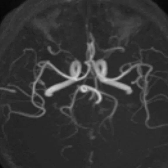
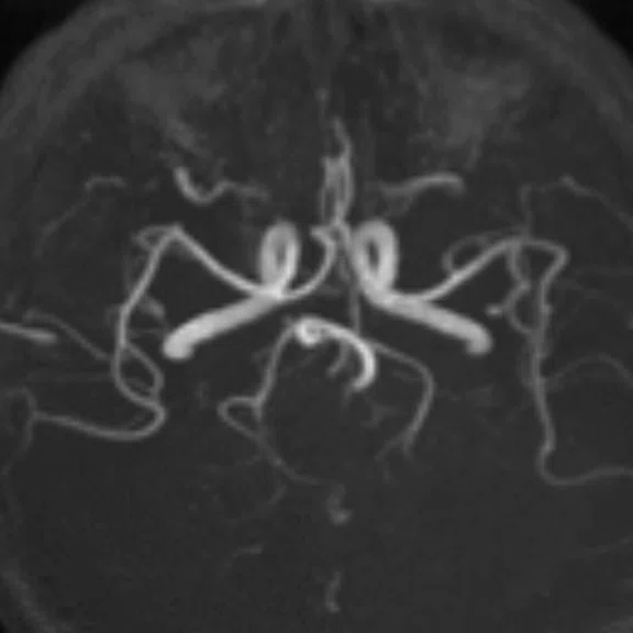
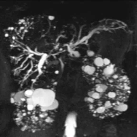
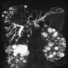
Head MRA and upper abdominal MRCP. (A) Without HyperSense, 0.6 x 0.6 x 0.8 mm, 4:06 min.; (B) HyperSense factor of 2, 0.6 x 0.5 x 0.8 mm, 1:41 min.; (C) MRCP with HyperSense and breath-hold, 0:16 min.; and (D) MRCP with HyperSense and free-breathing, 2:33 min.
B
Figure 1.
Head MRA and upper abdominal MRCP. (A) Without HyperSense, 0.6 x 0.6 x 0.8 mm, 4:06 min.; (B) HyperSense factor of 2, 0.6 x 0.5 x 0.8 mm, 1:41 min.; (C) MRCP with HyperSense and breath-hold, 0:16 min.; and (D) MRCP with HyperSense and free-breathing, 2:33 min.
C
Figure 1.
Head MRA and upper abdominal MRCP. (A) Without HyperSense, 0.6 x 0.6 x 0.8 mm, 4:06 min.; (B) HyperSense factor of 2, 0.6 x 0.5 x 0.8 mm, 1:41 min.; (C) MRCP with HyperSense and breath-hold, 0:16 min.; and (D) MRCP with HyperSense and free-breathing, 2:33 min.
D
Figure 1.
Head MRA and upper abdominal MRCP. (A) Without HyperSense, 0.6 x 0.6 x 0.8 mm, 4:06 min.; (B) HyperSense factor of 2, 0.6 x 0.5 x 0.8 mm, 1:41 min.; (C) MRCP with HyperSense and breath-hold, 0:16 min.; and (D) MRCP with HyperSense and free-breathing, 2:33 min.
E
Figure 3.
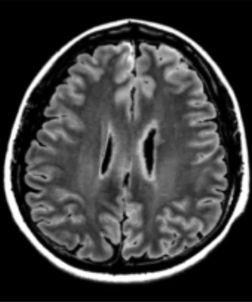
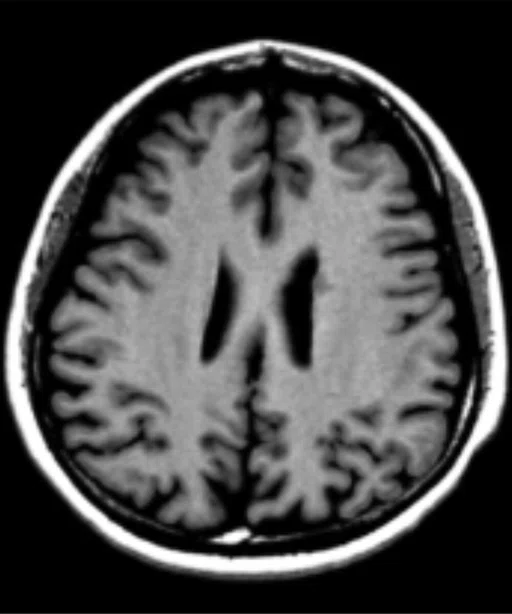
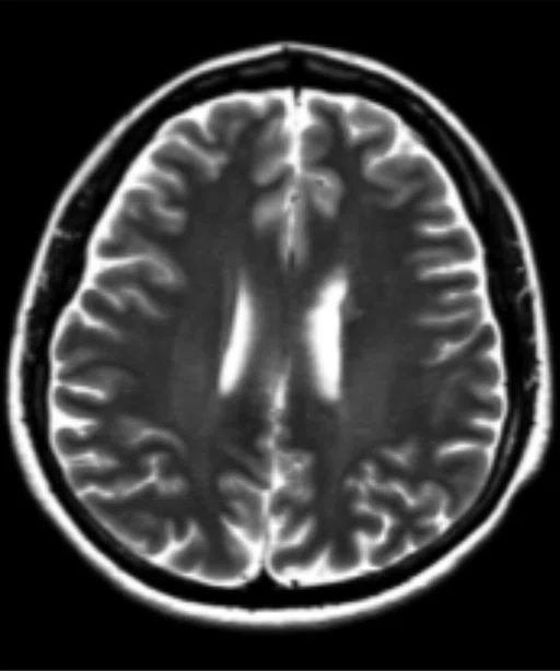
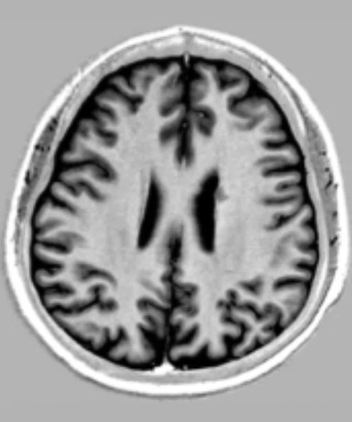
Lesion detected near the left lateral ventricle. Using MAGiC quantitative maps to measure the ROI of the lesion and ectocinerea, the values of T1, T2 and PD in these two tissues were completely consistent, enabling characterization of the lesion as heterotopic gray matter. (A) T2 FLAIR; (B) T1; (C) T2w; (D) PSIR; (E) T2 map, T1 1067 (40) ms, T2 86 (2) ms and PD 78.8 (2.1) pu; and (F) T2 map, T2 1083 (29) ms, T2 86 (2) ms and E F PD 77.6 (1.1) pu.
F
Figure 3.
Lesion detected near the left lateral ventricle. Using MAGiC quantitative maps to measure the ROI of the lesion and ectocinerea, the values of T1, T2 and PD in these two tissues were completely consistent, enabling characterization of the lesion as heterotopic gray matter. (A) T2 FLAIR; (B) T1; (C) T2w; (D) PSIR; (E) T2 map, T1 1067 (40) ms, T2 86 (2) ms and PD 78.8 (2.1) pu; and (F) T2 map, T2 1083 (29) ms, T2 86 (2) ms and E F PD 77.6 (1.1) pu.
A
Figure 2.
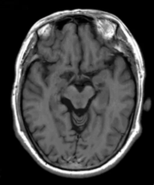
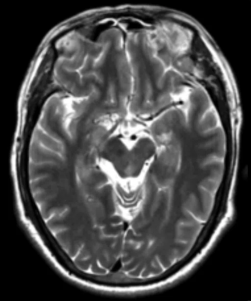
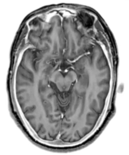
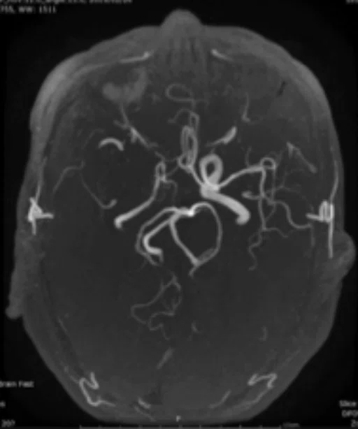
Male, 57 years old, with repeated dizziness for seven years. No clear lesions were detected on the (A) T1 and (B) T2 images. However, the (C) reconstructed PSIR VESSL demonstrated an absent right middle cerebral artery, confirmed by the (D) TOF-MRA.
B
Figure 2.
Male, 57 years old, with repeated dizziness for seven years. No clear lesions were detected on the (A) T1 and (B) T2 images. However, the (C) reconstructed PSIR VESSL demonstrated an absent right middle cerebral artery, confirmed by the (D) TOF-MRA.
C
Figure 2.
Male, 57 years old, with repeated dizziness for seven years. No clear lesions were detected on the (A) T1 and (B) T2 images. However, the (C) reconstructed PSIR VESSL demonstrated an absent right middle cerebral artery, confirmed by the (D) TOF-MRA.
D
Figure 2.
Male, 57 years old, with repeated dizziness for seven years. No clear lesions were detected on the (A) T1 and (B) T2 images. However, the (C) reconstructed PSIR VESSL demonstrated an absent right middle cerebral artery, confirmed by the (D) TOF-MRA.
A
Figure 3.
Lesion detected near the left lateral ventricle. Using MAGiC quantitative maps to measure the ROI of the lesion and ectocinerea, the values of T1, T2 and PD in these two tissues were completely consistent, enabling characterization of the lesion as heterotopic gray matter. (A) T2 FLAIR; (B) T1; (C) T2w; (D) PSIR; (E) T2 map, T1 1067 (40) ms, T2 86 (2) ms and PD 78.8 (2.1) pu; and (F) T2 map, T2 1083 (29) ms, T2 86 (2) ms and E F PD 77.6 (1.1) pu.
B
Figure 3.
Lesion detected near the left lateral ventricle. Using MAGiC quantitative maps to measure the ROI of the lesion and ectocinerea, the values of T1, T2 and PD in these two tissues were completely consistent, enabling characterization of the lesion as heterotopic gray matter. (A) T2 FLAIR; (B) T1; (C) T2w; (D) PSIR; (E) T2 map, T1 1067 (40) ms, T2 86 (2) ms and PD 78.8 (2.1) pu; and (F) T2 map, T2 1083 (29) ms, T2 86 (2) ms and E F PD 77.6 (1.1) pu.
C
Figure 3.
Lesion detected near the left lateral ventricle. Using MAGiC quantitative maps to measure the ROI of the lesion and ectocinerea, the values of T1, T2 and PD in these two tissues were completely consistent, enabling characterization of the lesion as heterotopic gray matter. (A) T2 FLAIR; (B) T1; (C) T2w; (D) PSIR; (E) T2 map, T1 1067 (40) ms, T2 86 (2) ms and PD 78.8 (2.1) pu; and (F) T2 map, T2 1083 (29) ms, T2 86 (2) ms and E F PD 77.6 (1.1) pu.
D
Figure 3.
Lesion detected near the left lateral ventricle. Using MAGiC quantitative maps to measure the ROI of the lesion and ectocinerea, the values of T1, T2 and PD in these two tissues were completely consistent, enabling characterization of the lesion as heterotopic gray matter. (A) T2 FLAIR; (B) T1; (C) T2w; (D) PSIR; (E) T2 map, T1 1067 (40) ms, T2 86 (2) ms and PD 78.8 (2.1) pu; and (F) T2 map, T2 1083 (29) ms, T2 86 (2) ms and E F PD 77.6 (1.1) pu.
result


PREVIOUS
${prev-page}
NEXT
${next-page}
Subscribe Now
Manage Subscription
FOLLOW US
Contact Us • Cookie Preferences • Privacy Policy • California Privacy PolicyDo Not Sell or Share My Personal Information • Terms & Conditions • Security
© 2024 GE HealthCare. GE is a trademark of General Electric Company. Used under trademark license.
IN PRACTICE
Hospital scans 100-plus patients in one 13-hour day on SIGNA Pioneer
Hospital scans 100-plus patients in one 13-hour day on SIGNA Pioneer
At The Second Hospital of Xi’an Jiaotong University, the demand for MR imaging has historically far exceeded capacity. Patient wait times were growing longer, leaving many people to wait one to two weeks to receive an MR examination.
At The Second Hospital of Xi’an Jiaotong University, the demand for MR imaging has historically far exceeded capacity. Patient wait times were growing longer, leaving many people to wait one to two weeks to receive an MR examination.
Yang Quanxin, MD, PhD, Director of the Radiology Department, knew that the two existing MR systems — a 3.0T SIGNA™ HDx and a 1.5T from another manufacturer — couldn’t keep up with the demand. As he began to explore purchasing a third MR system, he realized that in addition to addressing the clinical need he also wanted a system that could perform scientific research.
GE Healthcare has a strong relationship with the hospital and an excellent reputation for clinical performance across multiple systems and imaging modalities. According to Professor Quanxin, GE 3.0T MR systems are the largest installed base in Shaanxi Province. With GE’s assistance, many other sites have performed scientific research that has been published internationally in several peer-reviewed journals.
“My first impression of SIGNA™ Pioneer was the very fast scanning speed,” he explains.
100-plus scans in a day
The waiting time for MR exams was not only difficult on patients; the radiologists also felt the pressure to read through MR studies as quickly as possible.
“When we first began using SIGNA™ Pioneer, we noticed that it was very fast and also delivered a very high SNR,” says Professor Quanxin. “So, we had an idea to see if the system could scan more than 100 anatomic areas in a typical 13-hour day.”
The radiology team at The Second Hospital of Xi’an Jiaotong University collaborated with GE to embark on a project to scan as many high-quality MR exams as possible.
In February 2019, the radiology department performed 106 MR examinations from 7:30 am to 8:30 pm on SIGNA™ Pioneer in exams of the breast, abdomen, pelvis, various joints and fMRI. The team modified its protocols on SIGNA™ Pioneer, including utilizing HyperSense in MR angiography (MRA) and upper abdominal MRCP studies, and found that the scanning time for head MRA exams could be reduced by 45 percent. In patients who could not hold their breath for an MRCP exam, the speed of HyperSense enabled very short scan times — 16 seconds for breath-hold and 2:33 minutes for free-breathing exams (see Figure 1).
“With the same signal-to-noise-ratio, the scan time for each sequence on SIGNA™ Pioneer was nearly half the time as our sequence on the SIGNA™ HDx 3.0T system when scanning the same body area.”
Professor Yang Quanxin
Plus, he was intrigued by MAGiC‡ and its unique ability to output multiple image contrasts in a single 5-minute scan. With MAGiC, Professor Quanxin also has the potential to customize DIR for gray and white matter contrasts. Even after the acquisition, MAGiC allows the clinician to change contrast to provide more diagnostic information. Additionally, Professor Quanxin saw more opportunities to shorten scan time with HyperSense, an acceleration technique based on sparse data sampling and iterative reconstruction. For these reasons and the potential for research capabilities, Professor Quanxin chose SIGNA™ Pioneer for the hospital’s third MR system.
This volume was possible because in China exams are typically reduced to the most important acquisitions, typically three to five sequences. This is a common practice due to the large number of patients referred for an MR exam. At The Second Hospital of Xi’an Jiaotong University, the average study time on the prior system was 10 minutes. With SIGNA™ Pioneer it is now an average of 7 minutes per exam.
“The SNR was nearly unchanged in these shorter exams, so we could still make a confident clinical diagnosis,” Professor Quanxin says.
With MAGiC, the department performed one 4-minute scan to obtain the contrast and quantitative maps needed for diagnosis. In one case, a patient with repeated dizziness for seven years was found to have a missing right cerebral artery, detected by the MAGiC reconstructed PSIR and confirmed by time-of-flight MRA (see Figure 2).
“MAGiC quantitative mapping technology can also help us to characterize pathological findings,” Professor Quanxin adds (see Figure 3).
E
F
Figure 3.
Lesion detected near the left lateral ventricle. Using MAGiC quantitative maps to measure the ROI of the lesion and ectocinerea, the values of T1, T2 and PD in these two tissues were completely consistent, enabling characterization of the lesion as heterotopic gray matter. (A) T2 FLAIR; (B) T1; (C) T2w; (D) PSIR; (E) T2 map, T1 1067 (40) ms, T2 86 (2) ms and PD 78.8 (2.1) pu; and (F) T2 map, T2 1083 (29) ms, T2 86 (2) ms and E F PD 77.6 (1.1) pu.
DISCO was also utilized to improve the temporal resolution of dynamic contrast-enhanced imaging in the liver, breast and prostate, further enhancing image quality for more efficient and confident reads. In the liver, DISCO was instrumental in capturing the hepatic arterial phase, which can be difficult to clearly obtain with traditional MR sequences.
It’s not just the sequences that are helping to shorten and streamline MR scanning on the SIGNA™ Pioneer at The Second Hospital of Xi’an Jiaotong University. The system’s Auto Protocol Optimization (APX) simplifies the scanning process, and the touch bar on both sides of the MR table saves time in patient positioning. In just two steps, the technologist/radiographer can complete the positioning, and with APX they simply select between four and five options and then scan.
Today, SIGNA™ Pioneer is regularly utilized for a 15-hour day and often completes over 100 exams during that time frame. The other two MR systems are still used to capacity; however, it is SIGNA™ Pioneer that enables the heaviest workload.
“SIGNA™ Pioneer is a very good 3.0T MR system,” says Professor Quanxin. “It has many technologies that increase the scanning speed without losing the SNR of the image. It has definitely helped alleviate the issue of long appointment waiting times for our patients.”


