- Moser HW, Mahmood A, Raymond GV. Xlinked adrenoleukodystrophy. Nat Clin Pract Neurol. 2007;3(3):140-151.
- Engelen M, Kemp S, Poll-The BT. Xlinked adrenoleukodystrophy: pathogenesis and treatment. Curr Neurol Neurosci Rep. 2014;14(10):486.
- Miller WP, Rothman SM, Nascene D, et al. Outcomes after allogeneic hematopoietic cell transplantation for childhood cerebral adrenoleukodystrophy: the largest single-institution cohort report. Blood. 2011;118(7):1971-1978.
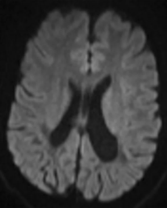
A
Figure 1.
Prior examination performed at 3.0T in 2017. (A-C) 2D FLAIR and (D, E) DTI.
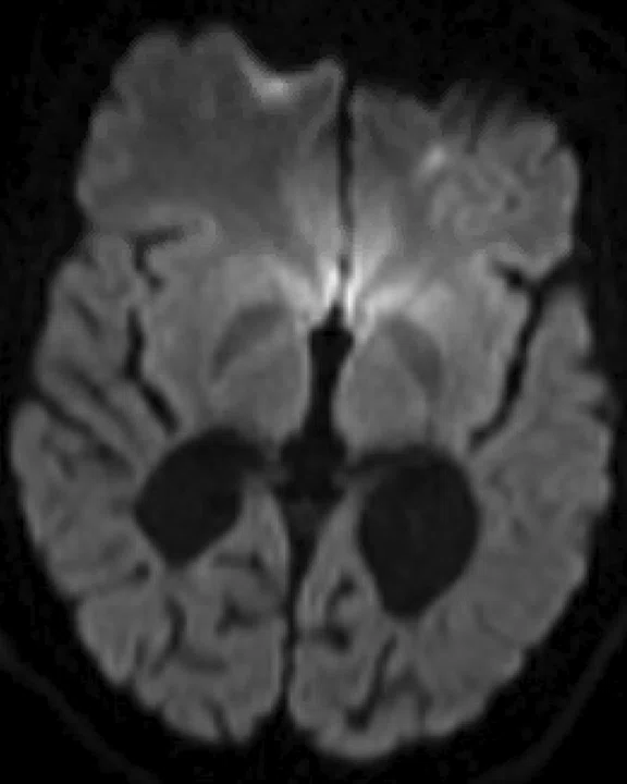
B
Figure 1.
Prior examination performed at 3.0T in 2017. (A-C) 2D FLAIR and (D, E) DTI.
C
Figure 1.
Prior examination performed at 3.0T in 2017. (A-C) 2D FLAIR and (D, E) DTI.
D
Figure 1.
Prior examination performed at 3.0T in 2017. (A-C) 2D FLAIR and (D, E) DTI.
E
Figure 1.
Prior examination performed at 3.0T in 2017. (A-C) 2D FLAIR and (D, E) DTI.
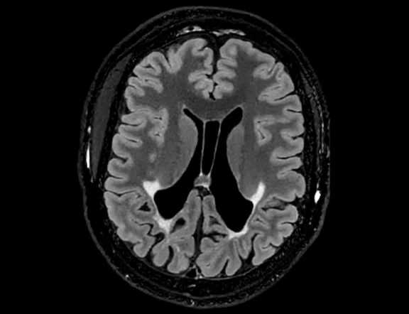
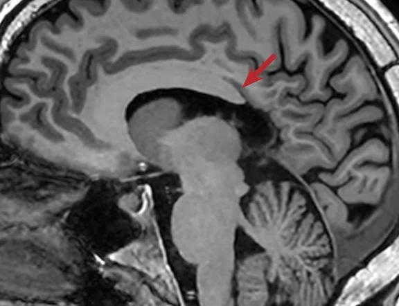
A
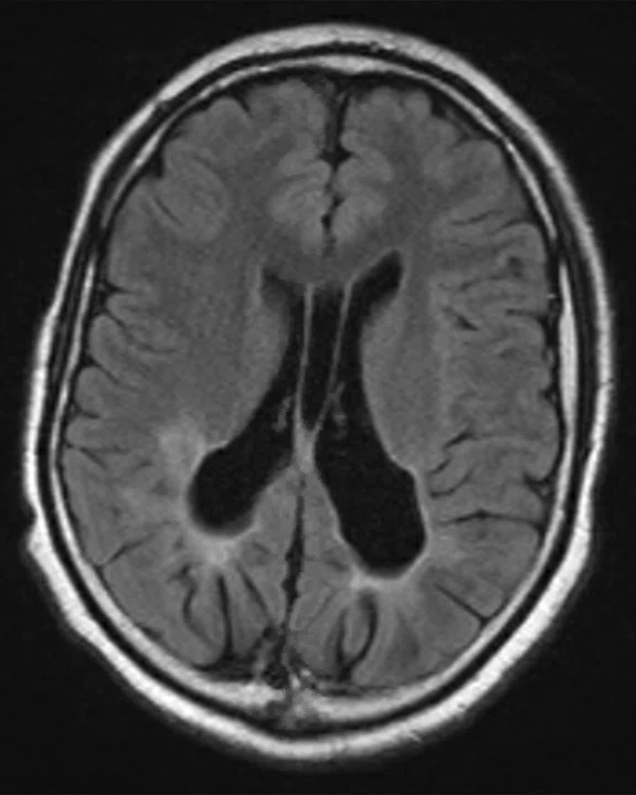
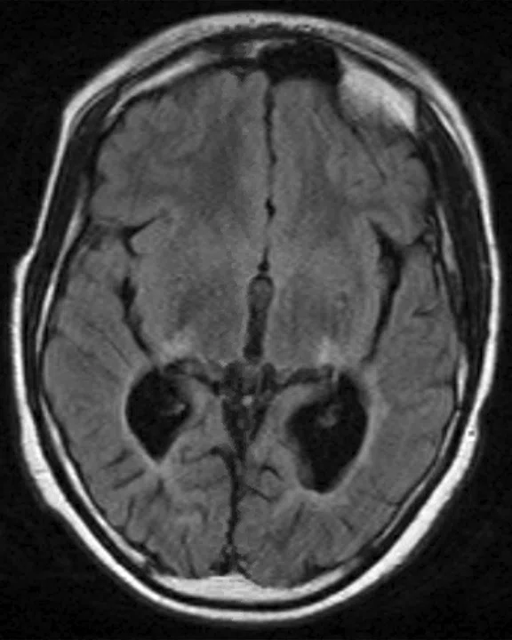
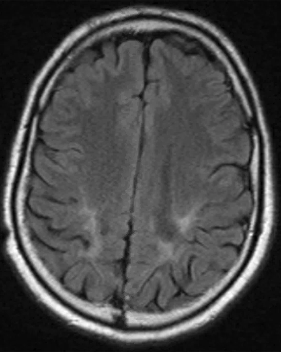
Figure 2.
Follow-up exam performed in 2023 on the SIGNA™ Premier with MR 30 for SIGNA™ and the 48-ch Head Coil. All sequences performed with AIR™ Recon DL. (A, B) 3D Cube FLAIR and (C, D) 3D MP-RAGE.
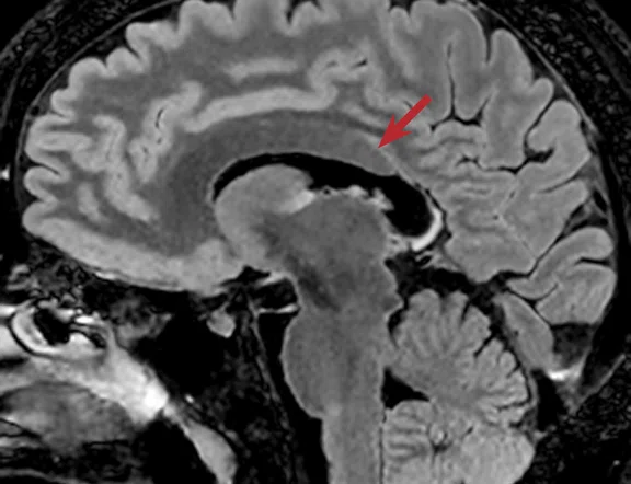
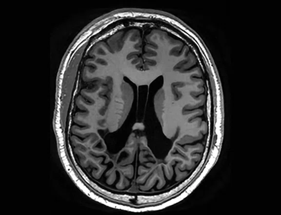
B
Figure 2.
Follow-up exam performed in 2023 on the SIGNA™ Premier with MR 30 for SIGNA™ and the 48-ch Head Coil. All sequences performed with AIR™ Recon DL. (A, B) 3D Cube FLAIR and (C, D) 3D MP-RAGE.
C
Figure 2.
Follow-up exam performed in 2023 on the SIGNA™ Premier with MR 30 for SIGNA™ and the 48-ch Head Coil. All sequences performed with AIR™ Recon DL. (A, B) 3D Cube FLAIR and (C, D) 3D MP-RAGE.
D
Figure 2.
Follow-up exam performed in 2023 on the SIGNA™ Premier with MR 30 for SIGNA™ and the 48-ch Head Coil. All sequences performed with AIR™ Recon DL. (A, B) 3D Cube FLAIR and (C, D) 3D MP-RAGE.
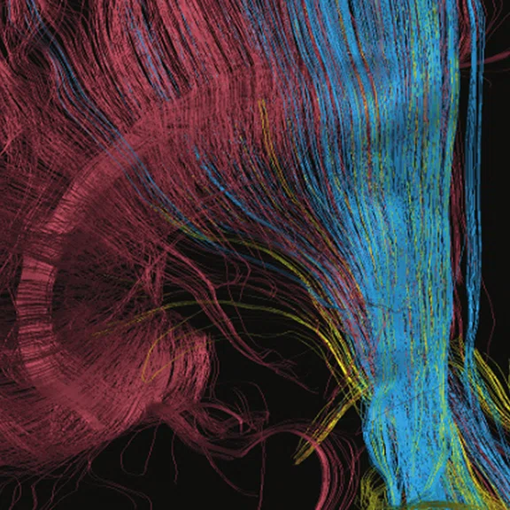
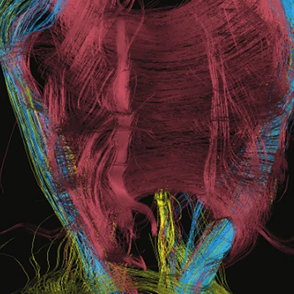
A
Figure 3.
(A-C) DTI in 64 directions with AIR™ Recon DL and the 48-ch Head Coil.
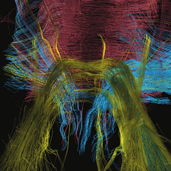
B
Figure 3.
(A-C) DTI in 64 directions with AIR™ Recon DL and the 48-ch Head Coil.
C
Figure 3.
(A-C) DTI in 64 directions with AIR™ Recon DL and the 48-ch Head Coil.
result


PREVIOUS
${prev-page}
NEXT
${next-page}
Subscribe Now
Manage Subscription
FOLLOW US
Contact Us • Cookie Preferences • Privacy Policy • California Privacy PolicyDo Not Sell or Share My Personal Information • Terms & Conditions • Security
© 2024 GE HealthCare. GE is a trademark of General Electric Company. Used under trademark license.
Case Studies
High spatial resolution and
SNR for brain volumetry and white-gray matter delineation
High spatial resolution and
SNR for brain volumetry and white-gray matter delineation
By Damien Galanaud, MD, Professor, Neuroimaging Department, La Pitié Salpêtriére University Hospital, Paris, France
Adrenoleukodystrophy (ALD) is a rare, genetic disease caused by mutations in the ABCD1 gene that results in a deficiency of the adrenoleukodystrophy protein (ALDP) and the accumulation of very-long-chain fatty acids in plasma and tissue — primarily of the nervous system and adrenal glands.
Without early diagnosis, the disease can develop into cerebral ALD, a severe, progressive and life-threatening neurodegenerative form of ALD that can cause behavioral, cognitive and neurological deficits. If left undiagnosed, ALD can lead to total disability and subsequent death within five years after initial symptoms. The severe form of this X-linked disease almost exclusively affects men.
Regular neuro monitoring of ALD patients using MR is important to diagnose cerebral ALD, which involves the destruction of the myelin sheath. In boys three to 12 years, MR should be performed every six months. After age 12, monitoring should continue, however, frequency of MR imaging is at the discretion of the physician.1,2,3
Patient history
A 27-year-old man with ALD presented for a neuro examination to evaluate possible progression of the disease. Patient received a hematopoietic marrow transplant in 1998. Patient continues to experience seizures.
In these cases, we must achieve good white-gray matter delineation with good spatial resolution in any plane that does not impede SNR to assess changes in brain volume and detect abnormalities in small areas of the brain. This is particularly important in patients with pre-existing white matter lesions.
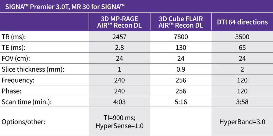
Because these patients are regularly monitored for the presence of cerebral ALD, we require good homogeneity and reproducible studies across multiple examinations. The use of AIR™ Recon DL 3D and the 48-channel Head Coil on the SIGNA™ Premier 3.0T system provides the homogeneity, high spatial resolution and SNR needed for these examinations. In addition, HyperSense is compatible with 3D MP-RAGE and HyperBand can be used with DTI.
Results
No evolution of ALD was detected.
Discussion
The combination of AIR™ Recon DL 3D and HyperSense provided excellent image quality in a comfortable scan time for the 3D acquisitions. AIR™ Recon DL DWI and HyperBand further improved SNR in the diffusion sequence. This resulted in more detailed images that improved reliability of the examination to detect any signs of clinical evolution to cerebral ALD and helped plan appropriate treatment.
The excellent image quality of the MP-RAGE allowed us to confidently assess the morphology of the brain for the presence of new lesions. With the increased SNR in the DWI, we were able to obtain DTI with a better visualization of deep gray matter than in prior examinations of this patient. The capability to perform 3D acquisitions with high spatial resolution in all required contrasts and DTI in 64 directions, all in a standard scan time of 13:17 minutes, was impressive.
DOWNLOAD ARTICLE HERE


