2. Holtås S, Olsson M, Romner B, Larsson EM, Säveland H, Brandt L. Comparison of MR imaging and CT in patients with intracranial aneurysm clips. AJNR Am J Neuroradiol. 1988;9(5):891-897.
1. Kivisaari RP, Salonen O, Servo A, Autti T, Hernesniemi J, Ohman J. MR imaging after aneurysmal subarachnoid hemorrhage and surgery: a long-term follow-up study. AJNR Am J Neuroradiol. 2001 Jun-Jul;22(6):1143-8.
A
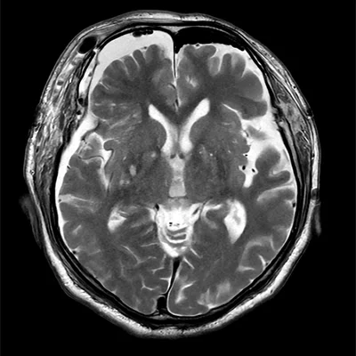
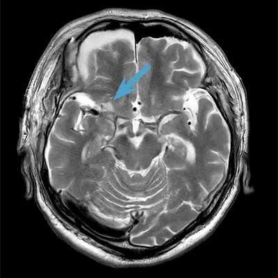
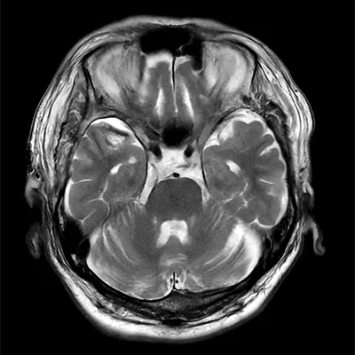
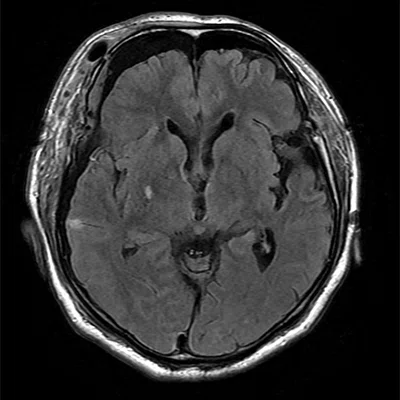
Figure 1.
Follow-up MR imaging in a 70-year-old male after a right MCA clipping surgery. Patient had an old small infarct. Clips and sinus lesions are difficult to evaluate with (J-L) single-shot EPI DWI, however, PROPELLER DWI with AIR™ Recon DL enabled visualization of the infarcted area (circles) while minimizing the effects of susceptibility artifacts (arrows, H and K). Note the difference in image quality due to the artifacts between the (G-I) PROPELLER DWI with AIR™ Recon DL and the (J-L) single-shot EPI DWI sequences. (A-C) Axial T2 FSE, 0.5 x 0.8 x 5.0 mm, 39 sec. with AIR™ Recon DL; (D-F) axial T2 FLAIR, 0.6 x 1.0 x 5.0 mm, 1:44 min. with AIR™ Recon DL; (G-I) axial PROPELLER DWI b1000, 1.7 x 1.7 x 5.0 mm, 2:23 min. with AIR™ Recon DL; and (J-L) axial single-shot EPI DWI b1000, 1.7 x 0.9 x 5.0 mm, 51 sec.
B
Figure 1.
Follow-up MR imaging in a 70-year-old male after a right MCA clipping surgery. Patient had an old small infarct. Clips and sinus lesions are difficult to evaluate with (J-L) single-shot EPI DWI, however, PROPELLER DWI with AIR™ Recon DL enabled visualization of the infarcted area (circles) while minimizing the effects of susceptibility artifacts (arrows, H and K). Note the difference in image quality due to the artifacts between the (G-I) PROPELLER DWI with AIR™ Recon DL and the (J-L) single-shot EPI DWI sequences. (A-C) Axial T2 FSE, 0.5 x 0.8 x 5.0 mm, 39 sec. with AIR™ Recon DL; (D-F) axial T2 FLAIR, 0.6 x 1.0 x 5.0 mm, 1:44 min. with AIR™ Recon DL; (G-I) axial PROPELLER DWI b1000, 1.7 x 1.7 x 5.0 mm, 2:23 min. with AIR™ Recon DL; and (J-L) axial single-shot EPI DWI b1000, 1.7 x 0.9 x 5.0 mm, 51 sec.
C
Figure 1.
Follow-up MR imaging in a 70-year-old male after a right MCA clipping surgery. Patient had an old small infarct. Clips and sinus lesions are difficult to evaluate with (J-L) single-shot EPI DWI, however, PROPELLER DWI with AIR™ Recon DL enabled visualization of the infarcted area (circles) while minimizing the effects of susceptibility artifacts (arrows, H and K). Note the difference in image quality due to the artifacts between the (G-I) PROPELLER DWI with AIR™ Recon DL and the (J-L) single-shot EPI DWI sequences. (A-C) Axial T2 FSE, 0.5 x 0.8 x 5.0 mm, 39 sec. with AIR™ Recon DL; (D-F) axial T2 FLAIR, 0.6 x 1.0 x 5.0 mm, 1:44 min. with AIR™ Recon DL; (G-I) axial PROPELLER DWI b1000, 1.7 x 1.7 x 5.0 mm, 2:23 min. with AIR™ Recon DL; and (J-L) axial single-shot EPI DWI b1000, 1.7 x 0.9 x 5.0 mm, 51 sec.
D
Figure 1.
Follow-up MR imaging in a 70-year-old male after a right MCA clipping surgery. Patient had an old small infarct. Clips and sinus lesions are difficult to evaluate with (J-L) single-shot EPI DWI, however, PROPELLER DWI with AIR™ Recon DL enabled visualization of the infarcted area (circles) while minimizing the effects of susceptibility artifacts (arrows, H and K). Note the difference in image quality due to the artifacts between the (G-I) PROPELLER DWI with AIR™ Recon DL and the (J-L) single-shot EPI DWI sequences. (A-C) Axial T2 FSE, 0.5 x 0.8 x 5.0 mm, 39 sec. with AIR™ Recon DL; (D-F) axial T2 FLAIR, 0.6 x 1.0 x 5.0 mm, 1:44 min. with AIR™ Recon DL; (G-I) axial PROPELLER DWI b1000, 1.7 x 1.7 x 5.0 mm, 2:23 min. with AIR™ Recon DL; and (J-L) axial single-shot EPI DWI b1000, 1.7 x 0.9 x 5.0 mm, 51 sec.
F
Figure 1.
Follow-up MR imaging in a 70-year-old male after a right MCA clipping surgery. Patient had an old small infarct. Clips and sinus lesions are difficult to evaluate with (J-L) single-shot EPI DWI, however, PROPELLER DWI with AIR™ Recon DL enabled visualization of the infarcted area (circles) while minimizing the effects of susceptibility artifacts (arrows, H and K). Note the difference in image quality due to the artifacts between the (G-I) PROPELLER DWI with AIR™ Recon DL and the (J-L) single-shot EPI DWI sequences. (A-C) Axial T2 FSE, 0.5 x 0.8 x 5.0 mm, 39 sec. with AIR™ Recon DL; (D-F) axial T2 FLAIR, 0.6 x 1.0 x 5.0 mm, 1:44 min. with AIR™ Recon DL; (G-I) axial PROPELLER DWI b1000, 1.7 x 1.7 x 5.0 mm, 2:23 min. with AIR™ Recon DL; and (J-L) axial single-shot EPI DWI b1000, 1.7 x 0.9 x 5.0 mm, 51 sec.
G
Figure 1.
Follow-up MR imaging in a 70-year-old male after a right MCA clipping surgery. Patient had an old small infarct. Clips and sinus lesions are difficult to evaluate with (J-L) single-shot EPI DWI, however, PROPELLER DWI with AIR™ Recon DL enabled visualization of the infarcted area (circles) while minimizing the effects of susceptibility artifacts (arrows, H and K). Note the difference in image quality due to the artifacts between the (G-I) PROPELLER DWI with AIR™ Recon DL and the (J-L) single-shot EPI DWI sequences. (A-C) Axial T2 FSE, 0.5 x 0.8 x 5.0 mm, 39 sec. with AIR™ Recon DL; (D-F) axial T2 FLAIR, 0.6 x 1.0 x 5.0 mm, 1:44 min. with AIR™ Recon DL; (G-I) axial PROPELLER DWI b1000, 1.7 x 1.7 x 5.0 mm, 2:23 min. with AIR™ Recon DL; and (J-L) axial single-shot EPI DWI b1000, 1.7 x 0.9 x 5.0 mm, 51 sec.
H
Figure 1.
Follow-up MR imaging in a 70-year-old male after a right MCA clipping surgery. Patient had an old small infarct. Clips and sinus lesions are difficult to evaluate with (J-L) single-shot EPI DWI, however, PROPELLER DWI with AIR™ Recon DL enabled visualization of the infarcted area (circles) while minimizing the effects of susceptibility artifacts (arrows, H and K). Note the difference in image quality due to the artifacts between the (G-I) PROPELLER DWI with AIR™ Recon DL and the (J-L) single-shot EPI DWI sequences. (A-C) Axial T2 FSE, 0.5 x 0.8 x 5.0 mm, 39 sec. with AIR™ Recon DL; (D-F) axial T2 FLAIR, 0.6 x 1.0 x 5.0 mm, 1:44 min. with AIR™ Recon DL; (G-I) axial PROPELLER DWI b1000, 1.7 x 1.7 x 5.0 mm, 2:23 min. with AIR™ Recon DL; and (J-L) axial single-shot EPI DWI b1000, 1.7 x 0.9 x 5.0 mm, 51 sec.
I
Figure 1.
Follow-up MR imaging in a 70-year-old male after a right MCA clipping surgery. Patient had an old small infarct. Clips and sinus lesions are difficult to evaluate with (J-L) single-shot EPI DWI, however, PROPELLER DWI with AIR™ Recon DL enabled visualization of the infarcted area (circles) while minimizing the effects of susceptibility artifacts (arrows, H and K). Note the difference in image quality due to the artifacts between the (G-I) PROPELLER DWI with AIR™ Recon DL and the (J-L) single-shot EPI DWI sequences. (A-C) Axial T2 FSE, 0.5 x 0.8 x 5.0 mm, 39 sec. with AIR™ Recon DL; (D-F) axial T2 FLAIR, 0.6 x 1.0 x 5.0 mm, 1:44 min. with AIR™ Recon DL; (G-I) axial PROPELLER DWI b1000, 1.7 x 1.7 x 5.0 mm, 2:23 min. with AIR™ Recon DL; and (J-L) axial single-shot EPI DWI b1000, 1.7 x 0.9 x 5.0 mm, 51 sec.
J
Figure 1.
Follow-up MR imaging in a 70-year-old male after a right MCA clipping surgery. Patient had an old small infarct. Clips and sinus lesions are difficult to evaluate with (J-L) single-shot EPI DWI, however, PROPELLER DWI with AIR™ Recon DL enabled visualization of the infarcted area (circles) while minimizing the effects of susceptibility artifacts (arrows, H and K). Note the difference in image quality due to the artifacts between the (G-I) PROPELLER DWI with AIR™ Recon DL and the (J-L) single-shot EPI DWI sequences. (A-C) Axial T2 FSE, 0.5 x 0.8 x 5.0 mm, 39 sec. with AIR™ Recon DL; (D-F) axial T2 FLAIR, 0.6 x 1.0 x 5.0 mm, 1:44 min. with AIR™ Recon DL; (G-I) axial PROPELLER DWI b1000, 1.7 x 1.7 x 5.0 mm, 2:23 min. with AIR™ Recon DL; and (J-L) axial single-shot EPI DWI b1000, 1.7 x 0.9 x 5.0 mm, 51 sec.
K
Figure 1.
Follow-up MR imaging in a 70-year-old male after a right MCA clipping surgery. Patient had an old small infarct. Clips and sinus lesions are difficult to evaluate with (J-L) single-shot EPI DWI, however, PROPELLER DWI with AIR™ Recon DL enabled visualization of the infarcted area (circles) while minimizing the effects of susceptibility artifacts (arrows, H and K). Note the difference in image quality due to the artifacts between the (G-I) PROPELLER DWI with AIR™ Recon DL and the (J-L) single-shot EPI DWI sequences. (A-C) Axial T2 FSE, 0.5 x 0.8 x 5.0 mm, 39 sec. with AIR™ Recon DL; (D-F) axial T2 FLAIR, 0.6 x 1.0 x 5.0 mm, 1:44 min. with AIR™ Recon DL; (G-I) axial PROPELLER DWI b1000, 1.7 x 1.7 x 5.0 mm, 2:23 min. with AIR™ Recon DL; and (J-L) axial single-shot EPI DWI b1000, 1.7 x 0.9 x 5.0 mm, 51 sec.
L
Figure 1.
Follow-up MR imaging in a 70-year-old male after a right MCA clipping surgery. Patient had an old small infarct. Clips and sinus lesions are difficult to evaluate with (J-L) single-shot EPI DWI, however, PROPELLER DWI with AIR™ Recon DL enabled visualization of the infarcted area (circles) while minimizing the effects of susceptibility artifacts (arrows, H and K). Note the difference in image quality due to the artifacts between the (G-I) PROPELLER DWI with AIR™ Recon DL and the (J-L) single-shot EPI DWI sequences. (A-C) Axial T2 FSE, 0.5 x 0.8 x 5.0 mm, 39 sec. with AIR™ Recon DL; (D-F) axial T2 FLAIR, 0.6 x 1.0 x 5.0 mm, 1:44 min. with AIR™ Recon DL; (G-I) axial PROPELLER DWI b1000, 1.7 x 1.7 x 5.0 mm, 2:23 min. with AIR™ Recon DL; and (J-L) axial single-shot EPI DWI b1000, 1.7 x 0.9 x 5.0 mm, 51 sec.
E
Figure 1.
Follow-up MR imaging in a 70-year-old male after a right MCA clipping surgery. Patient had an old small infarct. Clips and sinus lesions are difficult to evaluate with (J-L) single-shot EPI DWI, however, PROPELLER DWI with AIR™ Recon DL enabled visualization of the infarcted area (circles) while minimizing the effects of susceptibility artifacts (arrows, H and K). Note the difference in image quality due to the artifacts between the (G-I) PROPELLER DWI with AIR™ Recon DL and the (J-L) single-shot EPI DWI sequences. (A-C) Axial T2 FSE, 0.5 x 0.8 x 5.0 mm, 39 sec. with AIR™ Recon DL; (D-F) axial T2 FLAIR, 0.6 x 1.0 x 5.0 mm, 1:44 min. with AIR™ Recon DL; (G-I) axial PROPELLER DWI b1000, 1.7 x 1.7 x 5.0 mm, 2:23 min. with AIR™ Recon DL; and (J-L) axial single-shot EPI DWI b1000, 1.7 x 0.9 x 5.0 mm, 51 sec.


result
PREVIOUS
${prev-page}
NEXT
${next-page}
Subscribe Now
Manage Subscription
FOLLOW US
Contact Us • Cookie Preferences • Privacy Policy • California Privacy PolicyDo Not Sell or Share My Personal Information • Terms & Conditions • Security
© 2024 GE HealthCare. GE is a trademark of General Electric Company. Used under trademark license.

Masanori Kitase, MD
Kariya Toyota General Hospital
—
Kariya, Japan
Case Studies
PROPELLER DWI for minimizing susceptibility artifacts in MCA clip follow-up exams
PROPELLER DWI for minimizing susceptibility artifacts in MCA clip follow-up exams
by Masanori Kitase, MD, Department of Radiology, Kariya Toyota General Hospital, Kariya, Japan
Masanori Kitase, MD
Kariya Toyota General Hospital
—
Kariya, Japan
MR imaging is often used for follow-up imaging after surgical clipping of a cerebral aneurysm. The introduction of MR-Conditional metals (e.g., titanium) in clipping and coiling devices has led to MR often being the imaging study of choice for these cases. MR provides high contrast resolution and is superior to CT in revealing infarctions and detecting small lacunar lesions in the brain.1
While both CT and MR can be used for these types of evaluations, CT typically has stronger metal artifacts that may make the study difficult to evaluate. MR imaging may show susceptibility artifacts around the clips, particularly in the single-shot EPI DWI sequence; however, MR has been shown to be superior to CT in depicting anatomic details and lesions in patients with aneurysm clips.2
PROPELLER is a sequence that can be utilized to suppress susceptibility artifacts in DWI. However, with conventional imaging, a long scanning time is required to obtain high-quality images with PROPELLER. The introduction of AIR™ Recon DL PROPELLER DWI enables high-quality imaging in a short scan time. In this particular case, the sequence was acquired in 2:23 minutes, enabling completion of the entire examination within a 10-minute timeframe (as preferred by our hospital). We have now applied AIR™ Recon DL PROPELLER DWI to routine middle cerebral artery (MCA) clip imaging.

Patient History
A 78-year-old male weighing 63 kg (139 lbs.) with no specific medical history was referred for a regular follow-up MR examination after surgery for an unruptured cerebral aneurysm. CT performed one day after surgery showed no bleeding, no apparent cerebral infarction and no other gross complication.
Results
Five days after the MCA aneurysm clipping, the MR examination showed small infarcts of approximately 5 mm each in the right frontal and temporal lobes around the clip. The lesion in the frontal lobe was located near the clip and could not be evaluated by single-shot EPI DWI due to the strong susceptibility artifacts, while PROPELLER DWI showed the lesion with fewer artifacts. PROPELLER DWI also showed less distortion at the base of the skull and the lesions in the temporal lobe were clearly visualized.
A
B
C
D
E
F
G
H
I
J
K
L
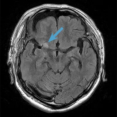
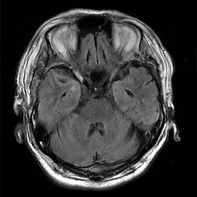
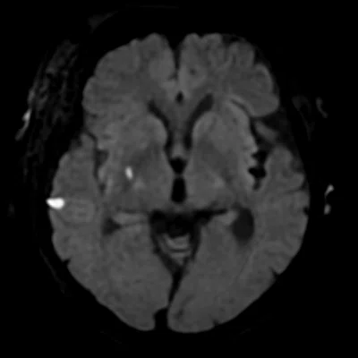
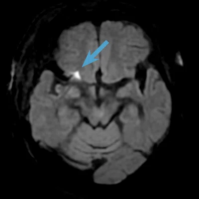
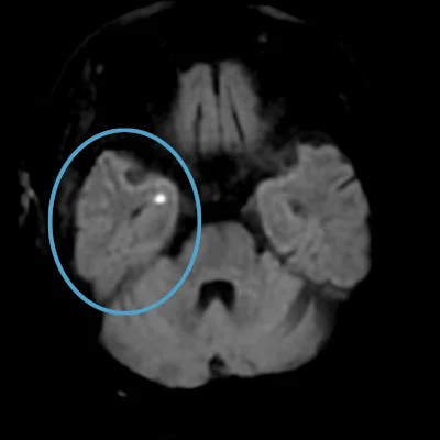
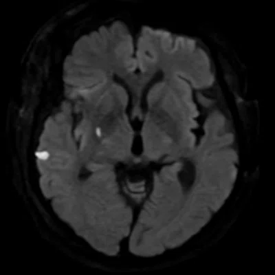
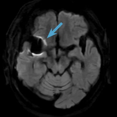
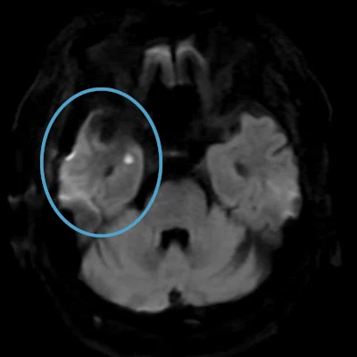
Figure 1.
Follow-up MR imaging in a 70-year-old male after a right MCA clipping surgery. Patient had an old small infarct. Clips and sinus lesions are difficult to evaluate with (J-L) single-shot EPI DWI, however, PROPELLER DWI with AIR™ Recon DL enabled visualization of the infarcted area (circles) while minimizing the effects of susceptibility artifacts (arrows, H and K). Note the difference in image quality due to the artifacts between the (G-I) PROPELLER DWI with AIR™ Recon DL and the (J-L) single-shot EPI DWI sequences. (A-C) Axial T2 FSE, 0.5 x 0.8 x 5.0 mm, 39 sec. with AIR™ Recon DL; (D-F) axial T2 FLAIR, 0.6 x 1.0 x 5.0 mm, 1:44 min. with AIR™ Recon DL; (G-I) axial PROPELLER DWI b1000, 1.7 x 1.7 x 5.0 mm, 2:23 min. with AIR™ Recon DL; and (J-L) axial single-shot EPI DWI b1000, 1.7 x 0.9 x 5.0 mm, 51 sec.
Discussion
PROPELLER DWI with AIR™ Recon DL is a useful sequence after aneurysm clipping. The area around the clip, in particular, is a high-risk area for infarction. Occasionally, asymptomatic infarcts are detected and the need for additional treatment is considered.
Lesions near aneurysm clips or the skull base can be difficult to assess with conventional single-shot EPI DWI due to artifacts. This is a case in which a new lesion, which could not be visualized before with single-shot EPI DWI, could be detected using PROPELLER DWI. The addition of AIR™ Recon DL provided better image quality and allowed us to shorten the PROPELLER DWI acquisition so there was a minimal increase in total examination time.
AIR™ Recon DL provided good image quality in a short scan time for both T2-weighted and FLAIR images. In diffusion-weighted images, good image quality was obtained in a short time with single-shot EPI DWI, but the artifacts in areas near the MR-Conditional clip were problematic. In addition, the brain parenchyma in areas close to air, such as the base of the skull, has strong susceptibility artifacts. PROPELLER DWI is useful to solve these problems, and the low-artifact images make reading easier and improve diagnostic confidence.
DOWNLOAD ARTICLE HERE
