A
Figure 1.
A 75-year-old woman with a right vestibular schwannoma (arrows) was referred for an MR exam on the SIGNA™ Artist 1.5T for radiosurgery planning. AIR™ Recon DL was employed, helping to improve spatial resolution in the 3D MP-RAGE and 3D FIESTA sequences and reduce scan time by 50% in the 3D FIESTA sequence. Note how the schwannoma is better visualized in the internal auditory canal in (F) the 3D FIESTA reformat reconstructed with AIR™ Recon DL. (A) Axial 3D MP-RAGE with conventional reconstruction, 1 x 1 x 1.2 mm, 9:20 min.; (B) axial 3D MP-RAGE reconstructed with AIR™ Recon DL, 1 x 1 x 1.2 mm, 8:55 min.; (C) axial 3D FIESTA with conventional reconstruction, 0.5 x 0.5 x 1 mm, 9:17 min.; (D) axial 3D FIESTA reconstructed with AIR™ Recon DL, 0.5 x 0.5 x 1 mm, 4:39 min.; (E) 3D FIESTA coronal reformat with conventional reconstruction; and (F) 3D FIESTA coronal reformat reconstructed with AIR™ Recon DL.
B
Figure 1.
A 75-year-old woman with a right vestibular schwannoma (arrows) was referred for an MR exam on the SIGNA™ Artist 1.5T for radiosurgery planning. AIR™ Recon DL was employed, helping to improve spatial resolution in the 3D MP-RAGE and 3D FIESTA sequences and reduce scan time by 50% in the 3D FIESTA sequence. Note how the schwannoma is better visualized in the internal auditory canal in (F) the 3D FIESTA reformat reconstructed with AIR™ Recon DL. (A) Axial 3D MP-RAGE with conventional reconstruction, 1 x 1 x 1.2 mm, 9:20 min.; (B) axial 3D MP-RAGE reconstructed with AIR™ Recon DL, 1 x 1 x 1.2 mm, 8:55 min.; (C) axial 3D FIESTA with conventional reconstruction, 0.5 x 0.5 x 1 mm, 9:17 min.; (D) axial 3D FIESTA reconstructed with AIR™ Recon DL, 0.5 x 0.5 x 1 mm, 4:39 min.; (E) 3D FIESTA coronal reformat with conventional reconstruction; and (F) 3D FIESTA coronal reformat reconstructed with AIR™ Recon DL.
C
Figure 1.
A 75-year-old woman with a right vestibular schwannoma (arrows) was referred for an MR exam on the SIGNA™ Artist 1.5T for radiosurgery planning. AIR™ Recon DL was employed, helping to improve spatial resolution in the 3D MP-RAGE and 3D FIESTA sequences and reduce scan time by 50% in the 3D FIESTA sequence. Note how the schwannoma is better visualized in the internal auditory canal in (F) the 3D FIESTA reformat reconstructed with AIR™ Recon DL. (A) Axial 3D MP-RAGE with conventional reconstruction, 1 x 1 x 1.2 mm, 9:20 min.; (B) axial 3D MP-RAGE reconstructed with AIR™ Recon DL, 1 x 1 x 1.2 mm, 8:55 min.; (C) axial 3D FIESTA with conventional reconstruction, 0.5 x 0.5 x 1 mm, 9:17 min.; (D) axial 3D FIESTA reconstructed with AIR™ Recon DL, 0.5 x 0.5 x 1 mm, 4:39 min.; (E) 3D FIESTA coronal reformat with conventional reconstruction; and (F) 3D FIESTA coronal reformat reconstructed with AIR™ Recon DL.
D
Figure 1.
A 75-year-old woman with a right vestibular schwannoma (arrows) was referred for an MR exam on the SIGNA™ Artist 1.5T for radiosurgery planning. AIR™ Recon DL was employed, helping to improve spatial resolution in the 3D MP-RAGE and 3D FIESTA sequences and reduce scan time by 50% in the 3D FIESTA sequence. Note how the schwannoma is better visualized in the internal auditory canal in (F) the 3D FIESTA reformat reconstructed with AIR™ Recon DL. (A) Axial 3D MP-RAGE with conventional reconstruction, 1 x 1 x 1.2 mm, 9:20 min.; (B) axial 3D MP-RAGE reconstructed with AIR™ Recon DL, 1 x 1 x 1.2 mm, 8:55 min.; (C) axial 3D FIESTA with conventional reconstruction, 0.5 x 0.5 x 1 mm, 9:17 min.; (D) axial 3D FIESTA reconstructed with AIR™ Recon DL, 0.5 x 0.5 x 1 mm, 4:39 min.; (E) 3D FIESTA coronal reformat with conventional reconstruction; and (F) 3D FIESTA coronal reformat reconstructed with AIR™ Recon DL.
E
Figure 1.
A 75-year-old woman with a right vestibular schwannoma (arrows) was referred for an MR exam on the SIGNA™ Artist 1.5T for radiosurgery planning. AIR™ Recon DL was employed, helping to improve spatial resolution in the 3D MP-RAGE and 3D FIESTA sequences and reduce scan time by 50% in the 3D FIESTA sequence. Note how the schwannoma is better visualized in the internal auditory canal in (F) the 3D FIESTA reformat reconstructed with AIR™ Recon DL. (A) Axial 3D MP-RAGE with conventional reconstruction, 1 x 1 x 1.2 mm, 9:20 min.; (B) axial 3D MP-RAGE reconstructed with AIR™ Recon DL, 1 x 1 x 1.2 mm, 8:55 min.; (C) axial 3D FIESTA with conventional reconstruction, 0.5 x 0.5 x 1 mm, 9:17 min.; (D) axial 3D FIESTA reconstructed with AIR™ Recon DL, 0.5 x 0.5 x 1 mm, 4:39 min.; (E) 3D FIESTA coronal reformat with conventional reconstruction; and (F) 3D FIESTA coronal reformat reconstructed with AIR™ Recon DL.
F
Figure 1.
A 75-year-old woman with a right vestibular schwannoma (arrows) was referred for an MR exam on the SIGNA™ Artist 1.5T for radiosurgery planning. AIR™ Recon DL was employed, helping to improve spatial resolution in the 3D MP-RAGE and 3D FIESTA sequences and reduce scan time by 50% in the 3D FIESTA sequence. Note how the schwannoma is better visualized in the internal auditory canal in (F) the 3D FIESTA reformat reconstructed with AIR™ Recon DL. (A) Axial 3D MP-RAGE with conventional reconstruction, 1 x 1 x 1.2 mm, 9:20 min.; (B) axial 3D MP-RAGE reconstructed with AIR™ Recon DL, 1 x 1 x 1.2 mm, 8:55 min.; (C) axial 3D FIESTA with conventional reconstruction, 0.5 x 0.5 x 1 mm, 9:17 min.; (D) axial 3D FIESTA reconstructed with AIR™ Recon DL, 0.5 x 0.5 x 1 mm, 4:39 min.; (E) 3D FIESTA coronal reformat with conventional reconstruction; and (F) 3D FIESTA coronal reformat reconstructed with AIR™ Recon DL.
result


PREVIOUS
${prev-page}
NEXT
${next-page}
Subscribe Now
Manage Subscription
FOLLOW US
Contact Us • Cookie Preferences • Privacy Policy • California Privacy PolicyDo Not Sell or Share My Personal Information • Terms & Conditions • Security
© 2024 GE HealthCare. GE is a trademark of General Electric Company. Used under trademark license.
Case Studies
High spatial resolution and SNR imaging of schwannoma for radiosurgery planning
High spatial resolution and SNR imaging of schwannoma for radiosurgery planning
By Natalia Shor, MD, Neuroimaging Department, La Pitié Salpêtriére University Hospital, Paris, France
Schwannoma is a tumor that develops from Schwann cells in the peripheral nervous system or nerve roots that is usually benign. Vestibular schwannomas develop from the balance and hearing nerves that supply the inner ear and can lead to hearing loss, tinnitus, dizziness or loss of balance. In some cases, the tumor can become large and press against nearby brain structures.
Radiosurgery is often employed in vestibular schwannomas to reduce the size or limit the growth of the tumor. MR imaging is typically the imaging exam of choice for radiosurgery planning in these cases. It is thus very important to have high spatial resolution in any reformat plane without impacting SNR to delineate the tumor correctly and to not harm the surrounding structures.
With the 1-channel head transmitter/receiver coil used in this exam, we cannot use parallel imaging to reduce scan time, which is desired. Furthermore, when aiming to reduce scan time, it is important to avoid increasing magnetic susceptibility artifacts and minimize distortion.
Patient history
A 75-year-old woman with a right vestibular schwannoma was referred for an MR exam for radiosurgery planning. A stereotactic frame was used for the exam to ensure images were captured in the same precise positioning as her treatment.
Results
The schwannoma was described as a Koos 3, Ohata B, with drop down of cochlear liquid signal.
Figure 1.
A 75-year-old woman with a right vestibular schwannoma (arrows) was referred for an MR exam on the SIGNA™ Artist 1.5T for radiosurgery planning. AIR™ Recon DL was employed, helping to improve spatial resolution in the 3D MP-RAGE and 3D FIESTA sequences and reduce scan time by 50% in the 3D FIESTA sequence. Note how the schwannoma is better visualized in the internal auditory canal in (B) the MP-RAGE with AIR™ Recon DL and (F) the 3D FIESTA reformat reconstructed with AIR™ Recon DL. (A) Axial 3D MP-RAGE with conventional reconstruction, 1 x 1 x 1.2 mm, 9:20 min.; (B) axial 3D MP-RAGE reconstructed with AIR™ Recon DL, 1 x 1 x 1.2 mm, 8:55 min.; (C) axial 3D FIESTA with conventional reconstruction, 0.5 x 0.5 x 1 mm, 9:17 min.; (D) axial 3D FIESTA reconstructed with AIR™ Recon DL, 0.5 x 0.5 x 1 mm, 4:39 min.; (E) 3D FIESTA coronal reformat with conventional reconstruction; and (F) 3D FIESTA coronal reformat reconstructed with AIR™ Recon DL.
Discussion
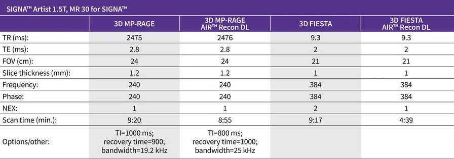
In this vestibular schwannoma examination, the addition of AIR™ Recon DL 3D has enabled a 50% reduction in scan time by reducing NEX from 2 to 1 on the 3D FIESTA sequence without degrading image quality.
The ability to use AIR™ Recon DL with the 3D FIESTA and 3D MP-RAGE sequences provided excellent image quality and high spatial resolution. The shorter scan time provided greater patient comfort, enabling the patient to remain still during the examination. The schwannoma was well depicted and easier to grade with AIR™ Recon DL. Also, AIR™ Recon DL does not require the use of a phased array and could therefore be used with the 1-channel head transmitter/receiver coil. The increased SNR also facilitated better visualization of the schwannoma in the internal auditory canal for more precise tumor contouring, crucial for Gamma Knife® radiosurgery treatment planning.
DOWNLOAD ARTICLE HERE


