A
Figure 1.
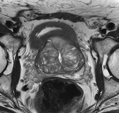
Patient referred for prostate MR exam. AIR™ Recon DL High, the 21-ch AIR™ MP Coil and 60-ch AIR™ PA Coil were used. (A, B) T2 FSE, 0.6 x 0.6 x 3 mm, (A) axial 3:03 min. and (B) coronal 2:39 min.; (C) sagittal T2 PROPELLER, 0.6 x 0.6 x 3 mm, 2:25 min.; and (D-F) T2 Cube, 0.7 x 0.7 x 0.4 mm, 3:42 min., shown in (D) axial, (E) coronal and (F) sagittal planes.
B
Figure 1.
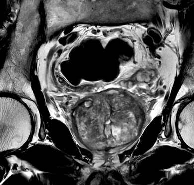
Patient referred for prostate MR exam. AIR™ Recon DL High, the 21-ch AIR™ MP Coil and 60-ch AIR™ PA Coil were used. (A, B) T2 FSE, 0.6 x 0.6 x 3 mm, (A) axial 3:03 min. and (B) coronal 2:39 min.; (C) sagittal T2 PROPELLER, 0.6 x 0.6 x 3 mm, 2:25 min.; and (D-F) T2 Cube, 0.7 x 0.7 x 0.4 mm, 3:42 min., shown in (D) axial, (E) coronal and (F) sagittal planes.
C
Figure 1.
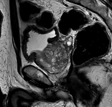
Patient referred for prostate MR exam. AIR™ Recon DL High, the 21-ch AIR™ MP Coil and 60-ch AIR™ PA Coil were used. (A, B) T2 FSE, 0.6 x 0.6 x 3 mm, (A) axial 3:03 min. and (B) coronal 2:39 min.; (C) sagittal T2 PROPELLER, 0.6 x 0.6 x 3 mm, 2:25 min.; and (D-F) T2 Cube, 0.7 x 0.7 x 0.4 mm, 3:42 min., shown in (D) axial, (E) coronal and (F) sagittal planes.
D
Figure 1.
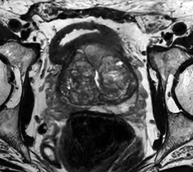
Patient referred for prostate MR exam. AIR™ Recon DL High, the 21-ch AIR™ MP Coil and 60-ch AIR™ PA Coil were used. (A, B) T2 FSE, 0.6 x 0.6 x 3 mm, (A) axial 3:03 min. and (B) coronal 2:39 min.; (C) sagittal T2 PROPELLER, 0.6 x 0.6 x 3 mm, 2:25 min.; and (D-F) T2 Cube, 0.7 x 0.7 x 0.4 mm, 3:42 min., shown in (D) axial, (E) coronal and (F) sagittal planes.
E
Figure 1.
Patient referred for prostate MR exam. AIR™ Recon DL High, the 21-ch AIR™ MP Coil and 60-ch AIR™ PA Coil were used. (A, B) T2 FSE, 0.6 x 0.6 x 3 mm, (A) axial 3:03 min. and (B) coronal 2:39 min.; (C) sagittal T2 PROPELLER, 0.6 x 0.6 x 3 mm, 2:25 min.; and (D-F) T2 Cube, 0.7 x 0.7 x 0.4 mm, 3:42 min., shown in (D) axial, (E) coronal and (F) sagittal planes.
F
Figure 1.
Patient referred for prostate MR exam. AIR™ Recon DL High, the 21-ch AIR™ MP Coil and 60-ch AIR™ PA Coil were used. (A, B) T2 FSE, 0.6 x 0.6 x 3 mm, (A) axial 3:03 min. and (B) coronal 2:39 min.; (C) sagittal T2 PROPELLER, 0.6 x 0.6 x 3 mm, 2:25 min.; and (D-F) T2 Cube, 0.7 x 0.7 x 0.4 mm, 3:42 min., shown in (D) axial, (E) coronal and (F) sagittal planes.


result
PREVIOUS
${prev-page}
NEXT
${next-page}
Subscribe Now
Manage Subscription
FOLLOW US
Contact Us • Cookie Preferences • Privacy Policy • California Privacy PolicyDo Not Sell or Share My Personal Information • Terms & Conditions • Security
© 2024 GE HealthCare. GE is a trademark of General Electric Company. Used under trademark license.

Manuel Recio Rodríguez, MD
Quirónsalud Madrid
University Hospital
—
Madrid, Spain

Vincente Martínez de Vega, MD
Quirónsalud Madrid
University Hospital
—
Madrid, Spain
Case Studies
Deep-learning-based technology improves multiparametric MR imaging in the prostate
Deep-learning-based technology improves multiparametric MR imaging in the prostate
By Manuel Recio Rodríguez, MD, Associate Head of the Diagnositic Imaging Service, and Vincente Martínez de Vega, MD, Head of the Division of Radiology, Quirónsalud Madrid University Hospital, Madrid, Spain
Manuel Recio Rodríguez, MD
Quirónsalud Madrid
University Hospital
—
Madrid, Spain

Vincente Martínez de Vega, MD
Quirónsalud Madrid
University Hospital
—
Madrid, Spain
MR is an essential diagnostic and staging tool for prostate cancer. Clinical challenges include the ability to improve the definition of the hypointense halo around adenomatous nodules and enhance spatial resolution while better visualizing the capsule and extraglandular extension.
The use of deep-learning-based tools, such as AIR™ Recon DL in 2D, 3D and PROPELLER acquisitions, provide the ability to gain SNR to improve image quality and facilitate interpretation, as well as shorten scan times to enhance patient comfort.
Patient history
A 172 cm (5 ft., 6 in.) patient weighing 62 kg (137 lbs.) with a history of transurethral resection and progressive increase in PSA levels was referred for a prostate MR exam.
Results
Prostatic hyperplasia with typical adenomatous nodules in the transitional zones and areas of prostatitis in the peripheral zones of both prostatic lobes.
A
B
C
D
E
F
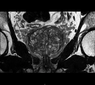
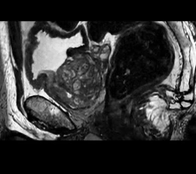
Figure 1.
Patient referred for prostate MR exam. AIR™ Recon DL High, the 21-ch AIR™ MP Coil and 60-ch AIR™ PA Coil were used. (A, B) T2 FSE, 0.6 x 0.6 x 3 mm, (A) axial 3:03 min. and (B) coronal 2:39 min.; (C) sagittal T2 PROPELLER, 0.6 x 0.6 x 3 mm, 2:25 min.; and (D-F) T2 Cube, 0.7 x 0.7 x 0.4 mm, 3:42 min., shown in (D) axial, (E) coronal and (F) sagittal planes.

Discussion
Prostate cancer was ruled out and the elevated PSA was found to be secondary to prostatitis, which was treated with antibiotics, reducing the patient’s PSA level.
With the 21-channel AIR™ Multi-Purpose Coil, we achieved a strong signal, patient comfort and speed in positioning, reducing the patient’s time in the MR system. AIR™ Recon DL has impacted all routine and advanced MR examinations, such as prostate, fetal, cardiac and breast studies. The good image quality we can achieve with these two technologies — AIR™ Recon DL and AIR™ Coils — facilitates the diagnosis of different pathologies and their longitudinal follow-up.
DOWNLOAD ARTICLE HERE
