Figure 1.
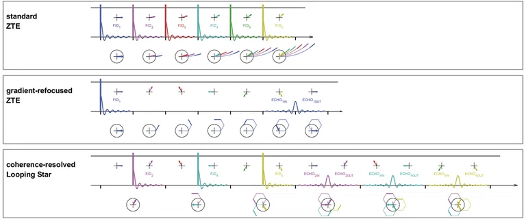
(Top row) Standard ZTE, (middle row) gradient-refocused ZTE and (bottom row) coherence-resolved Looping Star. In standard ZTE, one center-out radial spoke (colored arrows) is acquired after each RF excitation (colored thick vertical lines). Minimal gradient switching (horizontal gray line) in the form of small directional readout updates the results in silent imaging, which is used in Silenz. ZTE (i.e., TE=0) also allows the capturing of short-lived MR signals that are otherwise MR invisible, which is used in oZTEo bone imaging. Gradient-refocused ZTE achieves gradient refocusing by arranging subsequent spokes to refocus along a hexagonal trajectory. Looping Star can be considered as a time-multiplexed gradient-refocused ZTE, where multiple MR signal coherences are excited (one after the other during the excitation phase) and refocused (one after the other during the refocusing phase). To avoid overlap of subsequent gradient-echoes (during the refocusing phase) only every other spoke gets excited (during the initial excitation phase). While the FID spokes are of center-out (i.e. half spokes) nature, the gradient echo spokes are acquired full diameter.
3. Wiesinger F, Menini A, Solana AB. Looping Star. Magn Reson Med. 2019 Jan;81(1):57-68.
1. Dionisio-Parra B, Wiesinger F, Sämann PG, Czisch M, Solana AB. Looping Star fMRI in Cognitive Tasks and Resting State. J Magn Reson Imaging. 2020 Sep;52(3):739-751.
2. Damestani NL, O’Daly O, Solana AB, et al. Revealing the mechanisms behind novel auditory stimuli discrimination: An evaluation of silent functional MRI using looping star. Hum Brain Mapp. 2021 Jun 15;42(9):2833–2850.
‡ Technology in development that represents ongoing research and development efforts. These technologies are not products and may never become products. Not for sale. Not cleared or approved by the US FDA or any other global regulator for commercial availability.
‡ Technology in development that represents ongoing research and development efforts. These technologies are not products and may never become products. Not for sale. Not cleared or approved by the US FDA or any other global regulator for commercial availability.
4. ALS Therapy Development Institute. Limb and Bulbar Onset ALS. Available at: https://www.als.net/news/science-sunday-limb-and-bulbar-onset-als/.
Figure 2.
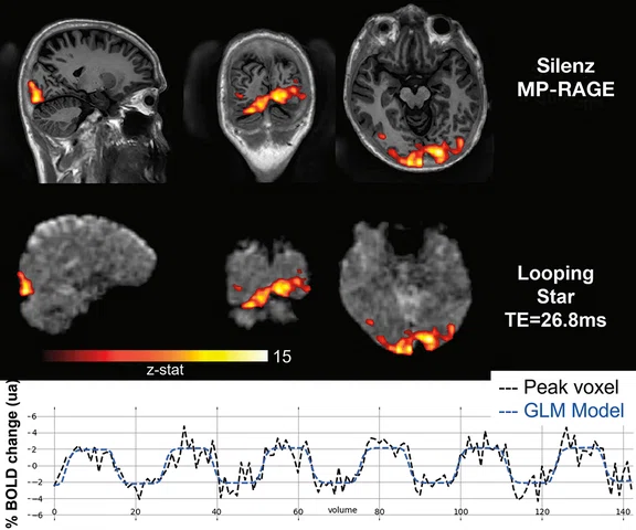
Block-design visual checkboard task statistical activation maps overlaid on (top row) Silenz MP-RAGE T1 anatomical scan after AIR™ Recon DL denoising and deblurring and (middle row) Looping Star echo image reconstructed using CG SENSE. (Bottom row) The timecourse at the most statistically significant voxel (peak voxel) in the visual cortex together with the GLM model is shown. BOLD signal change of 4-6% is achieved in this single subject experiment.
5. Leynes AP, Damestani NL, Lythgoe DJ, et al. Extreme Looping Star: Quiet fMRI at high spatiotemporal resolution. Proc ISMRM 2021, Abstract 0458. MagnaCumLaude Award.
6. Damestani NL, Solana AB, Wiesinger F, Fernandez B, Williams SCR, Lythgoe DJ. Quiet quantitative susceptibility mapping with Looping Star. Proc ISMRM 2022, abstract 2468.
1. Department of Neuroimaging, King’s College London, London, United Kingdom
2. GE Healthcare, Munich, Germany
2. GE Healthcare, Munich, Germany
3. GE Healthcare, Paris, France
1. Department of Neuroimaging, King’s College London, London, United Kingdom
1. Department of Neuroimaging, King’s College London, London, United Kingdom
‡ Technology in development that represents ongoing research and development efforts. These technologies are not products and may never become products. Not for sale. Not cleared or approved by the US FDA or any other global regulator for commercial availability.


result
PREVIOUS
${prev-page}
NEXT
${next-page}
Subscribe Now
Manage Subscription
FOLLOW US
Contact Us • Cookie Preferences • Privacy Policy • California Privacy PolicyDo Not Sell or Share My Personal Information • Terms & Conditions • Security
© 2024 GE HealthCare. GE is a trademark of General Electric Company. Used under trademark license.
SPOTLIGHT
Looping Star: Sounding out fMRI
Looping Star: Sounding out fMRI
A common patient complaint of MR imaging is the acoustic noise during scanning. It is caused by the intensive gradient switching and associated mechanical vibrations. The faster the imaging gradients and higher the field strength, the louder the acoustic noise.
A common patient complaint of MR imaging is the acoustic noise during scanning. It is caused by the intensive gradient switching and associated mechanical vibrations. The faster the imaging gradients and higher the field strength, the louder the acoustic noise.
Reducing the acoustic noise in an MR exam is particularly important in rapid fMRI studies of the brain. Steven Williams, PhD, Professor of Neuroimaging and Founder and Director of the Centre for Neuroimaging Sciences at the Institute of Psychiatry, Psychology & Neuroscience and Maudsley Hospital, King’s College London, has been pioneering the use of MR imaging in psychiatry and neurological disorders.
“There are a range of applications across the life span, from an infant who may no longer need sedation to an anxious elderly subject with dementia, where we can use an MR scan that is not only fast but incredibly quiet. If we don’t have this loud banging noise, then we can talk to the subject, hear what they are saying and even reassure them during the course of the scan, which will improve the scanning experience for everyone.”
Prof. Steven Williams
Thanks to the development of Looping Star‡, whole-brain coverage with sufficient spatiotemporal resolution and BOLD sensitivity at just 0.5 dB above ambient noise was demonstrated in fMRI experiments involving block-design, event-related and resting-state (rs) paradigms.1,2 This novel works-in-progress MR imaging method (which is now available to GE Healthcare research customers) was invented and spearheaded by Florian Wiesinger, PhD, Anne Menini, PhD, and Ana Beatriz Solana, PhD.3
Looping Star is based on the zero echo time (ZTE) pulse sequence that provides a robust and silent 3D MR acquisition, commercialized by GE as SilentScan (Silenz). Looping Star extends ZTE toward gradient echo (GRE) imaging via a time-multiplexed gradient refocusing mechanism to obtain a free-induction decay (FID) image with a nominal TE=0 (like standard ZTE) together with equidistant GRE images that contain additional T2* and susceptibility information (Figure 1). Hence, Looping Star has the potential to contribute to the current SilentScan acquisition with T2* mapping, susceptibility weighted imaging, quantitative susceptibility mapping‡ and fMRI applications.
Figure 1.
(Top row) Standard ZTE, (middle row) gradient-refocused ZTE and (bottom row) coherence-resolved Looping Star. In standard ZTE, one center-out radial spoke (colored arrows) is acquired after each RF excitation (colored thick vertical lines). Minimal gradient switching (horizontal gray line) in the form of small directional readout updates the results in silent imaging, which is used in Silenz. ZTE (i.e., TE=0) also allows the capturing of short-lived MR signals that are otherwise MR invisible, which is used in oZTEo bone imaging. Gradient-refocused ZTE achieves gradient refocusing by arranging subsequent spokes to refocus along a hexagonal trajectory. Looping Star can be considered as a time-multiplexed gradient-refocused ZTE, where multiple MR signal coherences are excited (one after the other during the excitation phase) and refocused (one after the other during the refocusing phase). To avoid overlap of subsequent gradient-echoes (during the refocusing phase) only every other spoke gets excited (during the initial excitation phase). While the FID spokes are of center-out (i.e. half spokes) nature, the gradient echo spokes are acquired full diameter.
A new era for fMRI
Looping Star may open up a new realm of neuroscience discoveries, as it may be possible to use fMRI imaging across a broad range of acoustic noise-averse subjects and research studies, including those with autism, schizophrenia and sleep deprivation.
“So often, we had to exclude some autistic participants from studies because of their extreme hypersensitivity to sound,” Professor Williams explains. “Silent fMRI may give us the ability to visualize the brain while someone with schizophrenia is hearing internal voices, conduct fundamental sensory studies of audition in patients suffering from tinnitus, evaluate language and speech in aphasic patients following a stroke, and even perform fundamental research on the impact of poor sleep. Now, we can examine these and many other diseases and conditions thanks to the development of Looping Star. The world is our oyster, as they say, with this capability.”
The ability to provide a more immersive experience by combining sound with visual stimuli may also help researchers better understand the underlying biology when a post-traumatic stress disorder (PTSD) subject has a flashback.
“If we can see that underlying network in the brain that occurs when a flashback is triggered, then we can start to explore behavioral talking therapy and pharmacological approaches to treat it,” Professor Williams adds.
As rs-fMRI is exploding in use within neurocognitive research, it’s only logical to have a scanner that is virtually silent. “For rs-fMRI, a quiet scan is really key to see how the brain is truly functioning in a state of rest,” says Dr. Solana.
Currently at King’s College London, Looping Star is being evaluated in healthy volunteers as Professor Williams, Dr. Fernando Zelaya and their colleagues begin to initiate clinical research to see how the brain responds to sounds and look at a fundamental process in the brain called prepulse inhibition of the acoustic startle reflex. Certain conditions such as Huntington’s disease and schizophrenia are known to have impairment of this startle response.
“What’s happening in these cases is disrupted sensorimotor gating,” Professor Williams explains. “It may be an early predictor of someone who will go on to develop psychosis or a prognostic marker in Huntington’s disease.”
Beyond exploring mental health or other neurological diseases, Looping Star could make a difference today in sleeping baby studies and dementia patients, potentially eliminating the need for sedation in both of these patient cohorts. With better understanding of neurodegenerative diseases, there are now sub-classes – from frontotemporal lobe disease to Parkinson’s to amyotrophic lateral sclerosis (ALS) to Alzheimer’s and beyond – that often have different treatments.
For example, it is estimated that 30% of people with ALS have bulbar onset, where symptoms first occur in the face and neck.4 Bulbar onset is considered more aggressive in disease progression and these people appear to have a frontal and temporal engagement in the disease process.
Figure 2.
Block-design visual checkboard task statistical activation maps overlaid on (top row) Silenz MP-RAGE T1 anatomical scan after AIR™ Recon DL denoising and deblurring and (middle row) Looping Star echo image reconstructed using CG SENSE. (Bottom row) The timecourse at the most statistically significant voxel (peak voxel) in the visual cortex together with the GLM model is shown. BOLD signal change of 4-6% is achieved in this single subject experiment.
The development of Looping Star
The discovery of Looping Star was truly experimental. Analyzing the k-space data of other ZTE experiments, there was a bright line, which was indicative of something not happening the way it was expected. This artifact was the result of an unexpected gradient refocusing in the pulse sequence. Detailed investigation and understanding of the source of the artifact was the start of a valuable, useful imaging sequence.
“The artifact looked exactly like a shooting star crossing all our k-space data,” says Dr. Wiesinger. “Following experimental implementation and in-vivo validation, we then adjusted the name to Looping Star which better reflects what the pulse sequence actually does.”
“The morning after observing the artifact, Dr. Wiesinger told us, ‘an artifact is an opportunity’,” adds Dr. Solana.
“One man’s food is another man’s poison, said the first century BC philosopher, Lucretius,” adds Professor Williams. “In MR, we do our best to remove artifacts as opposed to delving deeply into that artifact, and turning it to our advantage.”
“fMRI can also be seen as an artifact when you don’t want to see that alteration in your image due to T2*, yet this is precisely the information you use for BOLD-contrast based fMRI,” says Dr. Solana.
Dr. Solana credits the combination of expertise in physics, image reconstruction and neuro imaging in the development team as the reason why they saw the potential of this artifact as a new pulse sequence.
But one thing was missing. The ability to apply this new sequence in a clinical research environment.
“That is where the collaboration with Professor Williams and King’s College continued this technical exploration and added that critical piece in the sequence development,” says Dr. Wiesinger.
To explore the spatio-temporal limits of Looping-Star fMRI, the industry-academia collaboration between GE and King’s College London joined forces with collaborators at the University of California San Francisco and University of California, Berkeley to develop Extreme Looping Star‡. Sub-second temporal resolution and submillimeter spatial resolution fMRI was explored by using extreme MR reconstruction based on multi-scale low-rank decomposition with the Looping Star pulse sequence. This innovative collaboration was awarded the MagnaCumLaude Award by the Society of Magnetic Resonance in Medicine in 2021.5
GE and King’s College London are now working on a more comprehensive silent protocol using Looping Star, including silent susceptibility and T2*-weighted structural imaging.6 Additionally, several researchers have expressed interest to use the Looping Star research prototype for T2* mapping in MSK applications.
The future appears bright for Looping Star and other ZTE-based silent sequences, which can be seen as simple and efficient MRI pulse sequences. ZTE and Looping Star do not require any specific gradient performance (e.g., high field only), nor do they have any slew rate dependencies.
“This is all software on an existing MR system and infrastructure,” says Professor Williams. “There was no major, expensive modifications to use Looping Star, just some brilliant work in physics. I have said to our patients that if, in five years, you undergo an MR scan and it is noisy, have a word with your doctor, the radiologist and technologist, because there is no reason why we cannot address the noise in almost all MR exams by that time.”
Looping Star featured at ISMRM 2022
Researchers from King’s College London and GE Healthcare presented their latest work on Looping Star at ISMRM 2022.
Quiet quantitative susceptibility mapping with Looping Star
by Nikou Louise Damestani,1 Ana Beatriz Solana,2 Florian Wiesinger,2 Brice Fernandez,3 Steven Charles Rees Williams,1 and David John Lythgoe1
Looping Star is an acoustically quiet T2*-weighted acquisition technique, which has primarily been used to demonstrate functional sensitivity. We present the application of Looping Star to quantitative susceptibility mapping in a small cohort, directly comparing its outcomes with a conventional susceptibility-weighted imaging technique. We found that Looping Star produced comparable quantitative values in subcortical regions in comparison with conventional susceptibility-weighted imaging.


