1. Etkin CD, Springer BD. The American Joint Replacement Registry-the first 5 years. Arthroplast Today. 2017;3(2):67-69. Published 2017 Mar 14. doi:10.1016/j.artd.2017.02.002.
2. Bayliss LE, Culliford D, Monk AP, Glyn-Jones S, Prieto-Alhambra D, Judge A, Cooper C, Carr AJ, Arden NK, Beard DJ, Price AJ. The effect of patient age at intervention on risk of implant revision after total replacement of the hip or knee: a population-based cohort study. Lancet. 2017 Apr 8;389(10077):1424-1430. doi: 10.1016/S0140-6736(17)30059-4. Epub 2017 Feb 14. Erratum in: Lancet. 2017 Apr 8;389(10077):1398. PMID: 28209371; PMCID: PMC5522532.
3. AAOS: Volume of Primary and Revision Total Joint Replacement in U.S. Projected to Grow by Triple Digits. Orthopedic Design & Technology, March 2018. Available at: https://www.odtmag.com/contents/view_breaking-news/2018-03-09/volume-of-primary-and-revision-total-joint-replacement-in-us-projected-to-grow-by-triple-digits/.
4. How Many Spinal Fusions are Performed Each Year in the United States? iData Research, May 2018. Available at: https://idataresearch.com/how-many-instrumented-spinal-fusions-are-performed-each-year-in-the-united-states/.
5. Guerini H, Morvan G, Judet T, et al. IRM des prothèses de hanche. La Hanche, 2019. Proceedings of the 46th congress of the Societe D’Imagerie Musculo-squelettique.
A
Figure 1.
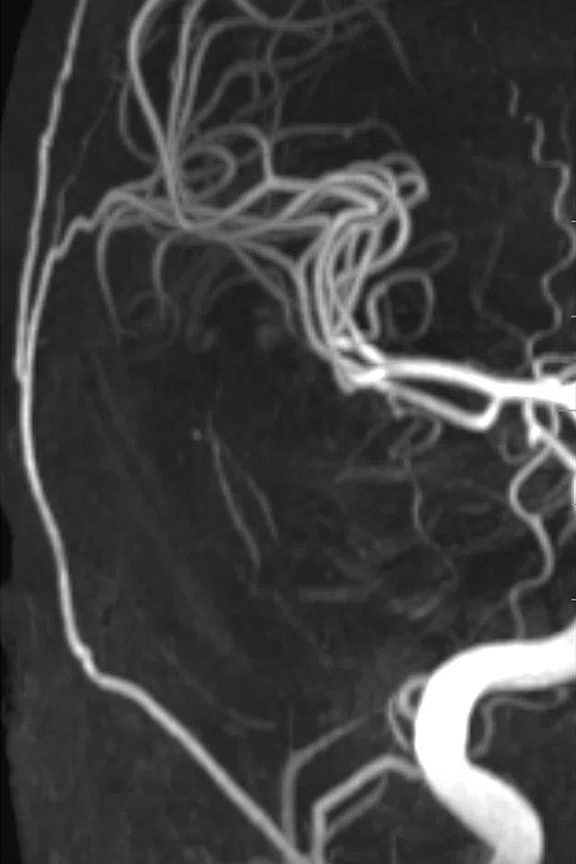
3D TOF MR angiography with HyperSense and AIR™ 48Ch Head Coil. (A) Conventional scan, (B) HyperSense factor of 1.4 for a 29 percent faster scan, (C) HyperSense factor of 1.8 for a 44 percent faster scan and (D) HyperSense factor of 1.5 utilized for 75 percent higher resolution with no change in scan time.
B
Figure 1.
3D TOF MR angiography with HyperSense and AIR™ 48Ch Head Coil. (A) Conventional scan, (B) HyperSense factor of 1.4 for a 29 percent faster scan, (C) HyperSense factor of 1.8 for a 44 percent faster scan and (D) HyperSense factor of 1.5 utilized for 75 percent higher resolution with no change in scan time.
C
Figure 1.
3D TOF MR angiography with HyperSense and AIR™ 48Ch Head Coil. (A) Conventional scan, (B) HyperSense factor of 1.4 for a 29 percent faster scan, (C) HyperSense factor of 1.8 for a 44 percent faster scan and (D) HyperSense factor of 1.5 utilized for 75 percent higher resolution with no change in scan time.
D
Figure 1.
3D TOF MR angiography with HyperSense and AIR™ 48Ch Head Coil. (A) Conventional scan, (B) HyperSense factor of 1.4 for a 29 percent faster scan, (C) HyperSense factor of 1.8 for a 44 percent faster scan and (D) HyperSense factor of 1.5 utilized for 75 percent higher resolution with no change in scan time.
A
Figure 2.
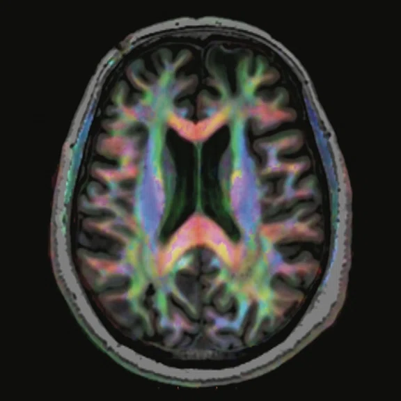
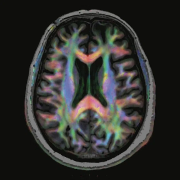
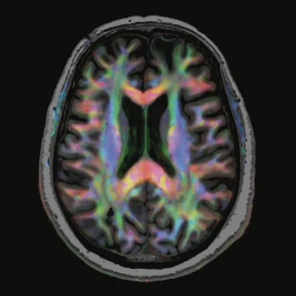
DTI with HyperBand and Distortion Correction using the AIR™ 48Ch Head Coil for fast, high resolution imaging. All images acquired with 24 directions and a resolution of 3 mm3. (A) Reference scan, TR/TE=7120/78.4 ms, 3:26 min; (B) 50 percent faster scan than reference scan at 1:54 min., using HyperBand x2, TR/TE=3945/79.0 ms; and (C) 65 percent faster scan time than reference scan at 1:11 min. using HyperBand x3, TR/TE=2440/79.5 ms.
B
Figure 2.
DTI with HyperBand and Distortion Correction using the AIR™ 48Ch Head Coil for fast, high resolution imaging. All images acquired with 24 directions and a resolution of 3 mm3. (A) Reference scan, TR/TE=7120/78.4 ms, 3:26 min; (B) 50 percent faster scan than reference scan at 1:54 min., using HyperBand x2, TR/TE=3945/79.0 ms; and (C) 65 percent faster scan time than reference scan at 1:11 min. using HyperBand x3, TR/TE=2440/79.5 ms.
C
Figure 2.
DTI with HyperBand and Distortion Correction using the AIR™ 48Ch Head Coil for fast, high resolution imaging. All images acquired with 24 directions and a resolution of 3 mm3. (A) Reference scan, TR/TE=7120/78.4 ms, 3:26 min; (B) 50 percent faster scan than reference scan at 1:54 min., using HyperBand x2, TR/TE=3945/79.0 ms; and (C) 65 percent faster scan time than reference scan at 1:11 min. using HyperBand x3, TR/TE=2440/79.5 ms.
A
Figure 3.
DTI with MUSE and distortion correction using the AIR™ 48Ch Head Coil. Note the difference between areas of distortion (arrows, A and B) and the improvement provided with distortion correction (arrows, C and D). (A) Conventional acquisition and reconstruction and (B) MUSE DTI with distortion correction of axial DTI acquisition reformatted to sagittal plane. (C, D) Sagittal MUSE DTI, 24 directions, 3 shots, TR/TE=6046/79.1 ms, 3 mm3 resolution, 8:10 min. (E) 3D tractography.
B
Figure 3.
DTI with MUSE and distortion correction using the AIR™ 48Ch Head Coil. Note the difference between areas of distortion (arrows, A and B) and the improvement provided with distortion correction (arrows, C and D). (A) Conventional acquisition and reconstruction and (B) MUSE DTI with distortion correction of axial DTI acquisition reformatted to sagittal plane. (C, D) Sagittal MUSE DTI, 24 directions, 3 shots, TR/TE=6046/79.1 ms, 3 mm3 resolution, 8:10 min. (E) 3D tractography.
E
Figure 3.
DTI with MUSE and distortion correction using the AIR™ 48Ch Head Coil. Note the difference between areas of distortion (arrows, A and B) and the improvement provided with distortion correction (arrows, C and D). (A) Conventional acquisition and reconstruction and (B) MUSE DTI with distortion correction of axial DTI acquisition reformatted to sagittal plane. (C, D) Sagittal MUSE DTI, 24 directions, 3 shots, TR/TE=6046/79.1 ms, 3 mm3 resolution, 8:10 min. (E) 3D tractography.
A
Figure 4.
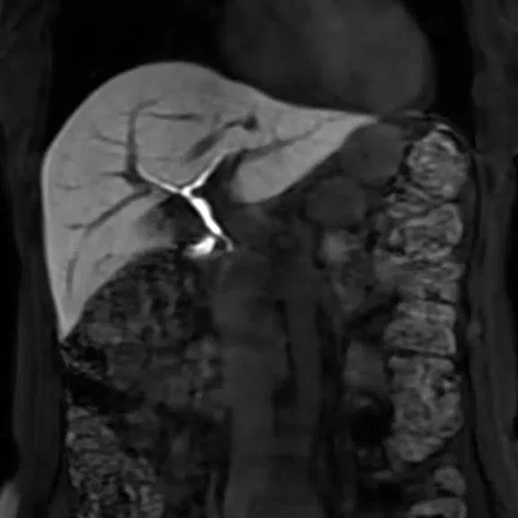
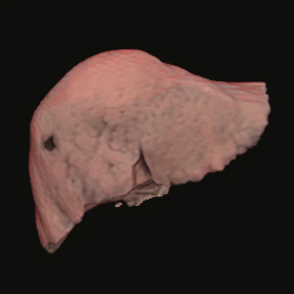
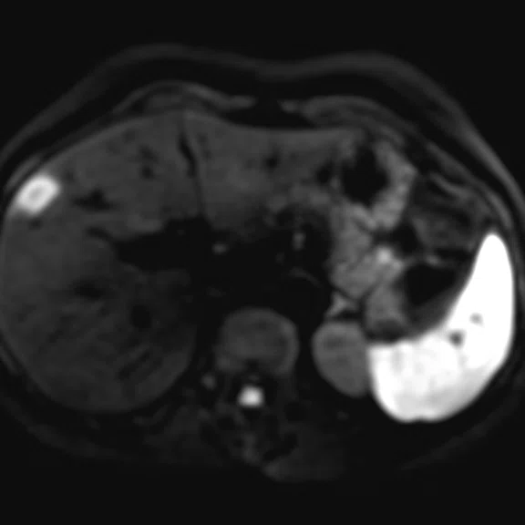
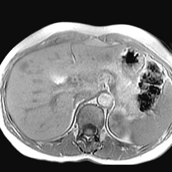
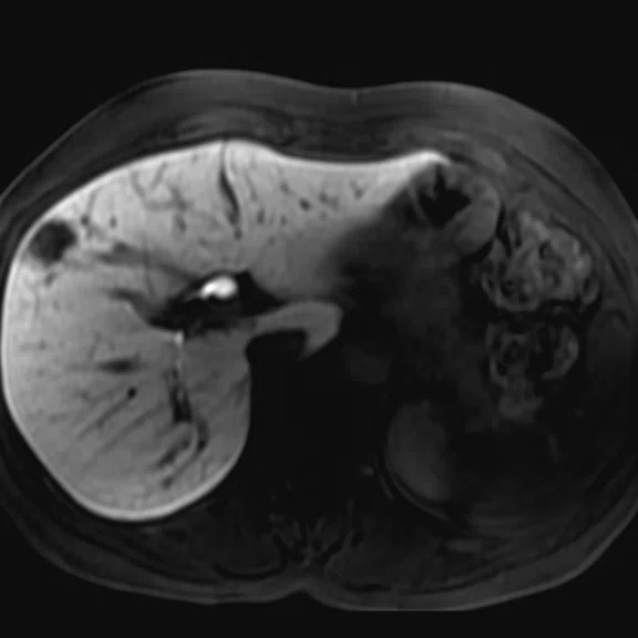
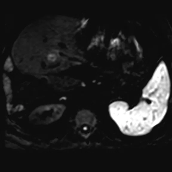
Liver imaging with the AIR™ AA Coil. (A) Coronal LAVA delayed-enhancement phase with gadolinium-ethoxybenzyl-diethylenetriamine pentaacetic acid (Gd-EOB-DTPA); (B) volume reconstruction of the liver; (C) DWI b800; (D) MUSE b800; (E) LAVA delayed-enhancement phase; and (F) MAGiC DWI b1500.
B
Figure 4.
Liver imaging with the AIR™ AA Coil. (A) Coronal LAVA delayed-enhancement phase with gadolinium-ethoxybenzyl-diethylenetriamine pentaacetic acid (Gd-EOB-DTPA); (B) volume reconstruction of the liver; (C) DWI b800; (D) MUSE b800; (E) LAVA delayed-enhancement phase; and (F) MAGiC DWI b1500.
C
Figure 4.
Liver imaging with the AIR™ AA Coil. (A) Coronal LAVA delayed-enhancement phase with gadolinium-ethoxybenzyl-diethylenetriamine pentaacetic acid (Gd-EOB-DTPA); (B) volume reconstruction of the liver; (C) DWI b800; (D) MUSE b800; (E) LAVA delayed-enhancement phase; and (F) MAGiC DWI b1500.
D
Figure 4.
Liver imaging with the AIR™ AA Coil. (A) Coronal LAVA delayed-enhancement phase with gadolinium-ethoxybenzyl-diethylenetriamine pentaacetic acid (Gd-EOB-DTPA); (B) volume reconstruction of the liver; (C) DWI b800; (D) MUSE b800; (E) LAVA delayed-enhancement phase; and (F) MAGiC DWI b1500.
E
Figure 4.
Liver imaging with the AIR™ AA Coil. (A) Coronal LAVA delayed-enhancement phase with gadolinium-ethoxybenzyl-diethylenetriamine pentaacetic acid (Gd-EOB-DTPA); (B) volume reconstruction of the liver; (C) DWI b800; (D) MUSE b800; (E) LAVA delayed-enhancement phase; and (F) MAGiC DWI b1500.
F
Figure 4.
Liver imaging with the AIR™ AA Coil. (A) Coronal LAVA delayed-enhancement phase with gadolinium-ethoxybenzyl-diethylenetriamine pentaacetic acid (Gd-EOB-DTPA); (B) volume reconstruction of the liver; (C) DWI b800; (D) MUSE b800; (E) LAVA delayed-enhancement phase; and (F) MAGiC DWI b1500.
A
Figure 5.
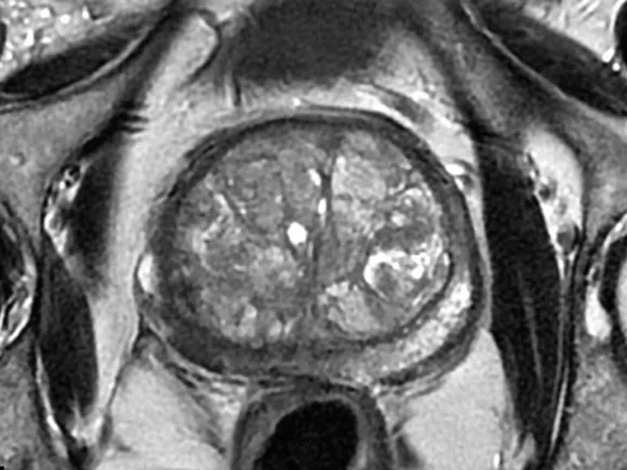
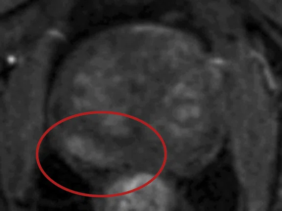
Prostate imaging with the AIR™ AA Coil. (A) T2 PROPELLER; (B) dynamic DISCO; (C) FOCUS b1400 acquired; and (D) FOCUS ADC.
B
Figure 5.
Prostate imaging with the AIR™ AA Coil. (A) T2 PROPELLER; (B) dynamic DISCO; (C) FOCUS b1400 acquired; and (D) FOCUS ADC.
C
Figure 5.
Prostate imaging with the AIR™ AA Coil. (A) T2 PROPELLER; (B) dynamic DISCO; (C) FOCUS b1400 acquired; and (D) FOCUS ADC.
D
Figure 5.
Prostate imaging with the AIR™ AA Coil. (A) T2 PROPELLER; (B) dynamic DISCO; (C) FOCUS b1400 acquired; and (D) FOCUS ADC.
A
Figure 6.
MR bone imaging scanned with the Silent Suite application (ZTE) with AIR™ AA Coil.
(A) Inverted and (B) inverted partial MinIP.
B
Figure 6.
MR bone imaging scanned with the Silent Suite application (ZTE) with AIR™ AA Coil.
(A) Inverted and (B) inverted partial MinIP.
A
Figure 7.
Cardiovascular imaging with AIR™ AA Coil. (A) T1 Map (MOLLI); (B) T2 Map; (C) FIESTA Cine; and (D) T2 FatSat FSE black blood.
B
Figure 7.
Cardiovascular imaging with AIR™ AA Coil. (A) T1 Map (MOLLI); (B) T2 Map; (C) FIESTA Cine; and (D) T2 FatSat FSE black blood.
C
Figure 7.
Cardiovascular imaging with AIR™ AA Coil. (A) T1 Map (MOLLI); (B) T2 Map; (C) FIESTA Cine; and (D) T2 FatSat FSE black blood.
D
Figure 7.
Cardiovascular imaging with AIR™ AA Coil. (A) T1 Map (MOLLI); (B) T2 Map; (C) FIESTA Cine; and (D) T2 FatSat FSE black blood.
C
Figure 3.
DTI with MUSE and distortion correction using the AIR™ 48Ch Head Coil. Note the difference between areas of distortion (arrows, A and B) and the improvement provided with distortion correction (arrows, C and D). (A) Conventional acquisition and reconstruction and (B) MUSE DTI with distortion correction of axial DTI acquisition reformatted to sagittal plane. (C, D) Sagittal MUSE DTI, 24 directions, 3 shots, TR/TE=6046/79.1 ms, 3 mm3 resolution, 8:10 min. (E) 3D tractography.
D
Figure 3.
DTI with MUSE and distortion correction using the AIR™ 48Ch Head Coil. Note the difference between areas of distortion (arrows, A and B) and the improvement provided with distortion correction (arrows, C and D). (A) Conventional acquisition and reconstruction and (B) MUSE DTI with distortion correction of axial DTI acquisition reformatted to sagittal plane. (C, D) Sagittal MUSE DTI, 24 directions, 3 shots, TR/TE=6046/79.1 ms, 3 mm3 resolution, 8:10 min. (E) 3D tractography.
A
Figure 7.
Cardiovascular imaging with AIR™ AA Coil. (A) T1 Map (MOLLI); (B) T2 Map; (C) FIESTA Cine; and (D) T2 FatSat FSE black blood.
B
Figure 7.
Cardiovascular imaging with AIR™ AA Coil. (A) T1 Map (MOLLI); (B) T2 Map; (C) FIESTA Cine; and (D) T2 FatSat FSE black blood.
C
Figure 7.
Cardiovascular imaging with AIR™ AA Coil. (A) T1 Map (MOLLI); (B) T2 Map; (C) FIESTA Cine; and (D) T2 FatSat FSE black blood.
D
Figure 7.
Cardiovascular imaging with AIR™ AA Coil. (A) T1 Map (MOLLI); (B) T2 Map; (C) FIESTA Cine; and (D) T2 FatSat FSE black blood.
result


PREVIOUS
${prev-page}
NEXT
${next-page}
Subscribe Now
Manage Subscription
FOLLOW US
Contact Us • Cookie Preferences • Privacy Policy • California Privacy PolicyDo Not Sell or Share My Personal Information • Terms & Conditions • Security
© 2024 GE HealthCare. GE is a trademark of General Electric Company. Used under trademark license.
IN PRACTICE
Making the move to 3.0T lets Hacettepe University push the boundaries of MR imaging
Making the move to 3.0T lets Hacettepe University push the boundaries of MR imaging
As one of the most respected hospitals and leading academic centers in Turkey, Hacettepe University Hospital (Ankara) has a long tradition of innovation, from implementing advanced technologies to adopting new concepts in the practice of medicine. The radiology department is no different. When it came time to move to 3.0T MR, hospital and radiology leaders looked for a company that would be a leader in technology, applications and clinical excellence. That company is GE Healthcare.
As one of the most respected hospitals and leading academic centers in Turkey, Hacettepe University Hospital (Ankara) has a long tradition of innovation, from implementing advanced technologies to adopting new concepts in the practice of medicine. The radiology department is no different. When it came time to move to 3.0T MR, hospital and radiology leaders looked for a company that would be a leader in technology, applications and clinical excellence. That company is GE Healthcare.
"Our work is system-based rather than modality-based, and that enables us to use all the different radiology methods during the diagnostic process," says Deniz Akata, MD, Professor and Chair of the Radiology Department. "We have become an institution that stays well on track with innovative technologies in every area."
With advanced technology providing the information needed for a confident and accurate diagnosis, the department is an essential and inseparable contributor to patient care. The department has evolved from a "classical" to a "clinical" radiology department, adds Mustafa Özmen, MD, Professor of Radiology.
"Specialization in radiology has become quite important within our institution," Professor Özmen adds. Whether it is musculoskeletal, abdominal or pediatrics, the sub-specialization of radiology has helped elevate not just diagnostic but also interventional radiology.
For many years, the hospital utilized 1.5T MR systems from different manufacturers. However, once Professors Akata and Özmen saw the difference in system performance and image quality between 1.5T and 3.0T, it became a top priority to acquire a 3.0T MR system.
"As an institution, we did not select a specific system," explains Professor Özmen. "GE Healthcare was awarded the contract by bidding the ultimate system with the highest technological features in the most budget-friendly offer. One of the most significant achievements of SIGNA™ Architect is that it gives us the opportunity to push boundaries. We can acquire detailed and elaborate images with higher visual quality. More importantly, our confidence in the data is higher."
Figure 1.
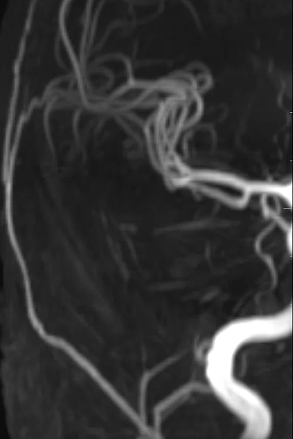
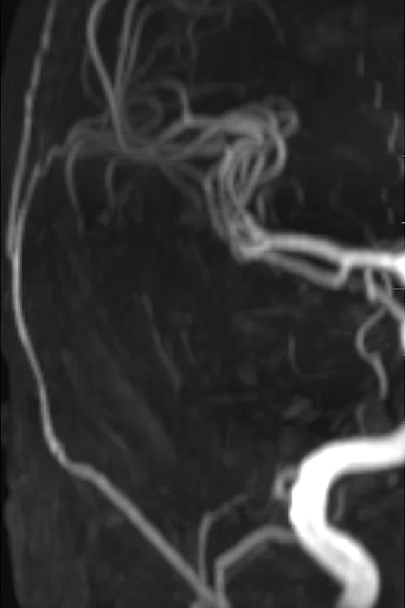
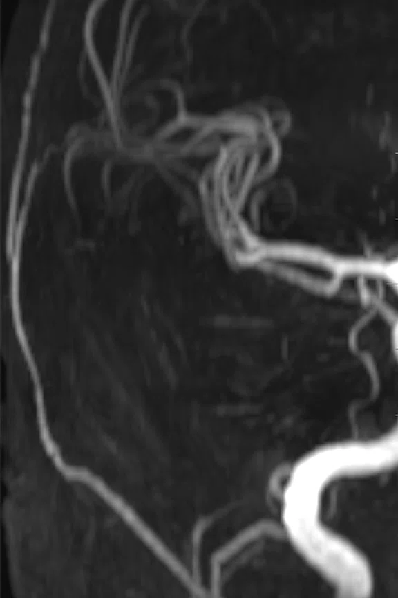
3D TOF MR angiography with HyperSense and AIR™ 48Ch Head Coil. (A) Conventional scan, (B) HyperSense factor of 1.4 for a 29 percent faster scan, (C) HyperSense factor of 1.8 for a 44 percent faster scan and (D) HyperSense factor of 1.5 utilized for 75 percent higher resolution with no change in scan time.
Figure 2.
DTI with HyperBand and Distortion Correction using the AIR™ 48Ch Head Coil for fast, high resolution imaging. All images acquired with 24 directions and a resolution of 3 mm3. (A) Reference scan, TR/TE=7120/78.4 ms, 3:26 min; (B) 50 percent faster scan than reference scan at 1:54 min., using HyperBand x2, TR/TE=3945/79.0 ms; and (C) 65 percent faster scan time than reference scan at 1:11 min. using HyperBand x3, TR/TE=2440/79.5 ms.
Advanced imaging with AIR™
In tandem with the SIGNA™ Architect installation, Hacettepe University Hospital also acquired the AIR™ 48Ch Head Coil, AIR™ Anterior Array (AA) Coil, AIR x™ intelligent MR slice prescription and AIR™ Recon smart reconstruction algorithm.
AIR x™ utilizes deep-learning algorithms to automatically detect anatomical landmarks and prescribe slices in the brain, providing reproducible planning for exam consistency for patient followup and longitudinal studies.
AIR x™ is perceived as extremely helpful, especially considering the SIGNA™ Architect is being used 24/7, performing a high volume of exams each day with continually rotating staff.
According to Kader Karlı Oğuz, MD, EDiNR, EDiPNR, Professor of Radiology, Division of Neuroradiology, Hacettepe University, the AIR™ 48Ch Head Coil is used for both routine diagnostic exams for lesion depiction and advanced neurologic studies for neurodegenerative diseases and functional MR imaging (fMRI).
"The high signal-to-noise ratio (SNR) and homogeneous signal penetration have made this coil the number one choice in our department," Professor Oğuz says. With an adaptable design, the coil fits 99.99 percent of the population.
Improved parallel imaging capabilities built into the AIR™ 48Ch Head Coil and the additional use of HyperSense enables faster acquisitions, especially 3D applications. In addition to faster imaging, HyperSense also allows for higher resolution or the possibility to balance between speed and resolution.
Another scanning acceleration achievement, HyperBand, was successfully implemented for DWI, DTI, fMRI and T2∗ DSC imaging. DWI applications are particularly sensitive to susceptibility artifacts in air-bone-tissue interfaces, in the skull base and in post-trauma patients. The utilization of Multi-plexed Sensitivity Encoding (MUSE), a multi-shot diffusion technique, and distortion correction has reduced these artifacts, resulting in higher, diagnostic-quality images (Figure 3).
Figure 3.
DTI with MUSE and distortion correction using the AIR™ 48Ch Head Coil. Note the difference between areas of distortion (arrows, A and B) and the improvement provided with distortion correction (arrows, C and D). (A) Conventional acquisition and reconstruction and (B) MUSE DTI with distortion correction of axial DTI acquisition reformatted to sagittal plane. (C, D) Sagittal MUSE DTI, 24 directions, 3 shots, TR/TE=6046/79.1 ms, 3 mm3 resolution, 8:10 min. (E) 3D tractography.
Figure 4.
Liver imaging with the AIR™ AA Coil. (A) Coronal LAVA delayed-enhancement phase with gadolinium-ethoxybenzyl-diethylenetriamine pentaacetic acid (Gd-EOB-DTPA); (B) volume reconstruction of the liver; (C) DWI b800; (D) MUSE b800; (E) LAVA delayed-enhancement phase; and (F) MAGiC DWI b1500.
Professor Oğuz uses Quantib® Brain for comparative analysis. With brain volume quantification and white matter lesion follow-up studies, she adds, "Not only is the fast and high-resolution imaging important, quantification of MR imaging data is essential for patient treatment and follow-up."
In body imaging, Muşturay Karçaaltıncaba, MD, FESGAR, Professor of Radiology, one of the liver imaging team leaders at Hacettepe University, relies on the AIR™ AA Coil for abdominal imaging. With the highest channel count and coverage in the industry, the AIR™ AA Coil delivers exquisite images of liver in eDWI and post-contrast LAVA sequences. Plus, with synthetic DWI, he can generate high b-value images without adding scan time.
"We can already see that the AIR™ AA Coil is creating significant differences in many areas," says Professor Özmen. "Exams performed with the AIR™ Coil demonstrate high-clarity images due to the high SNR as well as high speed elements."
Multi-parametric prostate imaging is another high-volume exam at Hacettepe University that utilizes the high-channel count and coverage afforded by the AIR™ AA Coil from the very first day it was implemented. Both radiologists and urologists appreciate the small FOV high-resolution results.
Preferred acquisition techniques for prostate imaging include the motion robustness of PROPELLER, now the first choice for high-resolution, T2–weighted imaging at Hacettepe University, and FOCUS DWI, which expands the radiologist’s ability for differential diagnosis with small FOV DWI imaging. Additionally, DISCO for DCE provides both high temporal and spatial resolution for the enhancement assessment.
The majority of oncologic imaging exams at Hacettepe University utilize DWI imaging, including MUSE. Üstün Aydıngöz, MD, and F. Bilge Ergen, MD, who are both Professors of Radiology, have found that MUSE is more immune to susceptibility artifacts, therefore they have added it to the routine protocol for oncology assessments.
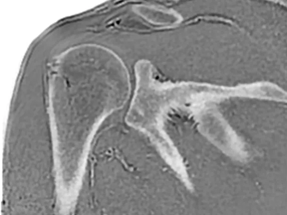
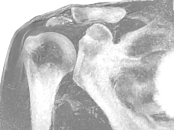
Using MR to visualize bony details is a relatively new capability with SIGNA™ Architect that has piqued the interest of Professors Aydıngöz and Ergen. They are planning to utilize the ZTE Silent Scan technique for their upcoming comparative research studies.
SIGNA™ Architect is also being used more often for cardiac imaging at Hacettepe University. Tuncay Hazırolan, MD, Professor of Radiology and the current president of the Turkish Radiology Society, specializes in cardiovascular imaging.
"Due to the higher SNR at 3.0T, we believe there is excellent potential to use SIGNA™ Architect for cardiac imaging," says Professor Hazırolan. "Regarding post-processing, we now have the cvi42® cardiac analysis software and after just a few weeks it quickly became a favorite application for our team."
Having the diagnostic tools to make a confident diagnosis is not an easy task, adds Professor Özmen. "When we compare images of the same patient acquired on different systems side-by-side with the SIGNA™ Architect, we can in fact see the difference and the gain in overall image quality. In their reporting process, our radiologists now have a higher clinical confidence. We can see how AI has the potential to advance MR imaging and workflow processes, and these improvements excite us more."


