A
Figure 1.
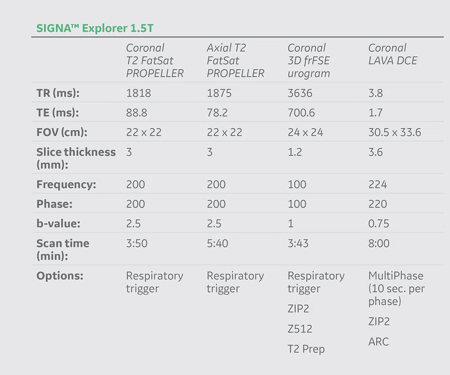
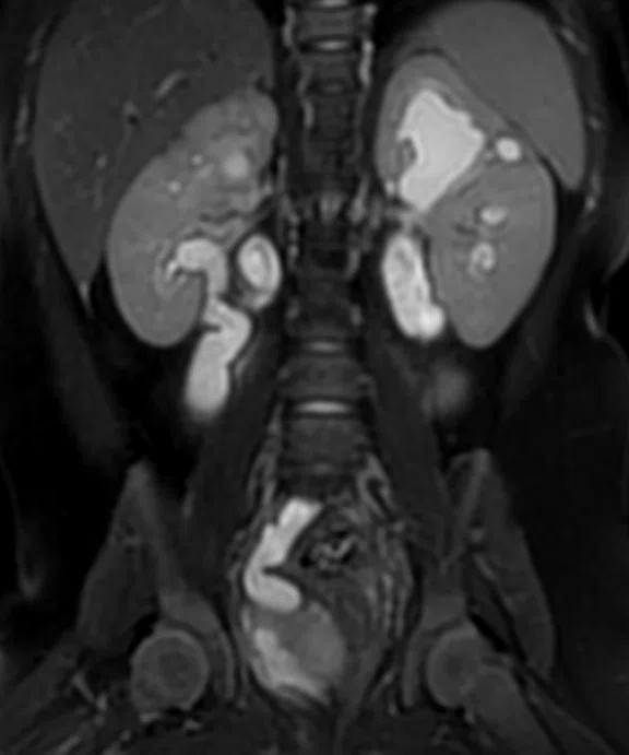
(A) Coronal T2 PROPELLER FatSat with respiratory triggering, 1.1 x 1.1 x 3 mm3, 3:50 min.; (B) volume-rendered coronal 3D frFSE urogram with respiratory triggering, 1 x 1.3 x 1.2/-0.6 mm3, 3:43 min., depicting the left and right lower collecting systems (yellow arrows) and left and right upper collecting systems (red arrows); and (C) fused image of (A) and (B).
B
Figure 1.
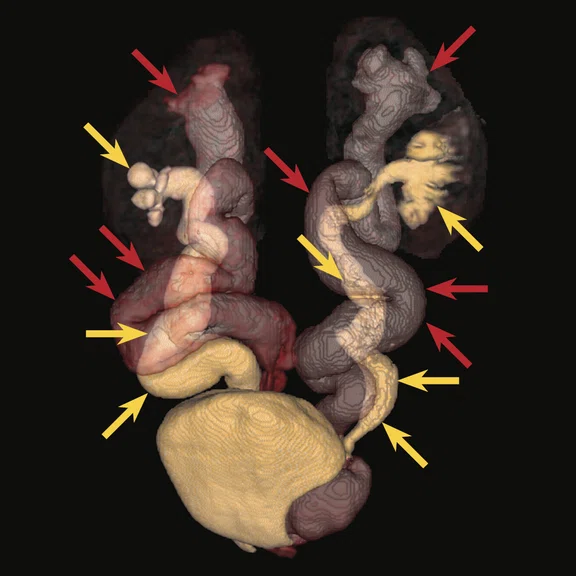
(A) Coronal T2 PROPELLER FatSat with respiratory triggering, 1.1 x 1.1 x 3 mm3, 3:50 min.; (B) volume-rendered coronal 3D frFSE urogram with respiratory triggering, 1 x 1.3 x 1.2/-0.6 mm3, 3:43 min., depicting the left and right lower collecting systems (yellow arrows) and left and right upper collecting systems (red arrows); and (C) fused image of (A) and (B).
C
Figure 1.
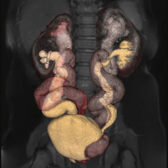
(A) Coronal T2 PROPELLER FatSat with respiratory triggering, 1.1 x 1.1 x 3 mm3, 3:50 min.; (B) volume-rendered coronal 3D frFSE urogram with respiratory triggering, 1 x 1.3 x 1.2/-0.6 mm3, 3:43 min., depicting the left and right lower collecting systems (yellow arrows) and left and right upper collecting systems (red arrows); and (C) fused image of (A) and (B).
1. Capone VP, Morello W, Taroni F, Montini G. Genetics of Congenital Anomalies of the Kidney and Urinary Tract: The Current State of Play. Int J Mol Sci. 2017 Apr 11;18(4):796.
2. National Library of Medicine. Congenital anomalies of kidney and urinary tract. Available at: https://medlineplus.gov/genetics/condition/congenital-anomalies-of-kidney-and-urinary-tract/
3. Damasio MB, Bodria M, Dolores M, et al.Comparative Study Between Functional MR Urography and Renal Scintigraphy to Evaluate Drainage Curves and Split Renal Function in Children With Congenital Anomalies of Kidney Urinary Tract (CAKUT). Front. Pediatr. 2020 Jan. 28;7:527.
A
Figure 2.
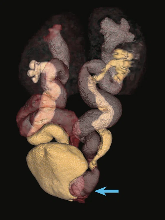
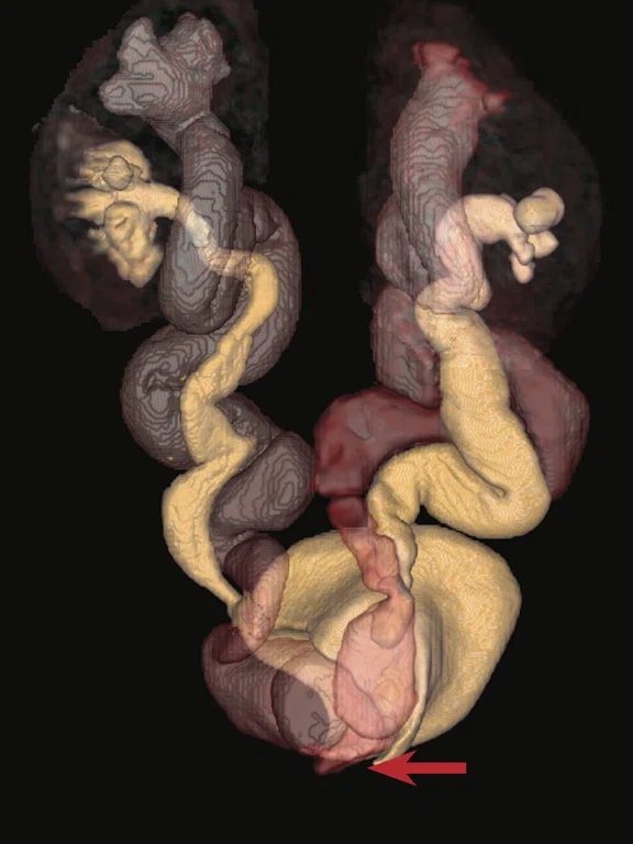
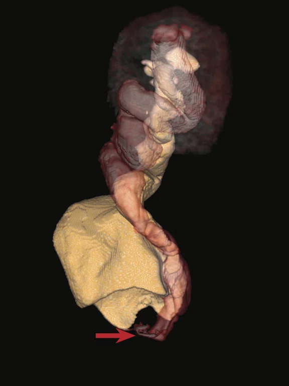
(A) Volume-rendered coronal 3D frFSE urogram, anterior view with left tilt demonstrating ureterocele on dilated left upper system (blue arrow); (B) volume-rendered coronal and (C) right lateral view of 3D frFSE urogram view demonstrating ectopic implantation of ureter of right upper system (red arrow).
B
Figure 2.
(A) Volume-rendered coronal 3D frFSE urogram, anterior view with left tilt demonstrating ureterocele on dilated left upper system (blue arrow); (B) volume-rendered coronal and (C) right lateral view of 3D frFSE urogram view demonstrating ectopic implantation of ureter of right upper system (red arrow).
C
Figure 2.
(A) Volume-rendered coronal 3D frFSE urogram, anterior view with left tilt demonstrating ureterocele on dilated left upper system (blue arrow); (B) volume-rendered coronal and (C) right lateral view of 3D frFSE urogram view demonstrating ectopic implantation of ureter of right upper system (red arrow).


result
PREVIOUS
${prev-page}
NEXT
${next-page}
Subscribe Now
Manage Subscription
FOLLOW US
Contact Us • Cookie Preferences • Privacy Policy • California Privacy PolicyDo Not Sell or Share My Personal Information • Terms & Conditions • Security
© 2024 GE HealthCare. GE is a trademark of General Electric Company. Used under trademark license.
CASE STUDIES
MR urography in congenital malformations of the urinary system
MR urography in congenital malformations of the urinary system
by Zouhir Berrou, MD, Radiologist, Centre d’Imagerie Médicale – Gué de Constantine, Algiers, Algeria
Congenital anomalies of the urinary tract system and kidneys are the most frequent malformation at birth.1 In addition to the kidneys, affected parts can include the bladder, ureters and urethra. The malformation varies in severity and can result in life-threatening kidney failure and end-stage renal disease.2
A kidney or urinary tract malformation can be detected prior to birth on routine prenatal ultrasound. If the problem is not detected until after birth, a renal scan (dynamic scintigraphy), ultrasound, CT or MR can be used to assess the structure and function of the kidneys and urinary tract. Determining relative renal function is also important for surgical planning.
Currently in our institution, MR urography is fully integrated into the diagnostic pathway in pediatric urology, including for the detection of obstructive malformation uropathies. Studies have shown an excellent correlation between results from dynamic scintigraphy and renal function MR for a differential or relative renal function study.3
With MR, post-processing tools such as segmentation and volume rendering are helpful for depicting the tracts from the kidney to the bladder. MR can provide both structural and functional (urography) imaging, making it an ideal imaging test for these type of cases. Our MR system was upgraded to SIGNA™ Explorer in 2020, providing new features, sequences and applications to our MR imaging services. In this patient case, the new version of
3D Viewer, available on both the MR console and the Advantage Workstation (AW), was crucial for surgical planning. The 16-channel Flex coil also helped deliver the image resolution needed to clearly depict the urinary system malformations for the pediatric surgeon.
Patient history
A five-month old female was referred for a MR urography exam for functional and anatomical evaluation of each reniculus for malformation-associated uropathy initially suspected during prenatal ultrasound exams.
Two days after birth, a neonatal ultrasound exam confirmed the malformation-associated uropathy as a bilateral double collecting system with left ureterocele. The infant suffered from repetitive urinary infections, which led to a cystography exam that revealed the absence of the vesicoureteral reflux (VUR).
Patient underwent decompression of the ureterocele by endoscopy at age two months and her first scintigraphy evaluation at age three months. Patient was referred for an MR urography exam for functional and anatomical evaluation of each reniculus at age five months.
Figure 1.
(A) Coronal T2 PROPELLER FatSat with respiratory triggering, 1.1 x 1.1 x 3 mm3, 3:50 min.; (B) volume-rendered coronal 3D frFSE urogram with respiratory triggering, 1 x 1.3 x 1.2/-0.6 mm3, 3:43 min., depicting the left and right lower collecting systems (yellow arrows) and left and right upper collecting systems (red arrows); and (C) fused image of (A) and (B).
Results
MR exam with advanced 3D Viewer post-processing demonstrated bilateral duplicated collecting systems with ectopic urethro-cervical implantation of the ureter on the right upper system and ureterocele on the dilated left upper system. The use of PROPELLER on pediatric urography examinations greatly improves image quality by reducing the effect of patient voluntary and physiologic motion (breathing, flow, peristalsis). 3D T2 frFSE allows for visualization and post processing of the renal collection system.
Figure 2.
(A) Volume-rendered coronal 3D frFSE urogram, anterior view with left tilt demonstrating ureterocele on dilated left upper system (blue arrow); (B) volume-rendered coronal and (C) right lateral view of 3D frFSE urogram view demonstrating ectopic implantation of ureter of right upper system (red arrow).
Discussion
The MR exam helped determine the patient had obstructive syndrome with a more pronounced urinary stasis on superior systems.
In addition to the lack of radiation exposure for the patient, MR allows for a high-quality morphological analysis (anatomical analysis) compared to standard X-ray techniques (intravenous urography) with the added value of angiographic imaging to depict vascular crossings. MR imaging is important for the surgical planning required in these cases of congenital anomalies of the urinary tract system.


