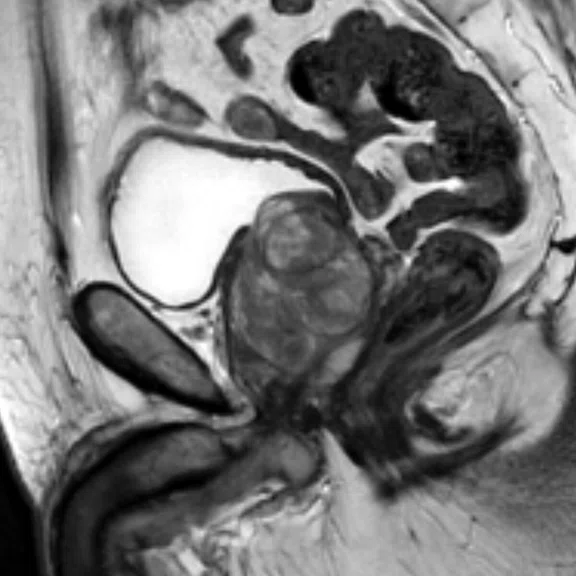
Sagittal T2 PROPELLER
0.7 x 0.7 x 3 mm
3:16 min.
Images courtesy of radiomed, Mainz, Germany
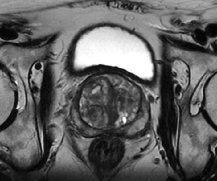
Axial T2 frFSE
0.6 x 0.8 x 3 mm
3:24 min.
Images courtesy of radiomed, Mainz, Germany
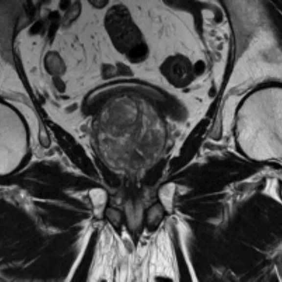
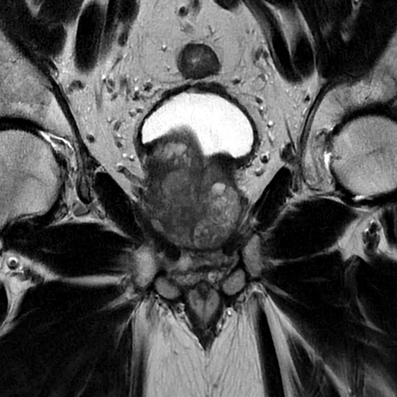
Coronal T2 frFSE
0.7 x 0.7 x 3 mm
3:50 min.
Images courtesy of radiomed, Mainz, Germany
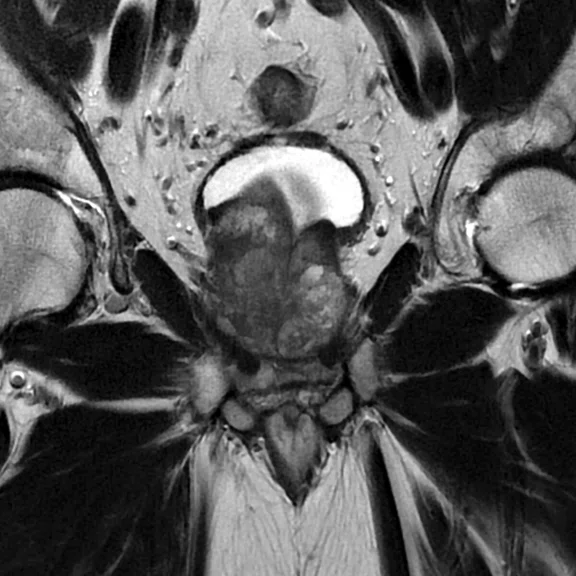
Without AIR™ Recon
4:35 min.
AIR™ Recon allows reduction in scan times and can lessen out of field-of-view artifacts to improve image quality and workflow. Both coronal T2 PROPELLER scans were acquired with the same resolution, however, the left image was acquired in 4:35 min. while the image on the right was acquired in only 3:37 min. using AIR™ Recon. Images courtesy of radiomed, Mainz, Germany
With AIR™ Recon
3:37 min.
AIR™ Recon allows reduction in scan times and can lessen out of field-of-view artifacts to improve image quality and workflow. Both coronal T2 PROPELLER scans were acquired with the same resolution, however, the left image was acquired in 4:35 min. while the image on the right was acquired in only 3:37 min. using AIR™ Recon. Images courtesy of radiomed, Mainz, Germany
Sagittal T2-weighted
0.6 x 0.8 x 5 mm
3:25 min.
Images courtesy of Osaka University Hospital, Osaka, Japan
Sagittal T2-weighted
0.6 x 0.8 x 5 mm
3:25 min.
Images courtesy of Osaka University Hospital, Osaka, Japan
Coronal T2 FSE
0.6 x 0.8 x 5 mm
3:58 min.
Images courtesy of Osaka University Hospital, Osaka, Japan
Coronal T2 FSE
0.6 x 0.8 x 5 mm
3:58 min.
Images courtesy of Osaka University Hospital, Osaka, Japan
result


PREVIOUS
${prev-page}
NEXT
${next-page}
Subscribe Now
Manage Subscription
FOLLOW US
Contact Us • Cookie Preferences • Privacy Policy • California Privacy PolicyDo Not Sell or Share My Personal Information • Terms & Conditions • Security
© 2024 GE HealthCare. GE is a trademark of General Electric Company. Used under trademark license.
AIR™ Recon allows reduction in scan times and can lessen out of field-of-view artifacts to improve image quality and workflow. Both coronal T2 PROPELLER scans were acquired with the same resolution, however, the left image was acquired in 4:35 min. while the image on the right was acquired in only 3:37 min. using AIR™ Recon. Images courtesy of radiomed, Mainz, Germany
Sagittal T2-weighted
0.6 x 0.8 x 5 mm
3:25 min.
Coronal T2 FSE
0.6 x 0.8 x 5 mm
3:58 min.
Images courtesy of Osaka University Hospital, Osaka, Japan


