result
A
Figure 1.
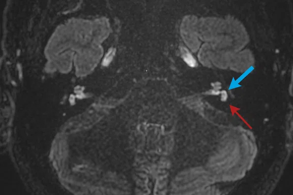
Case 1: A 59-year-old man with vertigo and suspicion of Ménière’s disease. (A) At the level of the lower part of the vestibule, the endolymphatic saccule (blue arrow) and utricle (red arrow) are seen as two dark, separate structures within an enhancing perilymphatic vestibule. (B) At the midmodiolar level, the cochlear duct is not dilated and barely visible within the cochlea.
B
Figure 1.
Case 1: A 59-year-old man with vertigo and suspicion of Ménière’s disease. (A) At the level of the lower part of the vestibule, the endolymphatic saccule (blue arrow) and utricle (red arrow) are seen as two dark, separate structures within an enhancing perilymphatic vestibule. (B) At the midmodiolar level, the cochlear duct is not dilated and barely visible within the cochlea.
1. Goebel JA. 2015 Equilibrium Committee Amendment to the 1995 AAO-HNS Guidelines for the Definition of Ménière’s Disease. Otolaryngol Head Neck Surg. 2016 Mar;154(3):403-4.
2. Hallpike C, Cairns H. Observations on the pathology of Meniere’s syndrome. Proc R Soc Med. 1938;31:1317–36.
3. Yamakawa K. Über die pathologische Veränderung bei einem Ménière-Kranken. J Otorhinolaryngol Soc Jpn. 1938;4:2310–2312.
4. Nakashima T, Naganawa S, Teranishi M, et al. Endolymphatic hydrops revealed by intravenous gadolinium injection in patients with Ménière’s disease. Acta Otolaryngol. 2010 Mar;130(3):338-43.
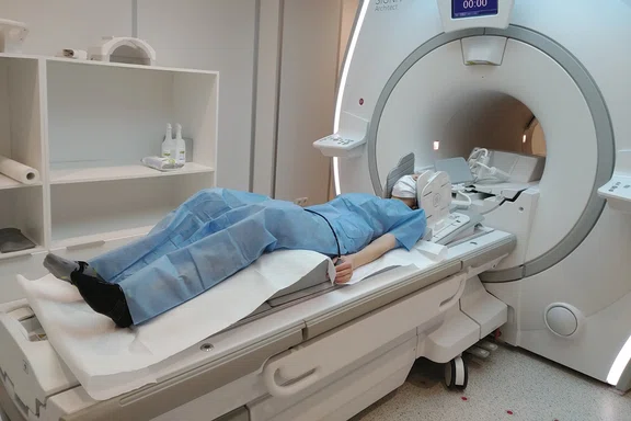
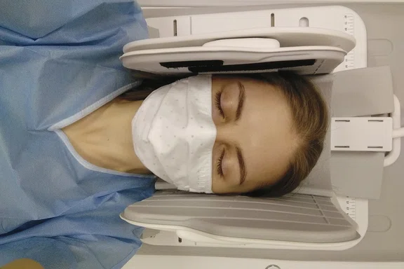
Patient positioning on SIGNA™ Architect with the Flex Coil positioner for imaging suspected Ménière’s disease. A 16ch Flex Coil is used to minimize the distance between the anatomy of interest and the coil.
Patient positioning on SIGNA™ Architect with the Flex Coil positioner for imaging suspected Ménière’s disease. A 16ch Flex Coil is used to minimize the distance between the anatomy of interest and the coil.
1. Goebel JA. 2015 Equilibrium Committee Amendment to the 1995 AAO-HNS Guidelines for the Definition of Ménière’s Disease. Otolaryngol Head Neck Surg. 2016 Mar;154(3):403-4.
A
Figure 2.
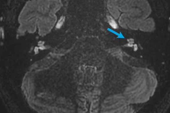
Case 2: A 44-year-old man with clinically diagnosed left-sided Ménière’s disease. (A) At the midmodiolar level, the cochlear duct partially obstructs the scala vestibuli in the cochlea, which is visible as multiple nonenhancing nodules (blue arrows). (B) At the level of the lower part of the vestibule, the endolymphatic saccule and utricle are dilated and confluent (thick yellow arrow) but a thin rim of perilymphatic space remains visible (thin yellow arrow). The normal right-side saccule (thick blue arrow) and utricle (thin blue arrow) are well discriminated and encompass less than 50% of the vestibule.
B
Figure 2.
Case 2: A 44-year-old man with clinically diagnosed left-sided Ménière’s disease. (A) At the midmodiolar level, the cochlear duct partially obstructs the scala vestibuli in the cochlea, which is visible as multiple nonenhancing nodules (blue arrows). (B) At the level of the lower part of the vestibule, the endolymphatic saccule and utricle are dilated and confluent (thick yellow arrow) but a thin rim of perilymphatic space remains visible (thin yellow arrow). The normal right-side saccule (thick blue arrow) and utricle (thin blue arrow) are well discriminated and encompass less than 50% of the vestibule.


PREVIOUS
${prev-page}
NEXT
${next-page}
Subscribe Now
Manage Subscription
FOLLOW US
Contact Us • Cookie Preferences • Privacy Policy • California Privacy PolicyDo Not Sell or Share My Personal Information • Terms & Conditions • Security
© 2024 GE HealthCare. GE is a trademark of General Electric Company. Used under trademark license.
CASE STUDIES
T2 FLAIR Cube for diagnosing Ménière’s disease
T2 FLAIR Cube for diagnosing Ménière’s disease
by Emilia Wnuk, MD, and Lucyna Grzybowska, (RT)(MR), radiographer, Medical University of Warsaw, Warszawa, Poland
Ménière’s disease is a disorder of the inner ear that can lead to spontaneous attacks of vertigo, tinnitus, aural fullness and low-frequency hearing loss1. More than 80 years ago, Hallpike and Cairns2 and Yamakawa3 revealed in postmortem temporal bone studies that the enlargement of the inner ear endolymphatic structures is present in patients with Ménière’s disease. For many years, it was impossible to assess endolymphatic hydrops in vivo. Advances in medical imaging, and in particular MR, enabled visualization of the inner ear structures. Nakashima et al. noticed that intravenously or intratympanically administered contrast penetrates only the structures that contain perilymph, whereas the endolymphatic space does not enhance4.
In most cases, Ménière’s disease affects only one ear. Ménière’s disease can occur at any age, but it usually starts between young and middle-aged adulthood. It is considered a chronic condition, although various treatments can help relieve symptoms and minimize the long-term impact on the patient’s life.
Case 1
Patient history
A 59-year-old man with vertigo and without hearing loss was referred for suspected Ménière’s disease.
Results
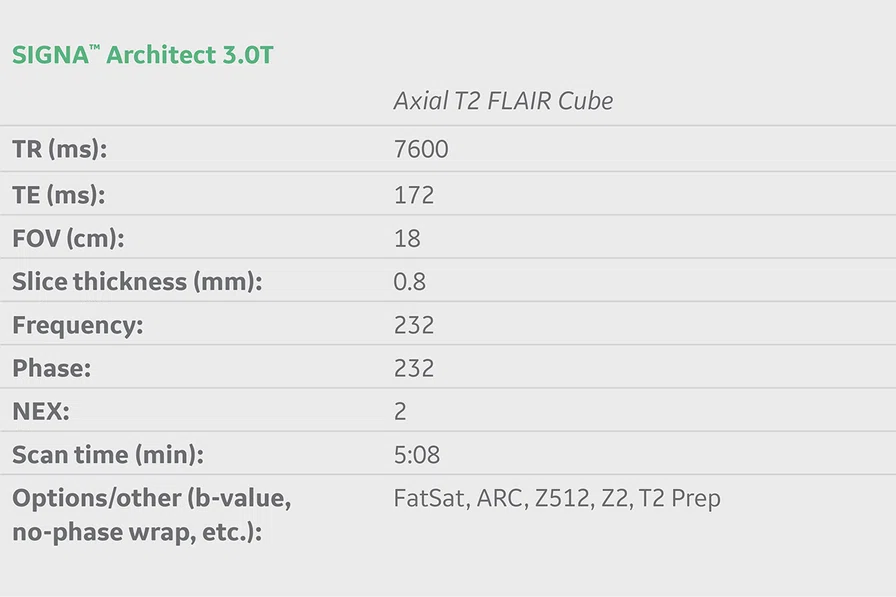
Four hours after a double dose of intravenous contrast administration, an axial T2 FLAIR Cube sequence of both ears was performed. Enhancement in the perilymphatic compartments of cochlea, vestibule and semicircular canals was present. The endolymphatic cochlear duct was not dilated and barely visible within the cochlea. In Figure 1A, an endolymphatic saccule (blue arrow) and utricle (red arrow) were visible as two separated dark dots within a vestibule. This case was diagnosed as normal with no sign of endolymphatic hydrops.
Figure 1.
Case 1: A 59-year-old man with vertigo and suspicion of Ménière’s disease. (A) At the level of the lower part of the vestibule, the endolymphatic saccule (blue arrow) and utricle (red arrow) are seen as two dark, separate structures within an enhancing perilymphatic vestibule. (B) At the midmodiolar level, the cochlear duct is not dilated and barely visible within the cochlea.
Case 2
Patient history
A 44-year-old man with clinically diagnosed left-sided Ménière’s disease according to Barany criteria1 (vertigo, audiometrically documented low frequency sensorineural hearing loss, tinnitus and fullness of the ear).
Results
Four hours after a double dose of intravenous contrast administration, an axial T2 FLAIR Cube sequence of both ears was performed. The enhancement of the perilymphatic compartments of cochlea, vestibule and semicircular canals was present. The endolymphatic cochlear duct was dilated, partially obstructing the scala vestibuli in the cochlea (Figure 2). The endolymphatic saccule and utricle were dilated and confluent; however, a thin rim of perilymphatic space remained visible (Figure 2). According to the Barath 3-stage grading system, the patient has a grade 1 cochlear hydrops and grade 1 vestibular hydrops.
Figure 2.
Case 2: A 44-year-old man with clinically diagnosed left-sided Ménière’s disease. (A) At the midmodiolar level, the cochlear duct partially obstructs the scala vestibuli in the cochlea, which is visible as multiple nonenhancing nodules (blue arrows). (B) At the level of the lower part of the vestibule, the endolymphatic saccule and utricle are dilated and confluent (thick yellow arrow) but a thin rim of perilymphatic space remains visible (thin yellow arrow). The normal right-side saccule (thick blue arrow) and utricle (thin blue arrow) are well discriminated and encompass less than 50% of the vestibule.
Discussion
The diagnosis of endolymphatic hydrops, a pathological hallmark of Ménière’s disease, is possible using a post-contrast T2 FLAIR Cube sequence. Previously, the diagnosis of Ménière’s disease was clinical with medical imaging (e.g., MR) used to exclude other pathologies that could have symptoms similar to Ménière’s disease. With the capability to acquire diagnostic- quality images of the inner ear, it is possible that certain radiological features will be the new diagnostic criteria for Ménière’s disease.


