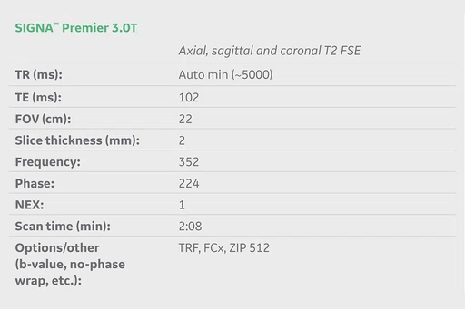
result
A
Figure 1.
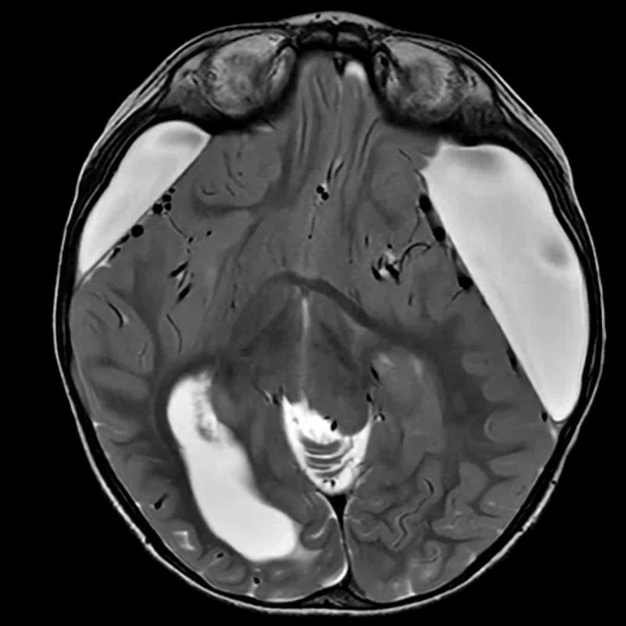
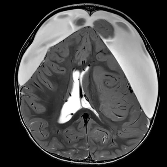
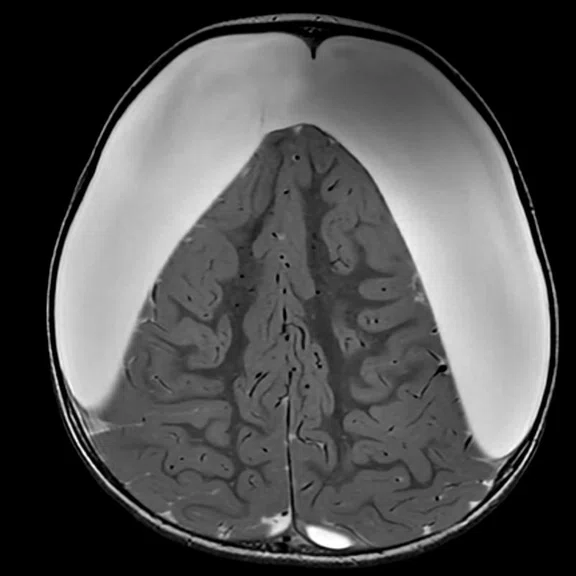
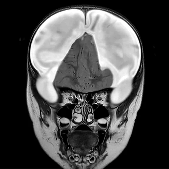
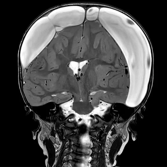
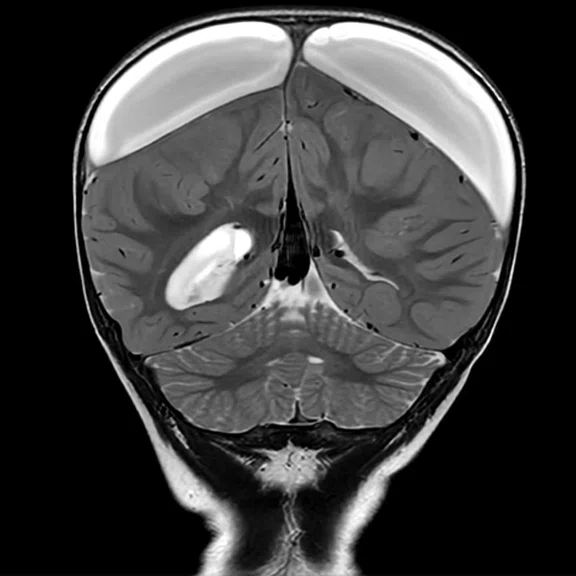
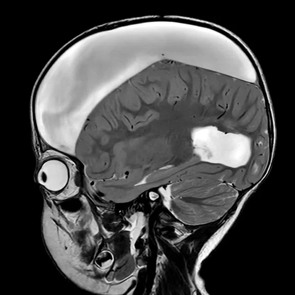
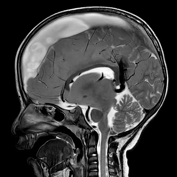
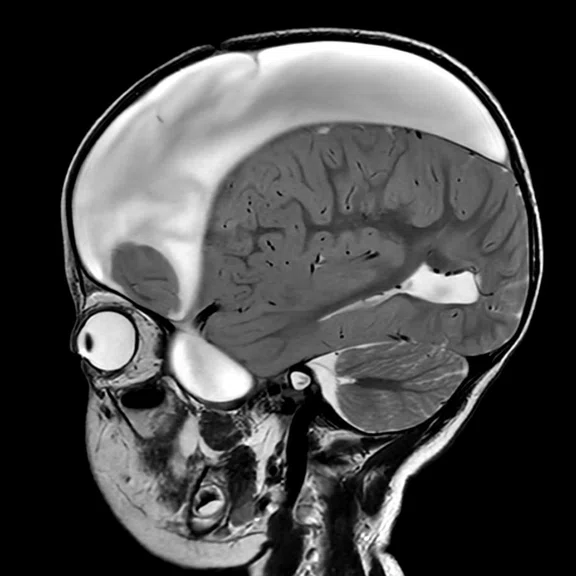
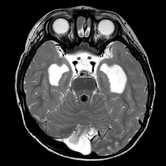
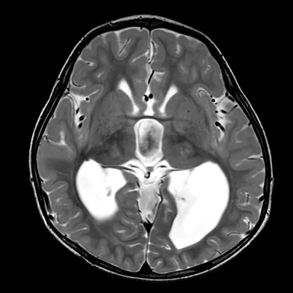
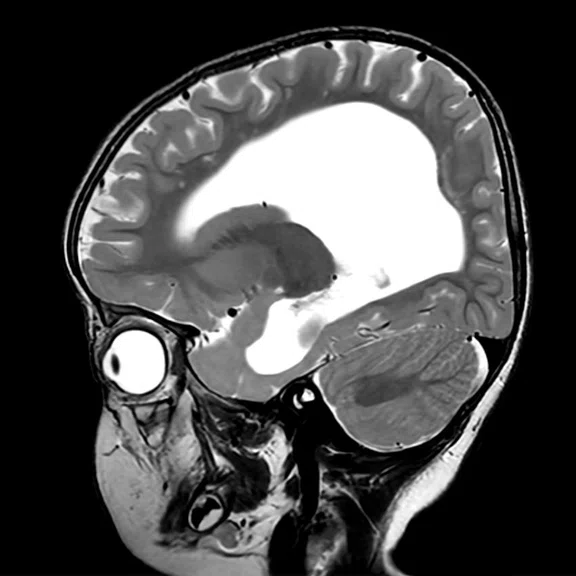
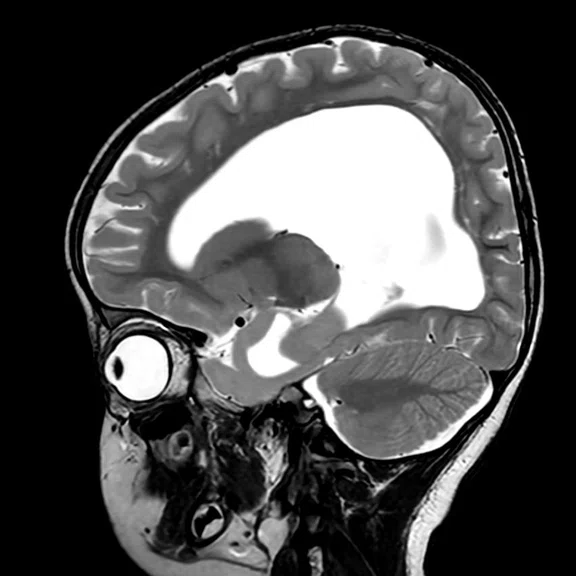
Case 1. Approximately more than 40% of the brain region is filled with hygroma. AIR x™ performed well to identify the standard axial, coronal and sagittal scan planes.
B
Figure 1.
Case 1. Approximately more than 40% of the brain region is filled with hygroma. AIR x™ performed well to identify the standard axial, coronal and sagittal scan planes.
C
Figure 1.
Case 1. Approximately more than 40% of the brain region is filled with hygroma. AIR x™ performed well to identify the standard axial, coronal and sagittal scan planes.
D
Figure 1.
Case 1. Approximately more than 40% of the brain region is filled with hygroma. AIR x™ performed well to identify the standard axial, coronal and sagittal scan planes.
E
Figure 1.
Case 1. Approximately more than 40% of the brain region is filled with hygroma. AIR x™ performed well to identify the standard axial, coronal and sagittal scan planes.
F
Figure 1.
Case 1. Approximately more than 40% of the brain region is filled with hygroma. AIR x™ performed well to identify the standard axial, coronal and sagittal scan planes.
G
Figure 1.
Case 1. Approximately more than 40% of the brain region is filled with hygroma. AIR x™ performed well to identify the standard axial, coronal and sagittal scan planes.
H
Figure 1.
Case 1. Approximately more than 40% of the brain region is filled with hygroma. AIR x™ performed well to identify the standard axial, coronal and sagittal scan planes.
I
Figure 1.
Case 1. Approximately more than 40% of the brain region is filled with hygroma. AIR x™ performed well to identify the standard axial, coronal and sagittal scan planes.
A
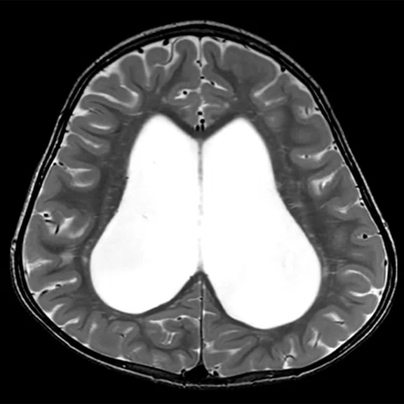
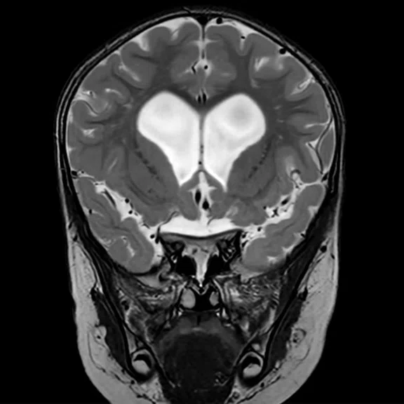
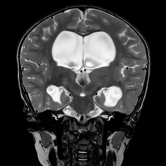
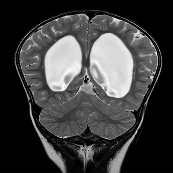
Figure 2.
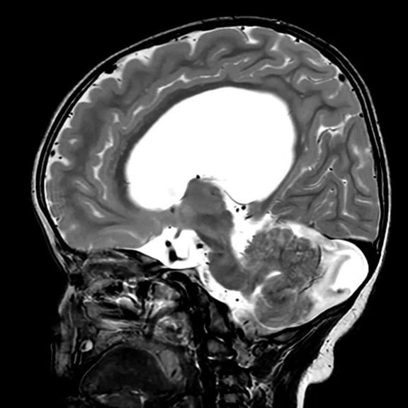
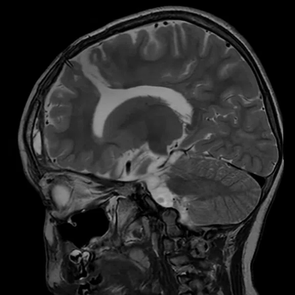
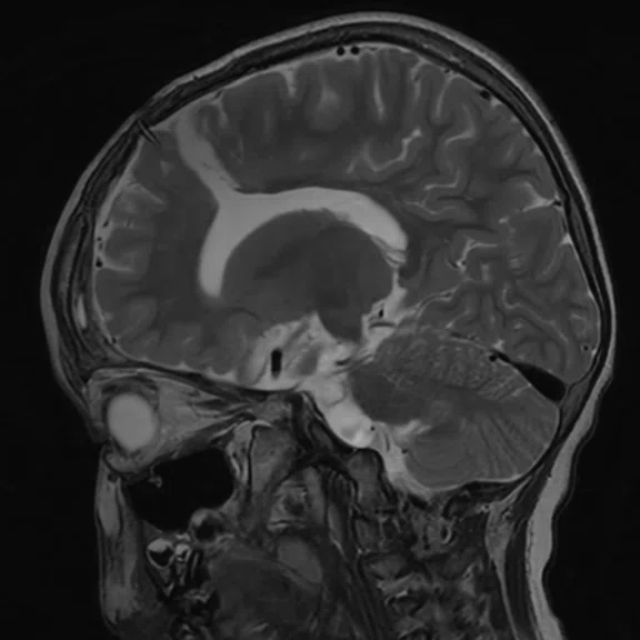
Case 2. Ventriculomegaly results in severe brain parenchyma deformation as well as overall cranial shape. AIR x™ works well in this kind of irregular case, helping the technologist when it may not be possible to locate an anatomical reference.
B
Figure 2.
Case 2. Ventriculomegaly results in severe brain parenchyma deformation as well as overall cranial shape. AIR x™ works well in this kind of irregular case, helping the technologist when it may not be possible to locate an anatomical reference.
C
Figure 2.
Case 2. Ventriculomegaly results in severe brain parenchyma deformation as well as overall cranial shape. AIR x™ works well in this kind of irregular case, helping the technologist when it may not be possible to locate an anatomical reference.
D
Figure 2.
Case 2. Ventriculomegaly results in severe brain parenchyma deformation as well as overall cranial shape. AIR x™ works well in this kind of irregular case, helping the technologist when it may not be possible to locate an anatomical reference.
E
Figure 2.
Case 2. Ventriculomegaly results in severe brain parenchyma deformation as well as overall cranial shape. AIR x™ works well in this kind of irregular case, helping the technologist when it may not be possible to locate an anatomical reference.
F
Figure 2.
Case 2. Ventriculomegaly results in severe brain parenchyma deformation as well as overall cranial shape. AIR x™ works well in this kind of irregular case, helping the technologist when it may not be possible to locate an anatomical reference.
G
Figure 2.
Case 2. Ventriculomegaly results in severe brain parenchyma deformation as well as overall cranial shape. AIR x™ works well in this kind of irregular case, helping the technologist when it may not be possible to locate an anatomical reference.
H
Figure 2.
Case 2. Ventriculomegaly results in severe brain parenchyma deformation as well as overall cranial shape. AIR x™ works well in this kind of irregular case, helping the technologist when it may not be possible to locate an anatomical reference.
I
Figure 2.
Case 2. Ventriculomegaly results in severe brain parenchyma deformation as well as overall cranial shape. AIR x™ works well in this kind of irregular case, helping the technologist when it may not be possible to locate an anatomical reference.
A
Figure 3.
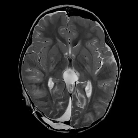
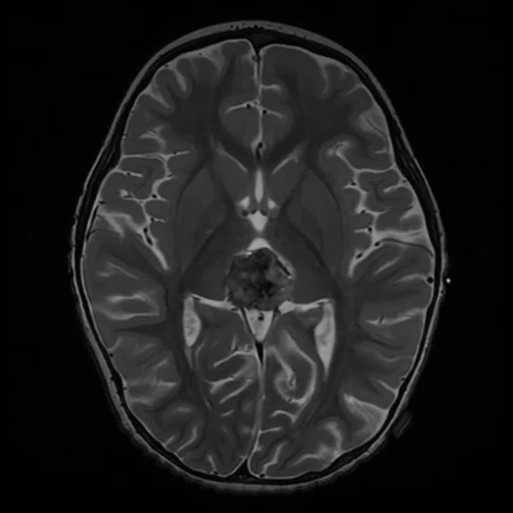
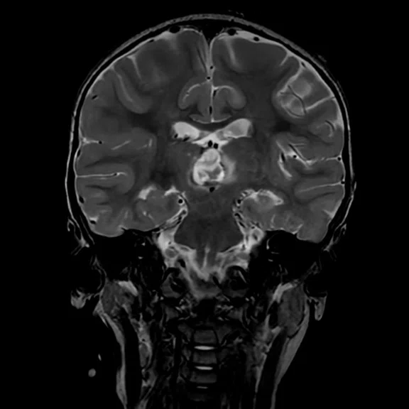
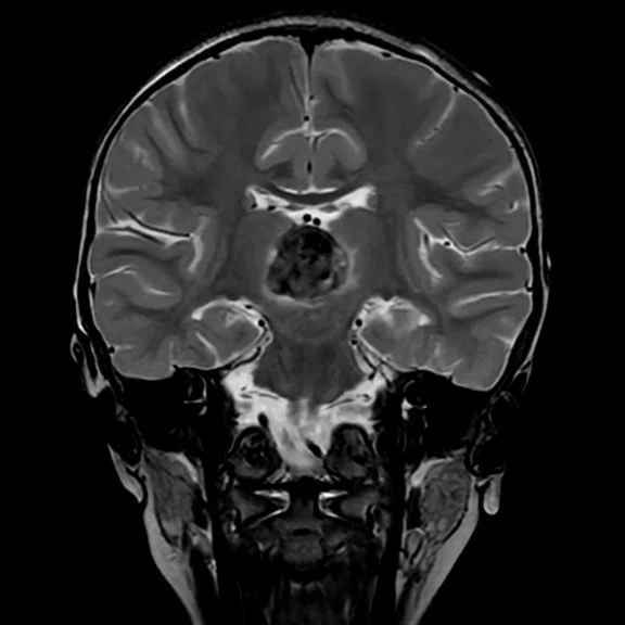
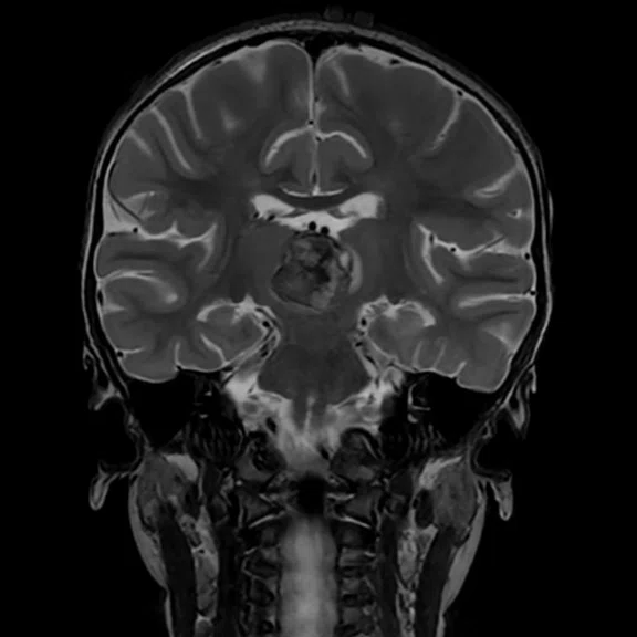
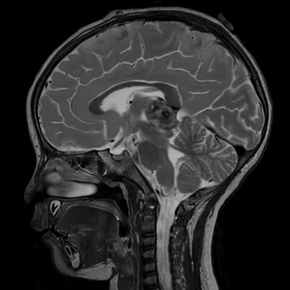
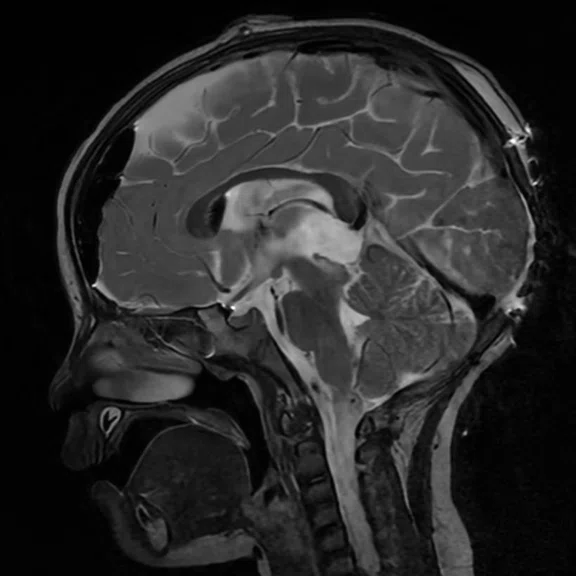
Case 3. A 2.7 x 2.0 x 2.5 cm sized pineal gland was detected in this case, and two additional exams were performed for further evaluation. AIR x™ continuously prescribed identical slice locations for the scans that occurred on different dates. (A, D, G) First exam, (B, E, H) second exam, six days after first exam and (C, F, I) third exam, eight days after first exam.
B
Figure 3.
Case 3. A 2.7 x 2.0 x 2.5 cm sized pineal gland was detected in this case, and two additional exams were performed for further evaluation. AIR x™ continuously prescribed identical slice locations for the scans that occurred on different dates. (A, D, G) First exam, (B, E, H) second exam, six days after first exam and (C, F, I) third exam, eight days after first exam.
C
Figure 3.
Case 3. A 2.7 x 2.0 x 2.5 cm sized pineal gland was detected in this case, and two additional exams were performed for further evaluation. AIR x™ continuously prescribed identical slice locations for the scans that occurred on different dates. (A, D, G) First exam, (B, E, H) second exam, six days after first exam and (C, F, I) third exam, eight days after first exam.
D
Figure 3.
Case 3. A 2.7 x 2.0 x 2.5 cm sized pineal gland was detected in this case, and two additional exams were performed for further evaluation. AIR x™ continuously prescribed identical slice locations for the scans that occurred on different dates. (A, D, G) First exam, (B, E, H) second exam, six days after first exam and (C, F, I) third exam, eight days after first exam.
E
Figure 3.
Case 3. A 2.7 x 2.0 x 2.5 cm sized pineal gland was detected in this case, and two additional exams were performed for further evaluation. AIR x™ continuously prescribed identical slice locations for the scans that occurred on different dates. (A, D, G) First exam, (B, E, H) second exam, six days after first exam and (C, F, I) third exam, eight days after first exam.
F
Figure 3.
Case 3. A 2.7 x 2.0 x 2.5 cm sized pineal gland was detected in this case, and two additional exams were performed for further evaluation. AIR x™ continuously prescribed identical slice locations for the scans that occurred on different dates. (A, D, G) First exam, (B, E, H) second exam, six days after first exam and (C, F, I) third exam, eight days after first exam.
G
Figure 3.
Case 3. A 2.7 x 2.0 x 2.5 cm sized pineal gland was detected in this case, and two additional exams were performed for further evaluation. AIR x™ continuously prescribed identical slice locations for the scans that occurred on different dates. (A, D, G) First exam, (B, E, H) second exam, six days after first exam and (C, F, I) third exam, eight days after first exam.
H
Figure 3.
Case 3. A 2.7 x 2.0 x 2.5 cm sized pineal gland was detected in this case, and two additional exams were performed for further evaluation. AIR x™ continuously prescribed identical slice locations for the scans that occurred on different dates. (A, D, G) First exam, (B, E, H) second exam, six days after first exam and (C, F, I) third exam, eight days after first exam.
I
Figure 3.
Case 3. A 2.7 x 2.0 x 2.5 cm sized pineal gland was detected in this case, and two additional exams were performed for further evaluation. AIR x™ continuously prescribed identical slice locations for the scans that occurred on different dates. (A, D, G) First exam, (B, E, H) second exam, six days after first exam and (C, F, I) third exam, eight days after first exam.
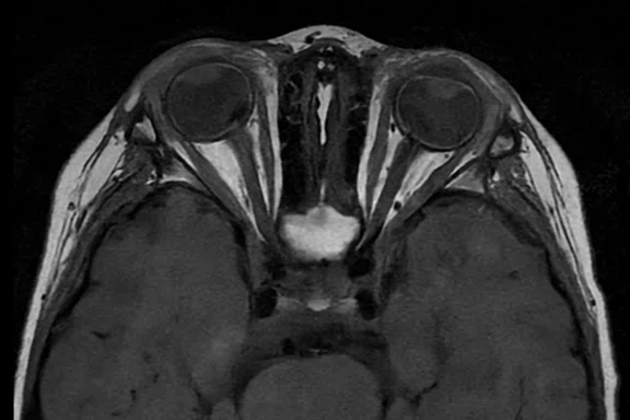
A
Figure 4.
Case 4. AIR x™ shows stable performance not only in the brain but in the optic nerve and optic-related anatomy even with lesions in both eyes, retinoblastoma in this case. (A, C) First exam and (B, D) second exam, 62 days later.
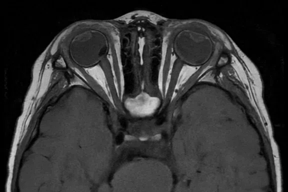
B
Figure 4.
Case 4. AIR x™ shows stable performance not only in the brain but in the optic nerve and optic-related anatomy even with lesions in both eyes, retinoblastoma in this case. (A, C) First exam and (B, D) second exam, 62 days later.
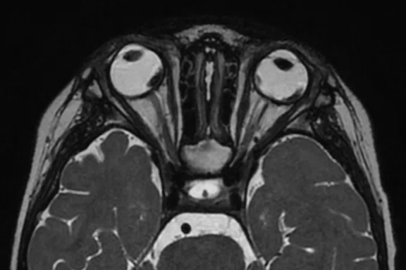
C
Figure 4.
Case 4. AIR x™ shows stable performance not only in the brain but in the optic nerve and optic-related anatomy even with lesions in both eyes, retinoblastoma in this case. (A, C) First exam and (B, D) second exam, 62 days later.
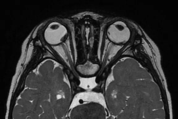
D
Figure 4.
Case 4. AIR x™ shows stable performance not only in the brain but in the optic nerve and optic-related anatomy even with lesions in both eyes, retinoblastoma in this case. (A, C) First exam and (B, D) second exam, 62 days later.
A
Figure 5.
Case 5. AIR x™ shows stable performance not only in the brain but in more complex regions such as the suprasellar. (A, C, E) First exam and (B, D, F) second exam, 15 days later.
B
Figure 5.
Case 5. AIR x™ shows stable performance not only in the brain but in more complex regions such as the suprasellar. (A, C, E) First exam and (B, D, F) second exam, 15 days later.
C
Figure 5.
Case 5. AIR x™ shows stable performance not only in the brain but in more complex regions such as the suprasellar. (A, C, E) First exam and (B, D, F) second exam, 15 days later.
D
Figure 5.
Case 5. AIR x™ shows stable performance not only in the brain but in more complex regions such as the suprasellar. (A, C, E) First exam and (B, D, F) second exam, 15 days later.
E
Figure 5.
Case 5. AIR x™ shows stable performance not only in the brain but in more complex regions such as the suprasellar. (A, C, E) First exam and (B, D, F) second exam, 15 days later.
F
Figure 5.
Case 5. AIR x™ shows stable performance not only in the brain but in more complex regions such as the suprasellar. (A, C, E) First exam and (B, D, F) second exam, 15 days later.


PREVIOUS
${prev-page}
NEXT
${next-page}
Subscribe Now
Manage Subscription
FOLLOW US
Contact Us • Cookie Preferences • Privacy Policy • California Privacy PolicyDo Not Sell or Share My Personal Information • Terms & Conditions • Security
© 2024 GE HealthCare. GE is a trademark of General Electric Company. Used under trademark license.
CASE STUDIES
Consistent slice prescription in irregular brain morphology and follow-up exams
Consistent slice prescription in irregular brain morphology and follow-up exams
by Young Hun Choi, MD, PhD, Associate Professor and Woo Jin Jeong, RT(R)(MR), Senior Technologist, Seoul National University Children’s Hospital, Seoul, Republic of Korea
Many children with various brain diseases, such as congenital brain malformations, brain tumors, and infectious and inflammatory brain diseases, are referred to the Seoul National University Children’s Hospital MR imaging center. Infants and young children cannot stay still inside the MR bore, therefore, in most cases they need to be sedated during the MR examination.
When using sedatives, it is important to complete the scan series as quickly as possible to limit sedation and avoid further injections, while maintaining high image quality. Productivity is important due to the large volume of patients.
Additionally, as a pediatric referral center, many patients often receive repeat MR exams to follow a particular disease, condition or injury. For scheduling, it is not possible to have the same technologist scan the same patient for each subsequent MR exam. This can lead to different scan prescriptions as each technologist may prescribe and scan a patient case differently, which can then impact the radiologist’s reading and reporting when comparing prior exams.
AIR x™, a deep-learning-based automated scan prescription tool, is now applied to all neuro exam protocols, from routine brain to more complicated anatomical regions such as the optic nerves, skull base, internal auditory canal (IAC), sella and peripheral nervous system. AIR x™ allows the technologist to rapidly acquire the brain images and also frees up their time to monitor the patient status.
Case 1
Patient history
A 29-month-old patient weighing 33 lbs. (15 kg) with subdural hygroma in bilateral cerebral convexities.
Case 2
Patient history
A 28-month-old patient weighing 29 lbs. (13 kg) with ventriculomegaly with retrocerebellar fluid collection.
Case 3
Patient history
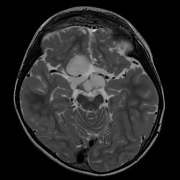
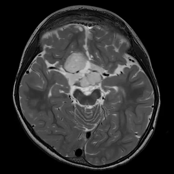
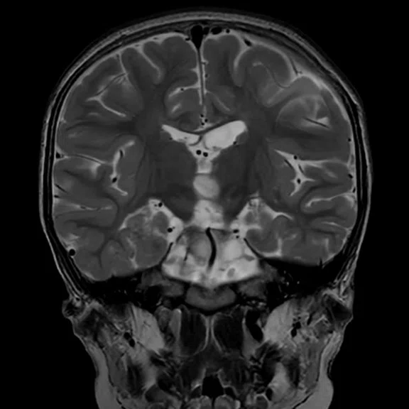
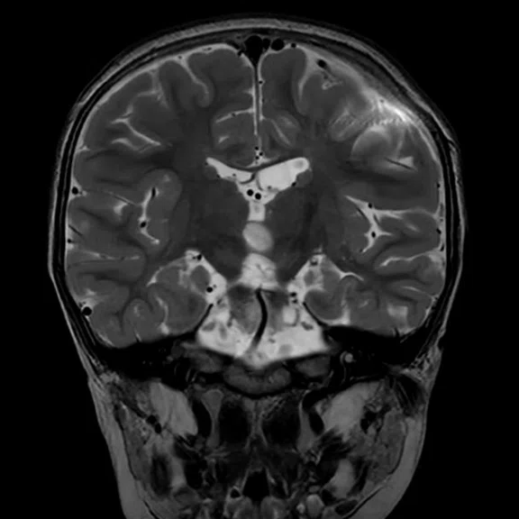
A 26-month-old patient weighing 24 lbs. (11 kg) with a pineal gland tumor with a high cellularity underwent follow-up exams.
Figure 3.
Case 3. A 2.7 x 2.0 x 2.5 cm sized pineal gland was detected in this case, and two additional exams were performed for further evaluation. AIR x™ continuously prescribed identical slice locations for the scans that occurred on different dates. (A, D, G) First exam, (B, E, H) second exam, six days after first exam and (C, F, I) third exam, eight days after first exam.
Case 4
Patient history
A 36-month-old patient weighing 37 lbs. (17 kg) with bilateral retinoblastoma underwent follow-up exams.
Case 5
Patient history
A 36-month-old patient weighing 42 lbs. (19 kg) with a glioma in the suprasellar area underwent follow-up exams.
Discussion
Even in cases of severely deformed brain structure, such as bulky subdural hygroma affecting brain parenchyma or enlarged ventricle with ventriculomegaly, AIR x™ can provide precise scan prescriptions. In follow-up studies, AIR x™ consistently provides an almost identical slice prescription across routine brain and more complicated anatomical exams, such as in the suprasellar and orbits, which assists in an accurate evaluation.


