A
Figure 1.
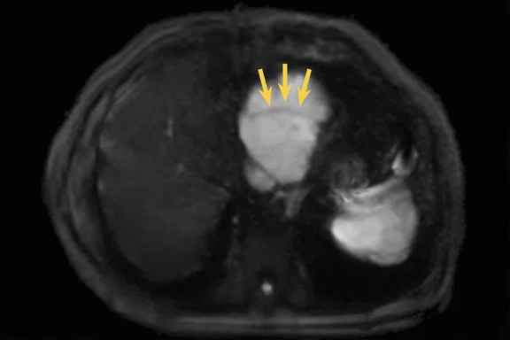
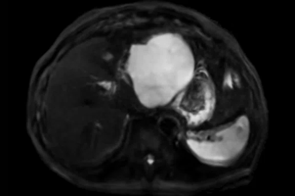
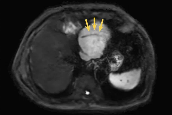
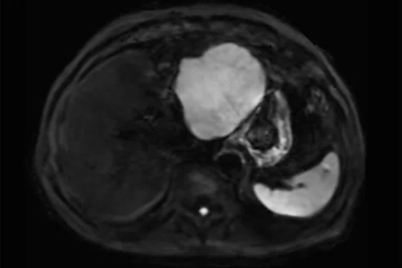
Comparison of image quality in DWI between conventional method and modified protocol. The conventional method with navigator gating has blurring in the shape of the liver tumor and the margin of the spleen. However, the modified 5 mm breath hold DWI depicted the liver tumor and spleen clearly and sharply. Intratumor blood vessels are blurred with conventional DWI, but can be clearly depicted with the modified protocol and AIR™ Recon DL (yellow arrows). (A, B) Conventional reconstruction of DWI with navigator; (C, D) modified breath hold DWI with 5 mm thin slices.
B
Figure 1.
Comparison of image quality in DWI between conventional method and modified protocol. The conventional method with navigator gating has blurring in the shape of the liver tumor and the margin of the spleen. However, the modified 5 mm breath hold DWI depicted the liver tumor and spleen clearly and sharply. Intratumor blood vessels are blurred with conventional DWI, but can be clearly depicted with the modified protocol and AIR™ Recon DL (yellow arrows). (A, B) Conventional reconstruction of DWI with navigator; (C, D) modified breath hold DWI with 5 mm thin slices.
C
Figure 1.
Comparison of image quality in DWI between conventional method and modified protocol. The conventional method with navigator gating has blurring in the shape of the liver tumor and the margin of the spleen. However, the modified 5 mm breath hold DWI depicted the liver tumor and spleen clearly and sharply. Intratumor blood vessels are blurred with conventional DWI, but can be clearly depicted with the modified protocol and AIR™ Recon DL (yellow arrows). (A, B) Conventional reconstruction of DWI with navigator; (C, D) modified breath hold DWI with 5 mm thin slices.
D
Figure 1.
Comparison of image quality in DWI between conventional method and modified protocol. The conventional method with navigator gating has blurring in the shape of the liver tumor and the margin of the spleen. However, the modified 5 mm breath hold DWI depicted the liver tumor and spleen clearly and sharply. Intratumor blood vessels are blurred with conventional DWI, but can be clearly depicted with the modified protocol and AIR™ Recon DL (yellow arrows). (A, B) Conventional reconstruction of DWI with navigator; (C, D) modified breath hold DWI with 5 mm thin slices.
A
Figure 4.
Stiffness map showed normal stiffness in the liver, but high stiffness in the tumor, consistent with HCC. And, IDEAL-IQ has confirmed about 5% mild fatty liver by fat-fraction and mild iron deposition by R2* map.(A) MR elastography stiffness map and IDEAL IQ (B) fat fraction and (C) R2* map of the liver.
B
Figure 4.
Stiffness map showed normal stiffness in the liver, but high stiffness in the tumor, consistent with HCC. And, IDEAL-IQ has confirmed about 5% mild fatty liver by fat-fraction and mild iron deposition by R2* map.(A) MR elastography stiffness map and IDEAL IQ (B) fat fraction and (C) R2* map of the liver.
C
Figure 4.
Stiffness map showed normal stiffness in the liver, but high stiffness in the tumor, consistent with HCC. And, IDEAL-IQ has confirmed about 5% mild fatty liver by fat-fraction and mild iron deposition by R2* map.(A) MR elastography stiffness map and IDEAL IQ (B) fat fraction and (C) R2* map of the liver.
A
Figure 2.
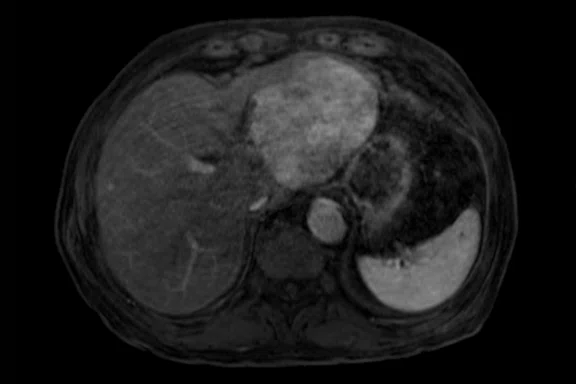
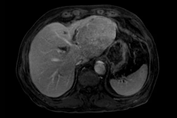
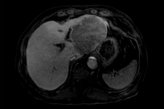
3D T1 with HyperSense and EOB protocol. Dynamic DISCO with HyperSense has a reduced temporal resolution of 3.4 sec. The hepatobiliary phase can be modified to high resolution using HyperSense to obtain detailed information. (A) Arterial phase, (B) portal phase and (C) late phase of the dynamic study using DISCO by EOB protocol, and (D) high-resolution navigated 3D T1 image with HyperSense at hepatobiliary phase.
B
Figure 2.
3D T1 with HyperSense and EOB protocol. Dynamic DISCO with HyperSense has a reduced temporal resolution of 3.4 sec. The hepatobiliary phase can be modified to high resolution using HyperSense to obtain detailed information. (A) Arterial phase, (B) portal phase and (C) late phase of the dynamic study using DISCO by EOB protocol, and (D) high-resolution navigated 3D T1 image with HyperSense at hepatobiliary phase.
C
Figure 2.
3D T1 with HyperSense and EOB protocol. Dynamic DISCO with HyperSense has a reduced temporal resolution of 3.4 sec. The hepatobiliary phase can be modified to high resolution using HyperSense to obtain detailed information. (A) Arterial phase, (B) portal phase and (C) late phase of the dynamic study using DISCO by EOB protocol, and (D) high-resolution navigated 3D T1 image with HyperSense at hepatobiliary phase.
D
Figure 2.
3D T1 with HyperSense and EOB protocol. Dynamic DISCO with HyperSense has a reduced temporal resolution of 3.4 sec. The hepatobiliary phase can be modified to high resolution using HyperSense to obtain detailed information. (A) Arterial phase, (B) portal phase and (C) late phase of the dynamic study using DISCO by EOB protocol, and (D) high-resolution navigated 3D T1 image with HyperSense at hepatobiliary phase.
A
Figure 3.
Whole liver imaging with SSFSE T2, 3 mm thin slices. T2 SSFSE without blurring, providing clear morphology of intrahepatic bile ducts and portal/hepatic veins. Although it is a 3 mm thin slice, AIR™ Recon DL ensures sufficient SNR. With the wide coverage, this sequence can also be used for evaluating gallbladder stones and pancreatic cysts.
B
Figure 3.
Whole liver imaging with SSFSE T2, 3 mm thin slices. T2 SSFSE without blurring, providing clear morphology of intrahepatic bile ducts and portal/hepatic veins. Although it is a 3 mm thin slice, AIR™ Recon DL ensures sufficient SNR. With the wide coverage, this sequence can also be used for evaluating gallbladder stones and pancreatic cysts.
C
Figure 3.
Whole liver imaging with SSFSE T2, 3 mm thin slices. T2 SSFSE without blurring, providing clear morphology of intrahepatic bile ducts and portal/hepatic veins. Although it is a 3 mm thin slice, AIR™ Recon DL ensures sufficient SNR. With the wide coverage, this sequence can also be used for evaluating gallbladder stones and pancreatic cysts.
1. Peters RD, Harris H, Lawson S. The clinical benefits of AIR Recon DL for MR image reconstruction. SIGNA Pulse of MR 2020. 29:77-80.


result
PREVIOUS
${prev-page}
NEXT
${next-page}
Subscribe Now
Manage Subscription
FOLLOW US
Contact Us • Cookie Preferences • Privacy Policy • California Privacy PolicyDo Not Sell or Share My Personal Information • Terms & Conditions • Security
© 2024 GE HealthCare. GE is a trademark of General Electric Company. Used under trademark license.
CASE STUDIES
Thin-slice, high SNR DWI for detection of left lobe liver tumors
Thin-slice, high SNR DWI for detection of left lobe liver tumors
by Kengo Yoshimitsu, MD, PhD, Chairman of the Radiology Department and Professor of Radiology, Fukuoka University Hospital, Fukuoka, Japan
The liver is one of the organs affected by respiratory (breathing) and cardiac (heartbeat) motion. Deterioration in MR image quality due to motion may make diagnosis difficult. Respiratory gating is commonly used to avoid the effects of breathing, however, irregular breathing can degrade image quality and interfere with diagnosis. Breath-hold imaging can also be considered to avoid imperfect respiratory synchronization, yet, it often results in lower SNR, making it difficult to implement with thin slices. In liver MR imaging, DWI is an important pulse sequence for diagnosing lesions, however it is also susceptible to cardiac motion artifacts and signal loss in the left hepatic lobe presenting a major diagnostic challenge.
Our institution recently upgraded a Discovery™ MR750w to SIGNA™Works AIR™ IQ Edition software. This upgrade, and particularly AIR™ Recon DL, improved the overall image quality of the liver examination. To enhance DWI image quality, we modified the scanning parameters to allow for 5 mm slice image acquisition using both the conventional liver navigation method and also breath hold DWI. With this modified DWI, it was possible to confirm that a black linear void in the large tumor of the left liver lobe is the dilated lateral branch of the left hepatic artery (A2), which is a nutrient artery for the tumor (Figure 1).
Figure 1.
Comparison of image quality in DWI between conventional method and modified protocol. The conventional method with navigator gating has blurring in the shape of the liver tumor and the margin of the spleen. However, the modified 5 mm breath hold DWI depicted the liver tumor and spleen clearly and sharply. Intratumor blood vessels are blurred with conventional DWI, but can be clearly depicted with the modified protocol and AIR™ Recon DL (yellow arrows). (A, B) Conventional reconstruction of DWI with navigator; (C, D) modified breath hold DWI with 5 mm thin slices.
At first, it was unclear whether this tumor originated from the caudate lobe (S1) or the lateral segment. Upon review of the breath hold DWI, we could confirm this finding and diagnose with high confidence that the tumor originated in the lateral segment.
Typically, this vessel is expected to be seen in the arterial phase of dynamic contrast enhancement with gadoxetate sodium (Gd-EOB). However, an artifact appeared that was specifically observed in EOB dynamic imaging. Unfortunately, this case could not be confirmed by dynamic study. Therefore, the modified breath hold DWI was very useful for diagnosis.
In the conventional DWI slice-thickness (8-10 mm), it is extremely difficult to visualize the left lobe of the liver due to the patient’s heartbeat. We believe that this diagnosis was made possible as a result of the AIR™ Recon DL and DWI enhancement introduced in this upgrade that allowed us to modify the DWI acquisition for thin-slice imaging to overcome both cardiac and respiratory motion. The SNR improvement achieved with AIR™ Recon DL improved visualization of the left hepatic lobe with DWI enhancement.
Patient history
A middle-aged male was referred for evaluation of a large, 10 cm tumor on the left lobe of the liver as depicted on CT.
Results
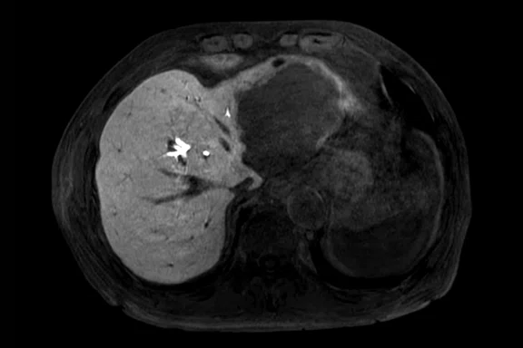
The morphology of the liver was within the normal range, and the main trunk to the right branch of the portal vein was patent. A hypervascular tumor approximately 10 cm was detected in the lateral segment of the left hepatic lobe (or S1 Spiegel), and hepatobiliary phase by EOB protocol confirmed that there was no uptake (Figure 2). The tumor has diffusion restrictions in DWI and tumor stiffness was confirmed by MR elastography.
Figure 2.
3D T1 with HyperSense and EOB protocol. Dynamic DISCO with HyperSense has a reduced temporal resolution of 3.4 sec. The hepatobiliary phase can be modified to high resolution using HyperSense to obtain detailed information. (A) Arterial phase, (B) portal phase and (C) late phase of the dynamic study using DISCO by EOB protocol, and (D) high-resolution navigated 3D T1 image with HyperSense at hepatobiliary phase.
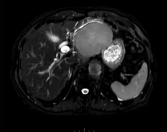
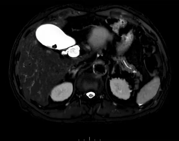
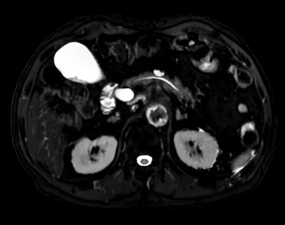
Multiple nodular EOB uptake reductions are observed at the edge of the large tumor, however intrahepatic metastasis (IM) was ruled out due to high ADC levels. There are multiple cysts in the lateral segment of the liver, but no findings suggestive of IM in other segments (Figure 3). Patient has a mildly fatty liver of about 5% using fat fraction, with very mild iron deposition and liver stiffness equivalent to F0 (Figure 4).
Corresponding laboratory results: high PIVKA II of 17078 and normal CEA/CA19-9/AFP.
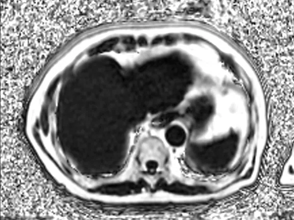
Figure 3.
Whole liver imaging with SSFSE T2, 3 mm thin slices. T2 SSFSE without blurring, providing clear morphology of intrahepatic bile ducts and portal/hepatic veins. Although it is a 3 mm thin slice, AIR™ Recon DL ensures sufficient SNR. With the wide coverage, this sequence can also be used for evaluating gallbladder stones and pancreatic cysts.
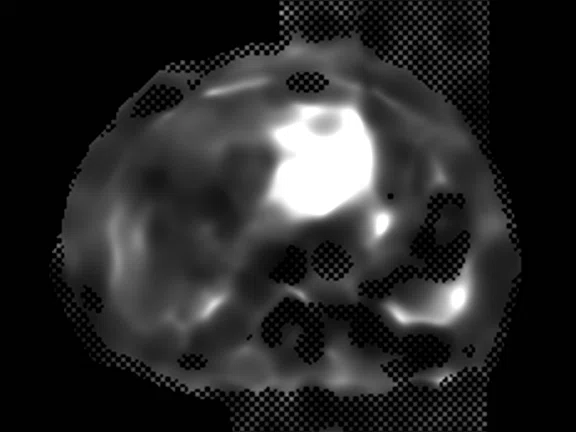
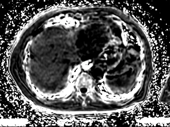
Figure 4.
Stiffness map showed normal stiffness in the liver, but high stiffness in the tumor, consistent with HCC. And, IDEAL-IQ has confirmed about 5% mild fatty liver by fat-fraction and mild iron deposition by R2* map.(A) MR elastography stiffness map and IDEAL IQ (B) fat fraction and (C) R2* map of the liver.
Discussion
A clinical diagnosis of hepatocellular carcinoma (HCC) without metastasis or vascular invasion was made, and the patient was scheduled for surgical resection. In this case, it was possible to identify the site of origin of the liver tumor with breath hold DWI using 5 mm thin slice. Although this tumor is located in the left lobe of the liver, it is not affected by cardiac motion. Due to the upgrade and DWI enhancement, we were able to obtain a very high-quality image. Furthermore, because this DWI acquisition covered the whole liver, the presence or absence of IM could also be diagnosed with certainty.
Additionally, T2 SSFSE was acquired using 3 mm thin slices; it is one of the imaging methods with image quality improved by AIR™ Recon DL. This SSFSE sequence reduces the matrix size to avoid T2 blurring, however the spatial resolution is maintained due to the sharpness improvement provided by AIR™ Recon DL.1 Thin slice SSFSE T2-weighted imaging facilitates the differentiation of peritumoral cysts from IM. With the wide coverage, this sequence can also be used for evaluating gallbladder stones and pancreatic cysts.
Furthermore, 3D T1 imaging improves the temporal resolution of dynamic studies using DISCO with HyperSense, which enables detailed diagnosis of hypervascular tumors. In addition, the hepatobiliary phase enables imaging with improved spatial/slice resolution, so detailed structures can be confirmed.
Due to the impact of HyperSense and AIR™ Recon DL in the SIGNA™Works AIR™ IQ Edition software, it is possible to obtain high-quality images without placing additional burden on the patient, which also impacts the quality and facilitates the radiologist’s interpretation.


