D
Figure #.
Figure Copy.
‡Not licensed in accordance with Canadian law. Not available for sale in Canada. Not CE marked. Not available for sale in all regions.
‡Not licensed in accordance with Canadian law. Not available for sale in Canada. Not CE marked. Not available for sale in all regions.
‡Not licensed in accordance with Canadian law. Not available for sale in Canada. Not CE marked. Not available for sale in all regions.
Not licensed in accordance with Canadian law. Not available for sale in Canada. Not CE marked. Not available for sale in all regions.
A
Figure 1.
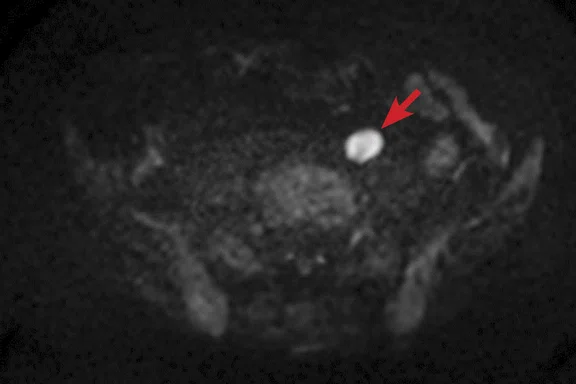
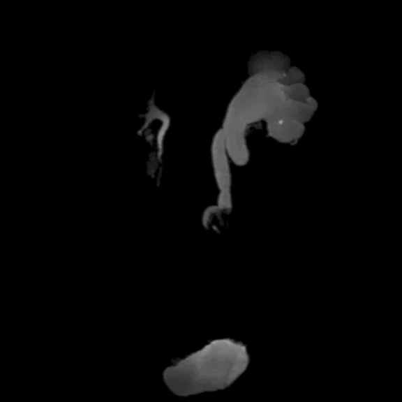
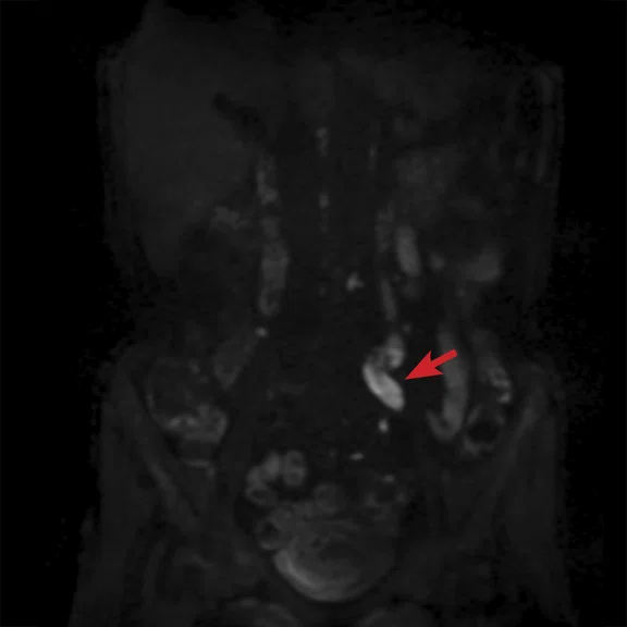
High-resolution MR Urography with reduced scan time is possible using HyperSense and HyperCube. A urethral tumor was visualized on the Coronal DWI using long axis. Even with an ASSET factor of 4.0, there was low distortion in the diffusion-weighted images. All images acquired with the AIR Technology™ AA. (A) Axial T2w SSFSE, 1.3 x 1.4 x 4 mm, 1:28 min.; (B) Axial DWI b1000, 2.7 x 2.7 x 4 mm, 3:57 min.; (C) MR urography, 0.7 x 1.2 x 1.4 mm, 2:40 min. (RTr); and (D) Coronal DWI b800, 3.1 x 1.6 x 4.5 mm, ASSET factor 4, 4:27 min. (RTr).
B
Figure 1.
High-resolution MR Urography with reduced scan time is possible using HyperSense and HyperCube. A urethral tumor was visualized on the Coronal DWI using long axis. Even with an ASSET factor of 4.0, there was low distortion in the diffusion-weighted images. All images acquired with the AIR Technology™ AA. (A) Axial T2w SSFSE, 1.3 x 1.4 x 4 mm, 1:28 min.; (B) Axial DWI b1000, 2.7 x 2.7 x 4 mm, 3:57 min.; (C) MR urography, 0.7 x 1.2 x 1.4 mm, 2:40 min. (RTr); and (D) Coronal DWI b800, 3.1 x 1.6 x 4.5 mm, ASSET factor 4, 4:27 min. (RTr).
C
Figure 1.
High-resolution MR Urography with reduced scan time is possible using HyperSense and HyperCube. A urethral tumor was visualized on the Coronal DWI using long axis. Even with an ASSET factor of 4.0, there was low distortion in the diffusion-weighted images. All images acquired with the AIR Technology™ AA. (A) Axial T2w SSFSE, 1.3 x 1.4 x 4 mm, 1:28 min.; (B) Axial DWI b1000, 2.7 x 2.7 x 4 mm, 3:57 min.; (C) MR urography, 0.7 x 1.2 x 1.4 mm, 2:40 min. (RTr); and (D) Coronal DWI b800, 3.1 x 1.6 x 4.5 mm, ASSET factor 4, 4:27 min. (RTr).
D
Figure 1.
High-resolution MR Urography with reduced scan time is possible using HyperSense and HyperCube. A urethral tumor was visualized on the Coronal DWI using long axis. Even with an ASSET factor of 4.0, there was low distortion in the diffusion-weighted images. All images acquired with the AIR Technology™ AA. (A) Axial T2w SSFSE, 1.3 x 1.4 x 4 mm, 1:28 min.; (B) Axial DWI b1000, 2.7 x 2.7 x 4 mm, 3:57 min.; (C) MR urography, 0.7 x 1.2 x 1.4 mm, 2:40 min. (RTr); and (D) Coronal DWI b800, 3.1 x 1.6 x 4.5 mm, ASSET factor 4, 4:27 min. (RTr).
A
Figure 2.
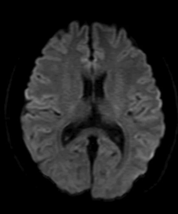
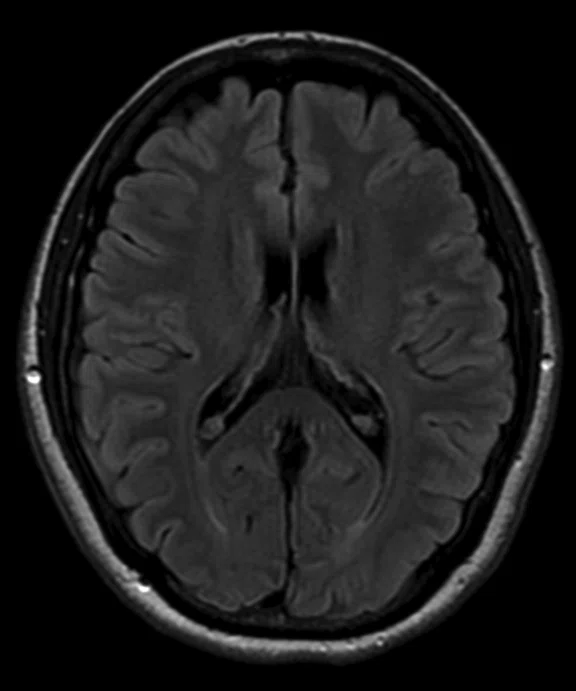
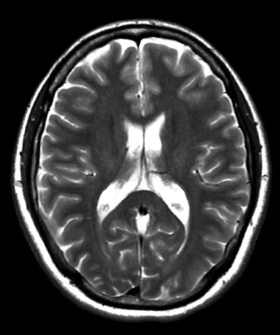
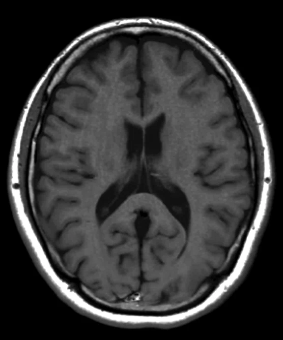
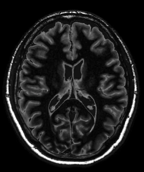
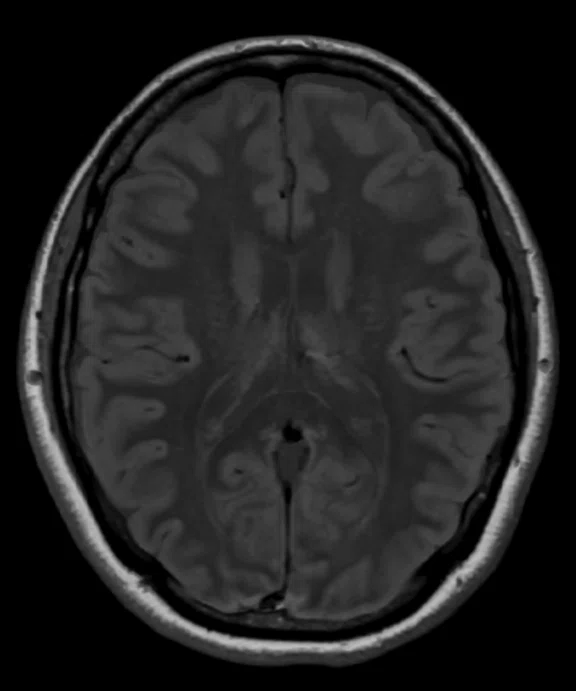
MAGiC is routinely used at Kawasaki Saiwai for neuro MR exams. The 48-channel Head Coil was used for this exam. (A) Axial DW-EPI in 45 sec. and (B) Axial T2 FLAIR in 3:07 min. Using MAGiC, from (A) and (B), the technologist can generate multiple contrasts, including (C) Axial T2w, (D) Axial T1w, (E) Axial DIR and (F) Axial PDw.
B
Figure 2.
MAGiC is routinely used at Kawasaki Saiwai for neuro MR exams. The 48-channel Head Coil was used for this exam. (A) Axial DW-EPI in 45 sec. and (B) Axial T2 FLAIR in 3:07 min. Using MAGiC, from (A) and (B), the technologist can generate multiple contrasts, including (C) Axial T2w, (D) Axial T1w, (E) Axial DIR and (F) Axial PDw.
C
Figure 2.
MAGiC is routinely used at Kawasaki Saiwai for neuro MR exams. The 48-channel Head Coil was used for this exam. (A) Axial DW-EPI in 45 sec. and (B) Axial T2 FLAIR in 3:07 min. Using MAGiC, from (A) and (B), the technologist can generate multiple contrasts, including (C) Axial T2w, (D) Axial T1w, (E) Axial DIR and (F) Axial PDw.
D
Figure 2.
MAGiC is routinely used at Kawasaki Saiwai for neuro MR exams. The 48-channel Head Coil was used for this exam. (A) Axial DW-EPI in 45 sec. and (B) Axial T2 FLAIR in 3:07 min. Using MAGiC, from (A) and (B), the technologist can generate multiple contrasts, including (C) Axial T2w, (D) Axial T1w, (E) Axial DIR and (F) Axial PDw.
E
Figure 2.
MAGiC is routinely used at Kawasaki Saiwai for neuro MR exams. The 48-channel Head Coil was used for this exam. (A) Axial DW-EPI in 45 sec. and (B) Axial T2 FLAIR in 3:07 min. Using MAGiC, from (A) and (B), the technologist can generate multiple contrasts, including (C) Axial T2w, (D) Axial T1w, (E) Axial DIR and (F) Axial PDw.
F
Figure 2.
MAGiC is routinely used at Kawasaki Saiwai for neuro MR exams. The 48-channel Head Coil was used for this exam. (A) Axial DW-EPI in 45 sec. and (B) Axial T2 FLAIR in 3:07 min. Using MAGiC, from (A) and (B), the technologist can generate multiple contrasts, including (C) Axial T2w, (D) Axial T1w, (E) Axial DIR and (F) Axial PDw.
A
Figure 3.
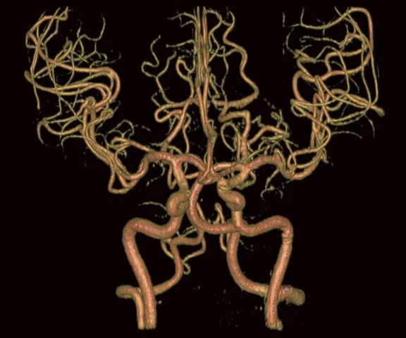
(A-C) TOF MRA with HyperSense factor of 2.5 and ARC factor of 2, 0.4 x 0.5 x 0.8 mm, 6:06 min.
B
Figure 3.
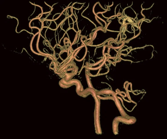
(A-C) TOF MRA with HyperSense factor of 2.5 and ARC factor of 2, 0.4 x 0.5 x 0.8 mm, 6:06 min.
C
Figure 3.
(A-C) TOF MRA with HyperSense factor of 2.5 and ARC factor of 2, 0.4 x 0.5 x 0.8 mm, 6:06 min.
A
Figure 4.
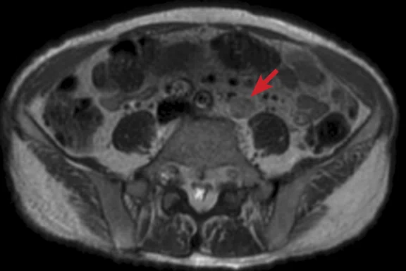
Patient weighed over 200 lbs. (120 kg). Abdominal kidney exam using the AIR Technology™ AA required a wide FOV, however, highly uniform images were acquired. (A) Axial T2w PROPELLER MB, 0.8 x 0.8 x 5 mm, ASSET 4.0, 5:39 min.; (B-D) Axial LAVA Flex; (B) water; (C) in-phase; (D) out-of-phase: 1.6 x 1.6 x 4 mm, 14 sec.; and (E) Coronal DWI b900, 3.1 x 1.6 x 5 mm, ASSET 4.0, 4:20 min.
B
Figure 4.
Patient weighed over 200 lbs. (120 kg). Abdominal kidney exam using the AIR Technology™ AA required a wide FOV, however, highly uniform images were acquired. (A) Axial T2w PROPELLER MB, 0.8 x 0.8 x 5 mm, ASSET 4.0, 5:39 min.; (B-D) Axial LAVA Flex; (B) water; (C) in-phase; (D) out-of-phase: 1.6 x 1.6 x 4 mm, 14 sec.; and (E) Coronal DWI b900, 3.1 x 1.6 x 5 mm, ASSET 4.0, 4:20 min.
E
Figure 4.
Patient weighed over 200 lbs. (120 kg). Abdominal kidney exam using the AIR Technology™ AA required a wide FOV, however, highly uniform images were acquired. (A) Axial T2w PROPELLER MB, 0.8 x 0.8 x 5 mm, ASSET 4.0, 5:39 min.; (B-D) Axial LAVA Flex; (B) water; (C) in-phase; (D) out-of-phase: 1.6 x 1.6 x 4 mm, 14 sec.; and (E) Coronal DWI b900, 3.1 x 1.6 x 5 mm, ASSET 4.0, 4:20 min.
C
Figure 4.
Patient weighed over 200 lbs. (120 kg). Abdominal kidney exam using the AIR Technology™ AA required a wide FOV, however, highly uniform images were acquired. (A) Axial T2w PROPELLER MB, 0.8 x 0.8 x 5 mm, ASSET 4.0, 5:39 min.; (B-D) Axial LAVA Flex; (B) water; (C) in-phase; (D) out-of-phase: 1.6 x 1.6 x 4 mm, 14 sec.; and (E) Coronal DWI b900, 3.1 x 1.6 x 5 mm, ASSET 4.0, 4:20 min.
D
Figure 4.
Patient weighed over 200 lbs. (120 kg). Abdominal kidney exam using the AIR Technology™ AA required a wide FOV, however, highly uniform images were acquired. (A) Axial T2w PROPELLER MB, 0.8 x 0.8 x 5 mm, ASSET 4.0, 5:39 min.; (B-D) Axial LAVA Flex; (B) water; (C) in-phase; (D) out-of-phase: 1.6 x 1.6 x 4 mm, 14 sec.; and (E) Coronal DWI b900, 3.1 x 1.6 x 5 mm, ASSET 4.0, 4:20 min.
A
Figure 5.
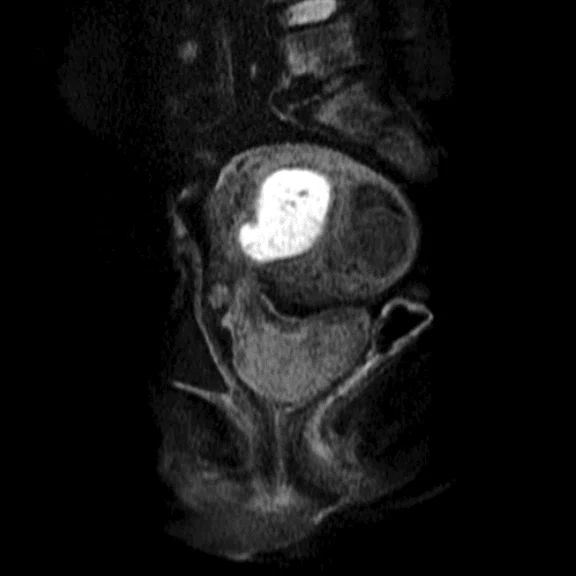
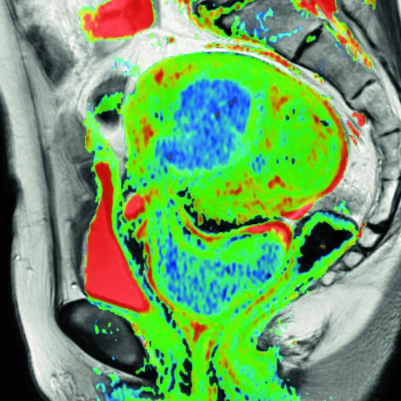
MUSE acquires high resolution DWI even at high b-values (b1000) and by using 4 shots with ARC acceleration of 1, distortion can be reduced. Fusing Sagittal T2w PROPELLER MB with the ADC map does not lead to distortion even in the presence of rectal gas. (A) Sagittal MUSE b1000, 1.5 x 0.9 x 6 mm, 4 shots with ARC acceleration of 1, 5 NEX, 4:39 min.; (B) Sagittal T2w PROPELLER MB, 0.8 x 0.8 x 6 mm, 4:12 min.; and (C) Fused ADC map with Sagittal T2w PROPELLER MB.
B
Figure 5.
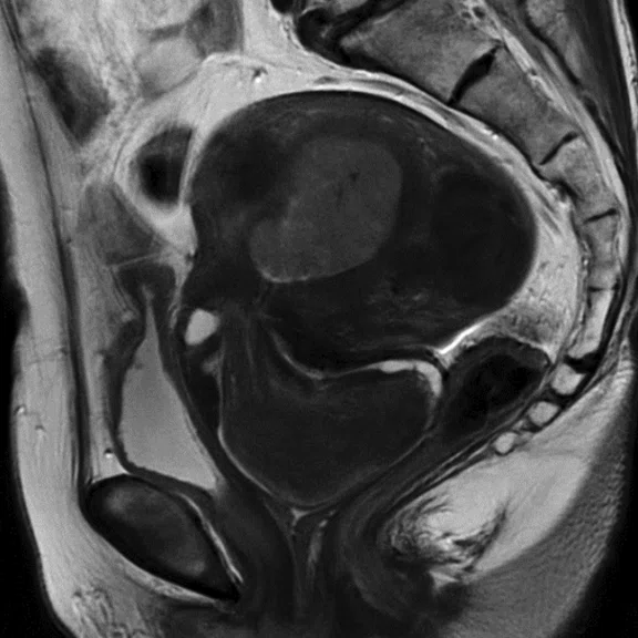
MUSE acquires high resolution DWI even at high b-values (b1000) and by using 4 shots with ARC acceleration of 1, distortion can be reduced. Fusing Sagittal T2w PROPELLER MB with the ADC map does not lead to distortion even in the presence of rectal gas. (A) Sagittal MUSE b1000, 1.5 x 0.9 x 6 mm, 4 shots with ARC acceleration of 1, 5 NEX, 4:39 min.; (B) Sagittal T2w PROPELLER MB, 0.8 x 0.8 x 6 mm, 4:12 min.; and (C) Fused ADC map with Sagittal T2w PROPELLER MB.
C
Figure 5.
MUSE acquires high resolution DWI even at high b-values (b1000) and by using 4 shots with ARC acceleration of 1, distortion can be reduced. Fusing Sagittal T2w PROPELLER MB with the ADC map does not lead to distortion even in the presence of rectal gas. (A) Sagittal MUSE b1000, 1.5 x 0.9 x 6 mm, 4 shots with ARC acceleration of 1, 5 NEX, 4:39 min.; (B) Sagittal T2w PROPELLER MB, 0.8 x 0.8 x 6 mm, 4:12 min.; and (C) Fused ADC map with Sagittal T2w PROPELLER MB.
A
Figure 6.
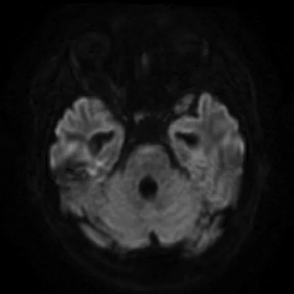
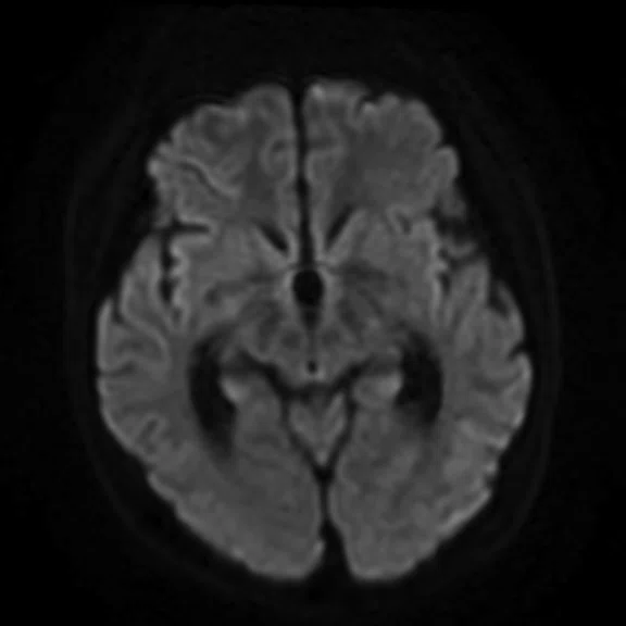
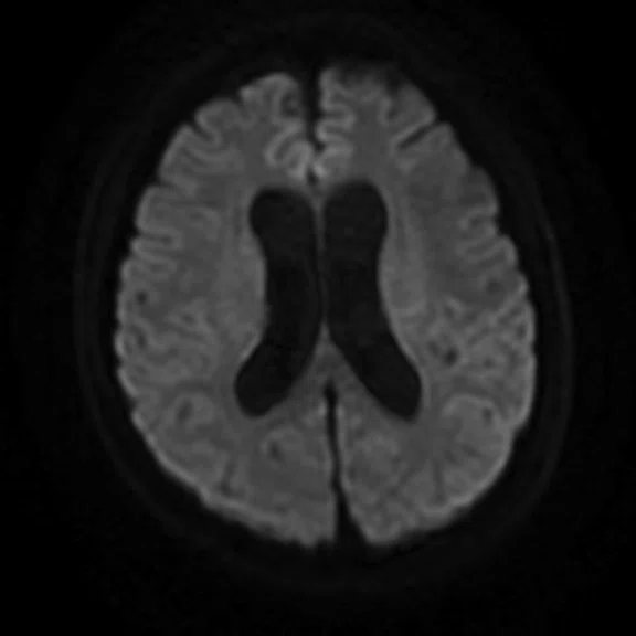
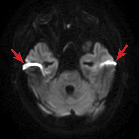
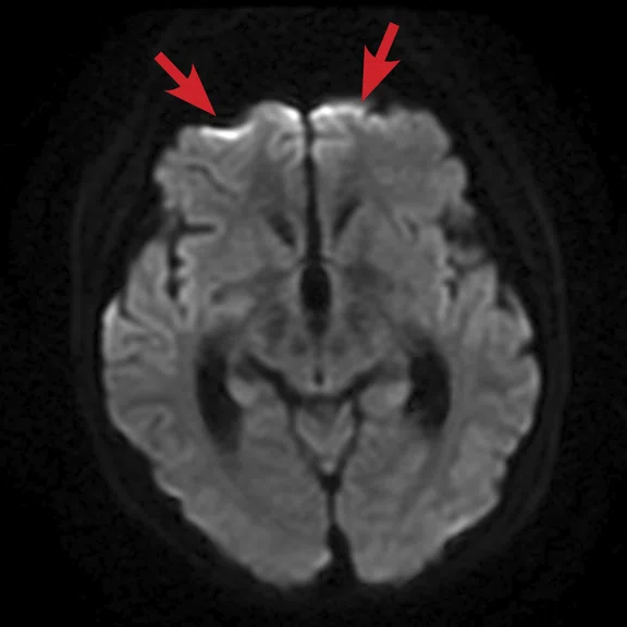
(A-C) DWI with PROGRES; and (D-F) conventional DWI. PROGRES provides less distortion than conventional DWI sequences, including fewer susceptibility artifacts (arrows) around the eye and inner ear.
B
Figure 6.
(A-C) DWI with PROGRES; and (D-F) conventional DWI. PROGRES provides less distortion than conventional DWI sequences, including fewer susceptibility artifacts (arrows) around the eye and inner ear.
C
Figure 6.
(A-C) DWI with PROGRES; and (D-F) conventional DWI. PROGRES provides less distortion than conventional DWI sequences, including fewer susceptibility artifacts (arrows) around the eye and inner ear.
D
Figure 6.
(A-C) DWI with PROGRES; and (D-F) conventional DWI. PROGRES provides less distortion than conventional DWI sequences, including fewer susceptibility artifacts (arrows) around the eye and inner ear.
E
Figure 6.
(A-C) DWI with PROGRES; and (D-F) conventional DWI. PROGRES provides less distortion than conventional DWI sequences, including fewer susceptibility artifacts (arrows) around the eye and inner ear.
F
Figure 6.
(A-C) DWI with PROGRES; and (D-F) conventional DWI. PROGRES provides less distortion than conventional DWI sequences, including fewer susceptibility artifacts (arrows) around the eye and inner ear.
Not licensed in accordance with Canadian law. Not available for sale in Canada. Not CE marked. Not available for sale in all regions.
A
Figure 2.
MAGiC is routinely used at Kawasaki Saiwai for neuro MR exams. The 48-channel Head Coil was used for this exam. (A) Axial DW-EPI in 45 sec. and (B) Axial T2 FLAIR in 3:07 min. Using MAGiC, from (A) and (B), the technologist can generate multiple contrasts, including (C) Axial T2w, (D) Axial T1w, (E) Axial DIR and (F) Axial PDw.
B
Figure 2.
MAGiC is routinely used at Kawasaki Saiwai for neuro MR exams. The 48-channel Head Coil was used for this exam. (A) Axial DW-EPI in 45 sec. and (B) Axial T2 FLAIR in 3:07 min. Using MAGiC, from (A) and (B), the technologist can generate multiple contrasts, including (C) Axial T2w, (D) Axial T1w, (E) Axial DIR and (F) Axial PDw.
C
Figure 2.
MAGiC is routinely used at Kawasaki Saiwai for neuro MR exams. The 48-channel Head Coil was used for this exam. (A) Axial DW-EPI in 45 sec. and (B) Axial T2 FLAIR in 3:07 min. Using MAGiC, from (A) and (B), the technologist can generate multiple contrasts, including (C) Axial T2w, (D) Axial T1w, (E) Axial DIR and (F) Axial PDw.
D
Figure 2.
MAGiC is routinely used at Kawasaki Saiwai for neuro MR exams. The 48-channel Head Coil was used for this exam. (A) Axial DW-EPI in 45 sec. and (B) Axial T2 FLAIR in 3:07 min. Using MAGiC, from (A) and (B), the technologist can generate multiple contrasts, including (C) Axial T2w, (D) Axial T1w, (E) Axial DIR and (F) Axial PDw.
E
Figure 2.
MAGiC is routinely used at Kawasaki Saiwai for neuro MR exams. The 48-channel Head Coil was used for this exam. (A) Axial DW-EPI in 45 sec. and (B) Axial T2 FLAIR in 3:07 min. Using MAGiC, from (A) and (B), the technologist can generate multiple contrasts, including (C) Axial T2w, (D) Axial T1w, (E) Axial DIR and (F) Axial PDw.
F
Figure 2.
MAGiC is routinely used at Kawasaki Saiwai for neuro MR exams. The 48-channel Head Coil was used for this exam. (A) Axial DW-EPI in 45 sec. and (B) Axial T2 FLAIR in 3:07 min. Using MAGiC, from (A) and (B), the technologist can generate multiple contrasts, including (C) Axial T2w, (D) Axial T1w, (E) Axial DIR and (F) Axial PDw.
result


PREVIOUS
${prev-page}
NEXT
${next-page}
Subscribe Now
Manage Subscription
FOLLOW US
Contact Us • Cookie Preferences • Privacy Policy • California Privacy PolicyDo Not Sell or Share My Personal Information • Terms & Conditions • Security
© 2024 GE HealthCare. GE is a trademark of General Electric Company. Used under trademark license.
IN PRACTICE
Ultra-flexible AIR Technology Suite making a difference in the technologist’s workflow
Ultra-flexible AIR Technology Suite making a difference in the technologist’s workflow
The AIR Technology™‡ Suite simplifies patient positioning and setup with AIR Touch™ automatic coil and element selection, a 60 percent lighter design than prior generations of coil technology and a flexible design that fits patients of various sizes and shapes.
The AIR Technology™‡ Suite simplifies patient positioning and setup with AIR Touch™ automatic coil and element selection, a 60 percent lighter design than prior generations of coil technology and a flexible design that fits patients of various sizes and shapes.
Kawasaki Saiwai Hospital in Kanagawa, Japan, installed the SIGNA™ Architect 3.0T MR system in late 2017. In February 2019, the hospital upgraded to the latest version of the SIGNA™Works productivity platform and acquired the AIR Technology™ Suite. As one of the main healthcare providers for the region, especially for acutely ill patients, the hospital embraces the concept of patient-centered healthcare. The AIR Technology™ Anterior Array (AA) conforms to the human body, is flexible to fit all shapes and sizes and has a 60 percent lighter design compared to previous generations of conventional coil technology—making it the ideal coil to deliver both patient comfort and high image quality at Kawasaki Saiwai Hospital.
Takafumi Naka, RT(R)(MR), Chief Technologist, evaluated the new system and coils and the impact on the technologist’s workflow and patient experience. He was most impressed that the AIR Technology™ AA is ultra-flexible and can be wrapped around the patient to facilitate positioning and fits a variety of patient body sizes.
In musculoskeletal (MSK) extremity imaging, coil selection for MR exams of the humerus and antebrachial bone would require two coils to image from the shoulder to the elbow. The AIR Technology™ AA, however, covers a larger region of interest (ROI) with a comfortable wrap-around fit and a higher SNR. Naka expects the same results in lower extremity imaging, particularly for patients with cellulitis and muscle contusions where large ROIs need to be acquired.
A conventional heavy, hard-shell coil on a patient’s chest could impact the respiratory detection device, so Naka would place a spacer between the coil and the patient.
"However, with the AIR Technology™ AA, we no longer have to do that," Naka says. "We now have better patient positioning workflow and also get an improvement in SNR because the coil is closer to the patient’s chest."
In addition, he does not have to worry about setting the coil center because AIR Touch™ automatically detects it, providing additional workflow improvements and removing the chance for human error.
Naka is also thrilled that he can use higher parallel imaging acceleration factors with the AIR Technology™ AA. In one case, he applied a factor of 4x for a body Coronal DWI and had less distortion and blurring than with a conventional coil (Figure 1).
Figure 2.
MAGiC is routinely used at Kawasaki Saiwai for neuro MR exams. The 48-channel Head Coil was used for this exam. (A) Axial DW-EPI in 45 sec. and (B) Axial T2 FLAIR in 3:07 min. Using MAGiC, from (A) and (B), the technologist can generate multiple contrasts, including (C) Axial T2w, (D) Axial T1w, (E) Axial DIR and (F) Axial PDw.
Figure 1.
High-resolution MR Urography with reduced scan time is possible using HyperSense and HyperCube. A urethral tumor was visualized on the Coronal DWI using long axis. Even with an ASSET factor of 4.0, there was low distortion in the diffusion-weighted images. All images acquired with the AIR Technology™ AA. (A) Axial T2w SSFSE, 1.3 x 1.4 x 4 mm, 1:28 min.; (B) Axial DWI b1000, 2.7 x 2.7 x 4 mm, 3:57 min.; (C) MR urography, 0.7 x 1.2 x 1.4 mm, 2:40 min. (RTr); and (D) Coronal DWI b800, 3.1 x 1.6 x 4.5 mm, ASSET factor 4, 4:27 min. (RTr).
IN PRACTICE
Ultra-flexible AIR Technology Suite making a difference in the technologist’s workflow
Ultra-flexible AIR Technology Suite making a difference in the technologist’s workflow
The AIR Technology™‡ Suite simplifies patient positioning and setup with AIR Touch™ automatic coil and element selection, a 60 percent lighter design than prior generations of coil technology and a flexible design that fits patients of various sizes and shapes.
The AIR Technology™‡ Suite simplifies patient positioning and setup with AIR Touch™ automatic coil and element selection, a 60 percent lighter design than prior generations of coil technology and a flexible design that fits patients of various sizes and shapes.
Kawasaki Saiwai Hospital in Kanagawa, Japan, installed the SIGNA™ Architect 3.0T MR system in late 2017. In February 2019, the hospital upgraded to the latest version of the SIGNA™Works productivity platform and acquired the AIR Technology™ Suite. As one of the main healthcare providers for the region, especially for acutely ill patients, the hospital embraces the concept of patient-centered healthcare. The AIR Technology™ Anterior Array (AA) conforms to the human body, is flexible to fit all shapes and sizes and has a 60 percent lighter design compared to previous generations of conventional coil technology—making it the ideal coil to deliver both patient comfort and high image quality at Kawasaki Saiwai Hospital.
Takafumi Naka, RT(R)(MR), Chief Technologist, evaluated the new system and coils and the impact on the technologist’s workflow and patient experience. He was most impressed that the AIR Technology™ AA is ultra-flexible and can be wrapped around the patient to facilitate positioning and fits a variety of patient body sizes.
In musculoskeletal (MSK) extremity imaging, coil selection for MR exams of the humerus and antebrachial bone would require two coils to image from the shoulder to the elbow. The AIR Technology™ AA, however, covers a larger region of interest (ROI) with a comfortable wrap-around fit and a higher SNR. Naka expects the same results in lower extremity imaging, particularly for patients with cellulitis and muscle contusions where large ROIs need to be acquired.
A conventional heavy, hard-shell coil on a patient’s chest could impact the respiratory detection device, so Naka would place a spacer between the coil and the patient.
"However, with the AIR Technology™ AA, we no longer have to do that," Naka says. "We now have better patient positioning workflow and also get an improvement in SNR because the coil is closer to the patient’s chest."
In addition, he does not have to worry about setting the coil center because AIR Touch™ automatically detects it, providing additional workflow improvements and removing the chance for human error.
Naka is also thrilled that he can use higher parallel imaging acceleration factors with the AIR Technology™ AA. In one case, he applied a factor of 4x for a body Coronal DWI and had less distortion and blurring than with a conventional coil (Figure 1).
"It was a single-shot EPI DWI, however, the anatomy detail and information was really amazing," he adds.
Naka has also used higher parallel imaging acceleration factors in body PROPELLER exams. He leveraged this capability for better image quality, such as refocusing the flip angle for higher T2 contrast. He sees the same impact in neuro imaging with the 48-channel Head Coil.
"We already use MAGiC in clinical routine neuro examinations to acquire excellent T1 contrast, which by principle is difficult to obtain at 3.0T," Naka says. "However, with the 48-channel Head Coil, we can reduce the scan time from six to three minutes because of the higher SNR," (Figure 2).
Plus, the 48-channel Head Coil allows Naka to use a higher HyperSense factor because of the high SNR and spatial resolution. As a result, he can now acquire a high-resolution MR angiography exam in six minutes—something that previously took approximately 20 minutes (Figure 3).
Figure 2.
MAGiC is routinely used at Kawasaki Saiwai for neuro MR exams. The 48-channel Head Coil was used for this exam. (A) Axial DW-EPI in 45 sec. and (B) Axial T2 FLAIR in 3:07 min. Using MAGiC, from (A) and (B), the technologist can generate multiple contrasts, including (C) Axial T2w, (D) Axial T1w, (E) Axial DIR and (F) Axial PDw.
He has noticed that patients are more relaxed with the AIR Technology™ AA than with conventional coils. Even large-sized patients weighing over 200 lbs. can fit comfortably inside the MR, with space between the coil and the bore (Figure 4).
Figure 4.
Patient weighed over 200 lbs. (120 kg). Abdominal kidney exam using the AIR Technology™ AA required a wide FOV, however, highly uniform images were acquired. (A) Axial T2w PROPELLER MB, 0.8 x 0.8 x 5 mm, ASSET 4.0, 5:39 min.; (B-D) Axial LAVA Flex; (B) water; (C) in-phase; (D) out-of-phase: 1.6 x 1.6 x 4 mm, 14 sec.; and (E) Coronal DWI b900, 3.1 x 1.6 x 5 mm, ASSET 4.0, 4:20 min.
Kawasaki Saiwai Hospital also installed the latest version of the SIGNA™Works productivity platform with SIGNA™ Architect. Naka loves the improvements in DWI, especially MUlti-plexed Sensitivity Encoding technique (MUSE) and PROGRES.
"The most impressive application is MUSE, a multi-shot DWI that allows us to achieve quite high spatial resolution compared to conventional DWI," he explains. "I find that MUSE DWI provides us completely different image quality versus the conventional sequence."
In particular, the improvement in female pelvis imaging is notable. MUSE DWI clearly depicts details of cervix and endometrial lesions when Naka sets the acquisition plane (slices) along the uterine axis (Figure 5).
Figure 5.
MUSE acquires high resolution DWI even at high b-values (b1000) and by using 4 shots with ARC acceleration of 1, distortion can be reduced. Fusing Sagittal T2w PROPELLER MB with the ADC map does not lead to distortion even in the presence of rectal gas. (A) Sagittal MUSE b1000, 1.5 x 0.9 x 6 mm, 4 shots with ARC acceleration of 1, 5 NEX, 4:39 min.; (B) Sagittal T2w PROPELLER MB, 0.8 x 0.8 x 6 mm, 4:12 min.; and (C) Fused ADC map with Sagittal T2w PROPELLER MB.
"We could see almost no distortion even in the Sagittal plane and there was less artifact from rectal gas," explains Naka. "Surprisingly, when we fuse MUSE DWI with T2-weighted images, we could not find misregistration caused by distortion. So, we think high-resolution MUSE DWI will have an advantage in detecting small lesions in the pancreas."
After investigating several sequences, Naka and his colleagues found PROGRES provided the best DWI image with the least distortion. Susceptibility artifacts around the eye and inner ear were decreased with no major impact on scan time when using PROGRES. As a result, PROGRES is being frequently used for neuro DWI at Kawasaki Saiwai Hospital (Figure 6).
From streamlined patient positioning to greater patient comfort, Naka sees the difference that AIR Technology™ has on the patient experience. For his department, the ability to use higher acceleration factors and save time in patient set-up will positively impact the technologist’s workflow, further improving staff satisfaction. And, with the new sequences available in SIGNA™Works, he and his team can deliver the excellent image quality clinicians need for a more confident diagnosis.


