A
Figure 3.
A 74-year-old male with known primary carcinoma, multiple myeloma. Post-treatment the patient was able to walk, however, he experienced pain in the lower spine. MR exam depicted (D, E) pronounced osteolytic lesions and (C, F) bone metastasis. AIR™ Recon DL, oZTEo and the 24-channel PA Coil were used for this exam. (A) Sagittal T2 FSE, 0.6 x 0.7 x 4.0 mm, 14 slices, 2:05 min.; (B) sagittal T1 FSE, 0.7 x 0.8 x 4.0 mm, 14 slices, 1:45 min.; (C) coronal 3D STIR with HyperCube, 1.6 x 1.6 x 1.0 mm, 150 slices, 2:31 min.; (D) coronal T1 FSE, 0.9 x 0.9 x 3.0 mm, 28 slices, 2:18 min.; (E) coronal STIR FSE, 1.0 x 1.0 x 3.0 mm, 28 slices, 2:42 min.; and (F) coronal 3D oZTEo, 1.4 x 1.4 x 1.4 mm, 140 slices, 4:57 min.
B
Figure 3.
A 74-year-old male with known primary carcinoma, multiple myeloma. Post-treatment the patient was able to walk, however, he experienced pain in the lower spine. MR exam depicted (D, E) pronounced osteolytic lesions and (C, F) bone metastasis. AIR™ Recon DL, oZTEo and the 24-channel PA Coil were used for this exam. (A) Sagittal T2 FSE, 0.6 x 0.7 x 4.0 mm, 14 slices, 2:05 min.; (B) sagittal T1 FSE, 0.7 x 0.8 x 4.0 mm, 14 slices, 1:45 min.; (C) coronal 3D STIR with HyperCube, 1.6 x 1.6 x 1.0 mm, 150 slices, 2:31 min.; (D) coronal T1 FSE, 0.9 x 0.9 x 3.0 mm, 28 slices, 2:18 min.; (E) coronal STIR FSE, 1.0 x 1.0 x 3.0 mm, 28 slices, 2:42 min.; and (F) coronal 3D oZTEo, 1.4 x 1.4 x 1.4 mm, 140 slices, 4:57 min.
C
Figure 3.
A 74-year-old male with known primary carcinoma, multiple myeloma. Post-treatment the patient was able to walk, however, he experienced pain in the lower spine. MR exam depicted (D, E) pronounced osteolytic lesions and (C, F) bone metastasis. AIR™ Recon DL, oZTEo and the 24-channel PA Coil were used for this exam. (A) Sagittal T2 FSE, 0.6 x 0.7 x 4.0 mm, 14 slices, 2:05 min.; (B) sagittal T1 FSE, 0.7 x 0.8 x 4.0 mm, 14 slices, 1:45 min.; (C) coronal 3D STIR with HyperCube, 1.6 x 1.6 x 1.0 mm, 150 slices, 2:31 min.; (D) coronal T1 FSE, 0.9 x 0.9 x 3.0 mm, 28 slices, 2:18 min.; (E) coronal STIR FSE, 1.0 x 1.0 x 3.0 mm, 28 slices, 2:42 min.; and (F) coronal 3D oZTEo, 1.4 x 1.4 x 1.4 mm, 140 slices, 4:57 min.
D
Figure 3.
A 74-year-old male with known primary carcinoma, multiple myeloma. Post-treatment the patient was able to walk, however, he experienced pain in the lower spine. MR exam depicted (D, E) pronounced osteolytic lesions and (C, F) bone metastasis. AIR™ Recon DL, oZTEo and the 24-channel PA Coil were used for this exam. (A) Sagittal T2 FSE, 0.6 x 0.7 x 4.0 mm, 14 slices, 2:05 min.; (B) sagittal T1 FSE, 0.7 x 0.8 x 4.0 mm, 14 slices, 1:45 min.; (C) coronal 3D STIR with HyperCube, 1.6 x 1.6 x 1.0 mm, 150 slices, 2:31 min.; (D) coronal T1 FSE, 0.9 x 0.9 x 3.0 mm, 28 slices, 2:18 min.; (E) coronal STIR FSE, 1.0 x 1.0 x 3.0 mm, 28 slices, 2:42 min.; and (F) coronal 3D oZTEo, 1.4 x 1.4 x 1.4 mm, 140 slices, 4:57 min.
E
Figure 3.
A 74-year-old male with known primary carcinoma, multiple myeloma. Post-treatment the patient was able to walk, however, he experienced pain in the lower spine. MR exam depicted (D, E) pronounced osteolytic lesions and (C, F) bone metastasis. AIR™ Recon DL, oZTEo and the 24-channel PA Coil were used for this exam. (A) Sagittal T2 FSE, 0.6 x 0.7 x 4.0 mm, 14 slices, 2:05 min.; (B) sagittal T1 FSE, 0.7 x 0.8 x 4.0 mm, 14 slices, 1:45 min.; (C) coronal 3D STIR with HyperCube, 1.6 x 1.6 x 1.0 mm, 150 slices, 2:31 min.; (D) coronal T1 FSE, 0.9 x 0.9 x 3.0 mm, 28 slices, 2:18 min.; (E) coronal STIR FSE, 1.0 x 1.0 x 3.0 mm, 28 slices, 2:42 min.; and (F) coronal 3D oZTEo, 1.4 x 1.4 x 1.4 mm, 140 slices, 4:57 min.
F
Figure 3.
A 74-year-old male with known primary carcinoma, multiple myeloma. Post-treatment the patient was able to walk, however, he experienced pain in the lower spine. MR exam depicted (D, E) pronounced osteolytic lesions and (C, F) bone metastasis. AIR™ Recon DL, oZTEo and the 24-channel PA Coil were used for this exam. (A) Sagittal T2 FSE, 0.6 x 0.7 x 4.0 mm, 14 slices, 2:05 min.; (B) sagittal T1 FSE, 0.7 x 0.8 x 4.0 mm, 14 slices, 1:45 min.; (C) coronal 3D STIR with HyperCube, 1.6 x 1.6 x 1.0 mm, 150 slices, 2:31 min.; (D) coronal T1 FSE, 0.9 x 0.9 x 3.0 mm, 28 slices, 2:18 min.; (E) coronal STIR FSE, 1.0 x 1.0 x 3.0 mm, 28 slices, 2:42 min.; and (F) coronal 3D oZTEo, 1.4 x 1.4 x 1.4 mm, 140 slices, 4:57 min.
A
Figure 4.
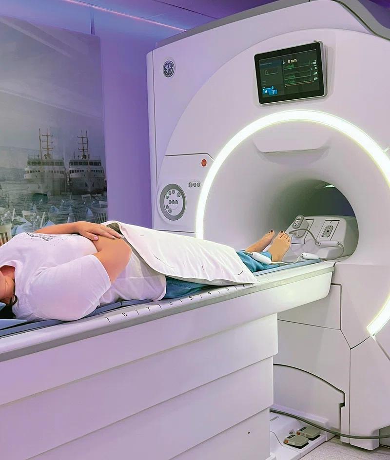
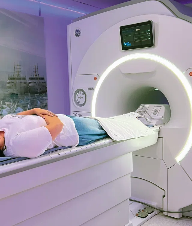
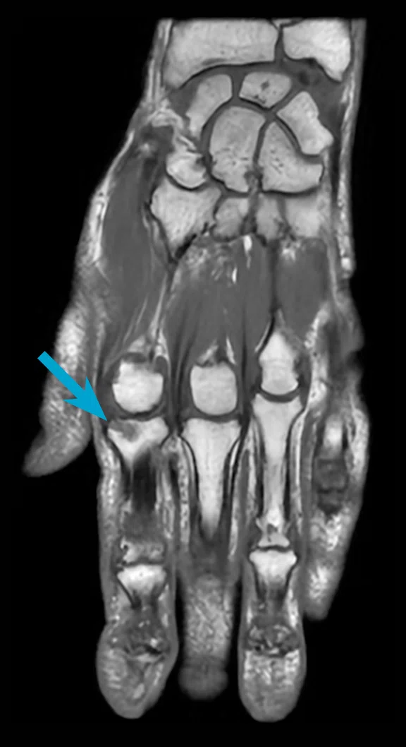
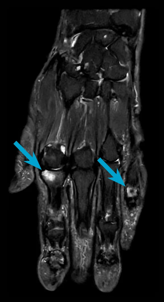
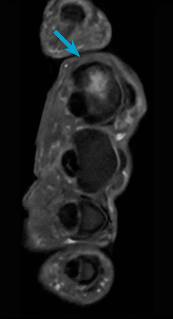
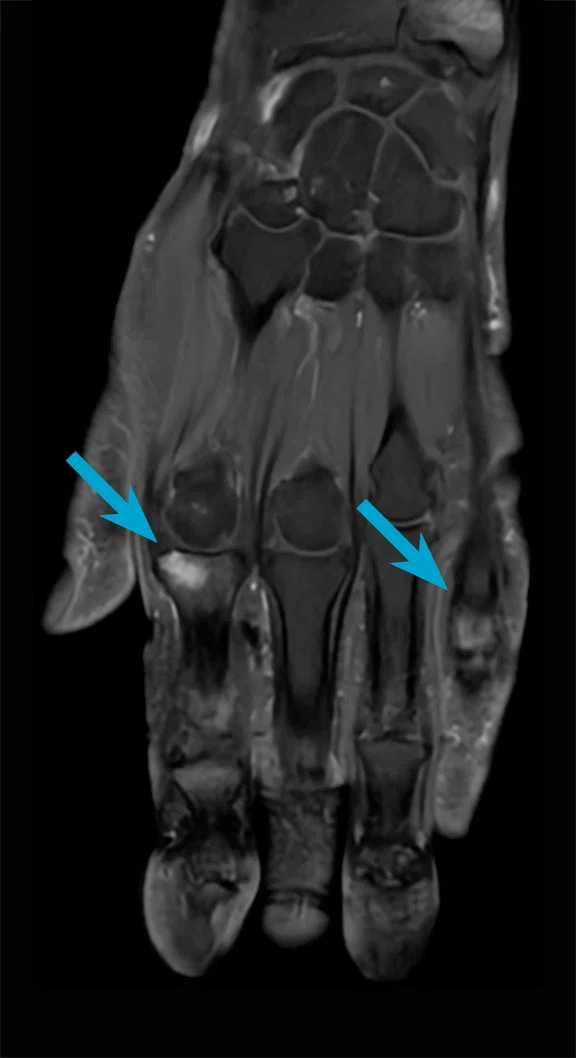
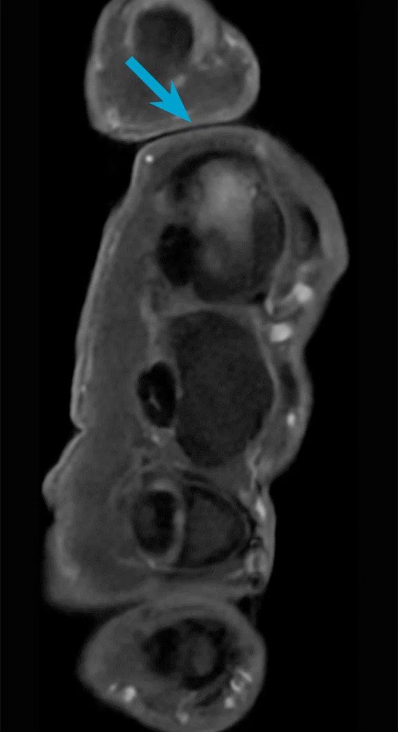
A 57-year-old female with chronic pain in the left metacarpophalangeal joint. MR exam depicted a defect zone of the radial extension hood at the level of the basal phalanx D II with accompanying stress reaction in the joint due to effusion formation. Rhizarthrosis in the thumb saddle joint with significant proximal malposition of the metacarpal D I was also depicted. AIR™ Recon DL and the 20-channel AIR™ MP Coil were used for this exam. The entire exam was 11:48 min., including contrast media administration. (A) Coronal T1 FSE, 0.8 x 0.8 x 2.0 mm, 18 slices, 1:11 min.; (B) coronal STIR FSE, 0.6 x 0.6 x 2.0 mm, 18 slices, 1:28 min.; (C) sagittal PD FS FSE, 0.8 x 0.8 x 2.0 mm, 42 slices, 2:32 min.; (D) axial PD FS FSE, 0.6 x 0.6 x 3.0 mm, 54 slices, 2:13 min.; (E) coronal T1 FS FSE +C, 0.8 x 0.8 x 2.0 mm, 18 slices, 1:33 min.; (F) axial T1 FS FSE +C, 0.5 x 0.5 x 3.0 mm, 54 slices, 2:51 min,; and (G) the 20-channel AIR™ MP Coil is wrapped around the patient’s hand, providing easy and comfortable patient positioning with very good SI coverage and homogeneous FatSat across the entire FOV.
B
Figure 4.
A 57-year-old female with chronic pain in the left metacarpophalangeal joint. MR exam depicted a defect zone of the radial extension hood at the level of the basal phalanx D II with accompanying stress reaction in the joint due to effusion formation. Rhizarthrosis in the thumb saddle joint with significant proximal malposition of the metacarpal D I was also depicted. AIR™ Recon DL and the 20-channel AIR™ MP Coil were used for this exam. The entire exam was 11:48 min., including contrast media administration. (A) Coronal T1 FSE, 0.8 x 0.8 x 2.0 mm, 18 slices, 1:11 min.; (B) coronal STIR FSE, 0.6 x 0.6 x 2.0 mm, 18 slices, 1:28 min.; (C) sagittal PD FS FSE, 0.8 x 0.8 x 2.0 mm, 42 slices, 2:32 min.; (D) axial PD FS FSE, 0.6 x 0.6 x 3.0 mm, 54 slices, 2:13 min.; (E) coronal T1 FS FSE +C, 0.8 x 0.8 x 2.0 mm, 18 slices, 1:33 min.; (F) axial T1 FS FSE +C, 0.5 x 0.5 x 3.0 mm, 54 slices, 2:51 min,; and (G) the 20-channel AIR™ MP Coil is wrapped around the patient’s hand, providing easy and comfortable patient positioning with very good SI coverage and homogeneous FatSat across the entire FOV.
C
Figure 4.
A 57-year-old female with chronic pain in the left metacarpophalangeal joint. MR exam depicted a defect zone of the radial extension hood at the level of the basal phalanx D II with accompanying stress reaction in the joint due to effusion formation. Rhizarthrosis in the thumb saddle joint with significant proximal malposition of the metacarpal D I was also depicted. AIR™ Recon DL and the 20-channel AIR™ MP Coil were used for this exam. The entire exam was 11:48 min., including contrast media administration. (A) Coronal T1 FSE, 0.8 x 0.8 x 2.0 mm, 18 slices, 1:11 min.; (B) coronal STIR FSE, 0.6 x 0.6 x 2.0 mm, 18 slices, 1:28 min.; (C) sagittal PD FS FSE, 0.8 x 0.8 x 2.0 mm, 42 slices, 2:32 min.; (D) axial PD FS FSE, 0.6 x 0.6 x 3.0 mm, 54 slices, 2:13 min.; (E) coronal T1 FS FSE +C, 0.8 x 0.8 x 2.0 mm, 18 slices, 1:33 min.; (F) axial T1 FS FSE +C, 0.5 x 0.5 x 3.0 mm, 54 slices, 2:51 min,; and (G) the 20-channel AIR™ MP Coil is wrapped around the patient’s hand, providing easy and comfortable patient positioning with very good SI coverage and homogeneous FatSat across the entire FOV.
D
Figure 4.
A 57-year-old female with chronic pain in the left metacarpophalangeal joint. MR exam depicted a defect zone of the radial extension hood at the level of the basal phalanx D II with accompanying stress reaction in the joint due to effusion formation. Rhizarthrosis in the thumb saddle joint with significant proximal malposition of the metacarpal D I was also depicted. AIR™ Recon DL and the 20-channel AIR™ MP Coil were used for this exam. The entire exam was 11:48 min., including contrast media administration. (A) Coronal T1 FSE, 0.8 x 0.8 x 2.0 mm, 18 slices, 1:11 min.; (B) coronal STIR FSE, 0.6 x 0.6 x 2.0 mm, 18 slices, 1:28 min.; (C) sagittal PD FS FSE, 0.8 x 0.8 x 2.0 mm, 42 slices, 2:32 min.; (D) axial PD FS FSE, 0.6 x 0.6 x 3.0 mm, 54 slices, 2:13 min.; (E) coronal T1 FS FSE +C, 0.8 x 0.8 x 2.0 mm, 18 slices, 1:33 min.; (F) axial T1 FS FSE +C, 0.5 x 0.5 x 3.0 mm, 54 slices, 2:51 min,; and (G) the 20-channel AIR™ MP Coil is wrapped around the patient’s hand, providing easy and comfortable patient positioning with very good SI coverage and homogeneous FatSat across the entire FOV.
E
Figure 4.
A 57-year-old female with chronic pain in the left metacarpophalangeal joint. MR exam depicted a defect zone of the radial extension hood at the level of the basal phalanx D II with accompanying stress reaction in the joint due to effusion formation. Rhizarthrosis in the thumb saddle joint with significant proximal malposition of the metacarpal D I was also depicted. AIR™ Recon DL and the 20-channel AIR™ MP Coil were used for this exam. The entire exam was 11:48 min., including contrast media administration. (A) Coronal T1 FSE, 0.8 x 0.8 x 2.0 mm, 18 slices, 1:11 min.; (B) coronal STIR FSE, 0.6 x 0.6 x 2.0 mm, 18 slices, 1:28 min.; (C) sagittal PD FS FSE, 0.8 x 0.8 x 2.0 mm, 42 slices, 2:32 min.; (D) axial PD FS FSE, 0.6 x 0.6 x 3.0 mm, 54 slices, 2:13 min.; (E) coronal T1 FS FSE +C, 0.8 x 0.8 x 2.0 mm, 18 slices, 1:33 min.; (F) axial T1 FS FSE +C, 0.5 x 0.5 x 3.0 mm, 54 slices, 2:51 min,; and (G) the 20-channel AIR™ MP Coil is wrapped around the patient’s hand, providing easy and comfortable patient positioning with very good SI coverage and homogeneous FatSat across the entire FOV.
F
Figure 4.
A 57-year-old female with chronic pain in the left metacarpophalangeal joint. MR exam depicted a defect zone of the radial extension hood at the level of the basal phalanx D II with accompanying stress reaction in the joint due to effusion formation. Rhizarthrosis in the thumb saddle joint with significant proximal malposition of the metacarpal D I was also depicted. AIR™ Recon DL and the 20-channel AIR™ MP Coil were used for this exam. The entire exam was 11:48 min., including contrast media administration. (A) Coronal T1 FSE, 0.8 x 0.8 x 2.0 mm, 18 slices, 1:11 min.; (B) coronal STIR FSE, 0.6 x 0.6 x 2.0 mm, 18 slices, 1:28 min.; (C) sagittal PD FS FSE, 0.8 x 0.8 x 2.0 mm, 42 slices, 2:32 min.; (D) axial PD FS FSE, 0.6 x 0.6 x 3.0 mm, 54 slices, 2:13 min.; (E) coronal T1 FS FSE +C, 0.8 x 0.8 x 2.0 mm, 18 slices, 1:33 min.; (F) axial T1 FS FSE +C, 0.5 x 0.5 x 3.0 mm, 54 slices, 2:51 min,; and (G) the 20-channel AIR™ MP Coil is wrapped around the patient’s hand, providing easy and comfortable patient positioning with very good SI coverage and homogeneous FatSat across the entire FOV.
G-1
Figure 4.
A 57-year-old female with chronic pain in the left metacarpophalangeal joint. MR exam depicted a defect zone of the radial extension hood at the level of the basal phalanx D II with accompanying stress reaction in the joint due to effusion formation. Rhizarthrosis in the thumb saddle joint with significant proximal malposition of the metacarpal D I was also depicted. AIR™ Recon DL and the 20-channel AIR™ MP Coil were used for this exam. The entire exam was 11:48 min., including contrast media administration. (A) Coronal T1 FSE, 0.8 x 0.8 x 2.0 mm, 18 slices, 1:11 min.; (B) coronal STIR FSE, 0.6 x 0.6 x 2.0 mm, 18 slices, 1:28 min.; (C) sagittal PD FS FSE, 0.8 x 0.8 x 2.0 mm, 42 slices, 2:32 min.; (D) axial PD FS FSE, 0.6 x 0.6 x 3.0 mm, 54 slices, 2:13 min.; (E) coronal T1 FS FSE +C, 0.8 x 0.8 x 2.0 mm, 18 slices, 1:33 min.; (F) axial T1 FS FSE +C, 0.5 x 0.5 x 3.0 mm, 54 slices, 2:51 min,; and (G) the 20-channel AIR™ MP Coil is wrapped around the patient’s hand, providing easy and comfortable patient positioning with very good SI coverage and homogeneous FatSat across the entire FOV.
G-2
Figure 4.
A 57-year-old female with chronic pain in the left metacarpophalangeal joint. MR exam depicted a defect zone of the radial extension hood at the level of the basal phalanx D II with accompanying stress reaction in the joint due to effusion formation. Rhizarthrosis in the thumb saddle joint with significant proximal malposition of the metacarpal D I was also depicted. AIR™ Recon DL and the 20-channel AIR™ MP Coil were used for this exam. The entire exam was 11:48 min., including contrast media administration. (A) Coronal T1 FSE, 0.8 x 0.8 x 2.0 mm, 18 slices, 1:11 min.; (B) coronal STIR FSE, 0.6 x 0.6 x 2.0 mm, 18 slices, 1:28 min.; (C) sagittal PD FS FSE, 0.8 x 0.8 x 2.0 mm, 42 slices, 2:32 min.; (D) axial PD FS FSE, 0.6 x 0.6 x 3.0 mm, 54 slices, 2:13 min.; (E) coronal T1 FS FSE +C, 0.8 x 0.8 x 2.0 mm, 18 slices, 1:33 min.; (F) axial T1 FS FSE +C, 0.5 x 0.5 x 3.0 mm, 54 slices, 2:51 min,; and (G) the 20-channel AIR™ MP Coil is wrapped around the patient’s hand, providing easy and comfortable patient positioning with very good SI coverage and homogeneous FatSat across the entire FOV.
A
Figure 1.
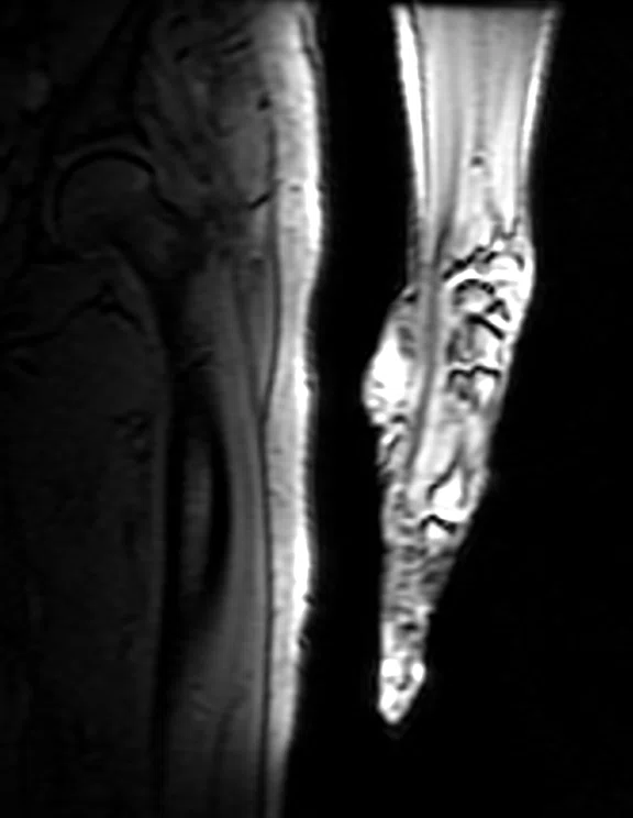
A 67-year-old female with a fatty mass in the distal thigh (A, arrow) between the femur and the quadriceps femoris muscle (B-D, arrows). MR exam showed signs of lipoma (F, arrow). AIR™ Recon DL, the 16-channel AIR™ AA Coil and the 24-channel PA Coil were used for this exam. (A) Coronal STIR FSE, 1.0 x 1.0 x 5.0 mm, 36 slices, 4:25 min.; (B) sagittal STIR FSE, 1.0 x 1.0 x 5.0 mm, 36 slices, 3:52 min.; (C) sagittal T1 FSE, 0.9 x 0.9 x 5.0 mm, 36 slices, 3:58 min.; (D) axial PD FS FSE, 1.0 x 1.0 x 5.0 mm, 42 slices, 1:22 min.; (E) coronal 3D LAVA Flex +C, 1.4 x 1.4 x 3.0 mm, 66 slices, 3:33 min.; and (F) sagittal 3D LAVA Flex +C, 1.0 x 1.0 x 2.0 mm, 100 slices, 3:08 min.
B-1
Figure 1.
A 67-year-old female with a fatty mass in the distal thigh (A, arrow) between the femur and the quadriceps femoris muscle (B-D, arrows). MR exam showed signs of lipoma (F, arrow). AIR™ Recon DL, the 16-channel AIR™ AA Coil and the 24-channel PA Coil were used for this exam. (A) Coronal STIR FSE, 1.0 x 1.0 x 5.0 mm, 36 slices, 4:25 min.; (B) sagittal STIR FSE, 1.0 x 1.0 x 5.0 mm, 36 slices, 3:52 min.; (C) sagittal T1 FSE, 0.9 x 0.9 x 5.0 mm, 36 slices, 3:58 min.; (D) axial PD FS FSE, 1.0 x 1.0 x 5.0 mm, 42 slices, 1:22 min.; (E) coronal 3D LAVA Flex +C, 1.4 x 1.4 x 3.0 mm, 66 slices, 3:33 min.; and (F) sagittal 3D LAVA Flex +C, 1.0 x 1.0 x 2.0 mm, 100 slices, 3:08 min.
C-1
Figure 1.
A 67-year-old female with a fatty mass in the distal thigh (A, arrow) between the femur and the quadriceps femoris muscle (B-D, arrows). MR exam showed signs of lipoma (F, arrow). AIR™ Recon DL, the 16-channel AIR™ AA Coil and the 24-channel PA Coil were used for this exam. (A) Coronal STIR FSE, 1.0 x 1.0 x 5.0 mm, 36 slices, 4:25 min.; (B) sagittal STIR FSE, 1.0 x 1.0 x 5.0 mm, 36 slices, 3:52 min.; (C) sagittal T1 FSE, 0.9 x 0.9 x 5.0 mm, 36 slices, 3:58 min.; (D) axial PD FS FSE, 1.0 x 1.0 x 5.0 mm, 42 slices, 1:22 min.; (E) coronal 3D LAVA Flex +C, 1.4 x 1.4 x 3.0 mm, 66 slices, 3:33 min.; and (F) sagittal 3D LAVA Flex +C, 1.0 x 1.0 x 2.0 mm, 100 slices, 3:08 min.
D
Figure 1.
A 67-year-old female with a fatty mass in the distal thigh (A, arrow) between the femur and the quadriceps femoris muscle (B-D, arrows). MR exam showed signs of lipoma (F, arrow). AIR™ Recon DL, the 16-channel AIR™ AA Coil and the 24-channel PA Coil were used for this exam. (A) Coronal STIR FSE, 1.0 x 1.0 x 5.0 mm, 36 slices, 4:25 min.; (B) sagittal STIR FSE, 1.0 x 1.0 x 5.0 mm, 36 slices, 3:52 min.; (C) sagittal T1 FSE, 0.9 x 0.9 x 5.0 mm, 36 slices, 3:58 min.; (D) axial PD FS FSE, 1.0 x 1.0 x 5.0 mm, 42 slices, 1:22 min.; (E) coronal 3D LAVA Flex +C, 1.4 x 1.4 x 3.0 mm, 66 slices, 3:33 min.; and (F) sagittal 3D LAVA Flex +C, 1.0 x 1.0 x 2.0 mm, 100 slices, 3:08 min.
E
Figure 1.
A 67-year-old female with a fatty mass in the distal thigh (A, arrow) between the femur and the quadriceps femoris muscle (B-D, arrows). MR exam showed signs of lipoma (F, arrow). AIR™ Recon DL, the 16-channel AIR™ AA Coil and the 24-channel PA Coil were used for this exam. (A) Coronal STIR FSE, 1.0 x 1.0 x 5.0 mm, 36 slices, 4:25 min.; (B) sagittal STIR FSE, 1.0 x 1.0 x 5.0 mm, 36 slices, 3:52 min.; (C) sagittal T1 FSE, 0.9 x 0.9 x 5.0 mm, 36 slices, 3:58 min.; (D) axial PD FS FSE, 1.0 x 1.0 x 5.0 mm, 42 slices, 1:22 min.; (E) coronal 3D LAVA Flex +C, 1.4 x 1.4 x 3.0 mm, 66 slices, 3:33 min.; and (F) sagittal 3D LAVA Flex +C, 1.0 x 1.0 x 2.0 mm, 100 slices, 3:08 min.
F-1
Figure 1.
A 67-year-old female with a fatty mass in the distal thigh (A, arrow) between the femur and the quadriceps femoris muscle (B-D, arrows). MR exam showed signs of lipoma (F, arrow). AIR™ Recon DL, the 16-channel AIR™ AA Coil and the 24-channel PA Coil were used for this exam. (A) Coronal STIR FSE, 1.0 x 1.0 x 5.0 mm, 36 slices, 4:25 min.; (B) sagittal STIR FSE, 1.0 x 1.0 x 5.0 mm, 36 slices, 3:52 min.; (C) sagittal T1 FSE, 0.9 x 0.9 x 5.0 mm, 36 slices, 3:58 min.; (D) axial PD FS FSE, 1.0 x 1.0 x 5.0 mm, 42 slices, 1:22 min.; (E) coronal 3D LAVA Flex +C, 1.4 x 1.4 x 3.0 mm, 66 slices, 3:33 min.; and (F) sagittal 3D LAVA Flex +C, 1.0 x 1.0 x 2.0 mm, 100 slices, 3:08 min.
F-2
Figure 1.
A 67-year-old female with a fatty mass in the distal thigh (A, arrow) between the femur and the quadriceps femoris muscle (B-D, arrows). MR exam showed signs of lipoma (F, arrow). AIR™ Recon DL, the 16-channel AIR™ AA Coil and the 24-channel PA Coil were used for this exam. (A) Coronal STIR FSE, 1.0 x 1.0 x 5.0 mm, 36 slices, 4:25 min.; (B) sagittal STIR FSE, 1.0 x 1.0 x 5.0 mm, 36 slices, 3:52 min.; (C) sagittal T1 FSE, 0.9 x 0.9 x 5.0 mm, 36 slices, 3:58 min.; (D) axial PD FS FSE, 1.0 x 1.0 x 5.0 mm, 42 slices, 1:22 min.; (E) coronal 3D LAVA Flex +C, 1.4 x 1.4 x 3.0 mm, 66 slices, 3:33 min.; and (F) sagittal 3D LAVA Flex +C, 1.0 x 1.0 x 2.0 mm, 100 slices, 3:08 min.
B-2
Figure 1.
A 67-year-old female with a fatty mass in the distal thigh (A, arrow) between the femur and the quadriceps femoris muscle (B-D, arrows). MR exam showed signs of lipoma (F, arrow). AIR™ Recon DL, the 16-channel AIR™ AA Coil and the 24-channel PA Coil were used for this exam. (A) Coronal STIR FSE, 1.0 x 1.0 x 5.0 mm, 36 slices, 4:25 min.; (B) sagittal STIR FSE, 1.0 x 1.0 x 5.0 mm, 36 slices, 3:52 min.; (C) sagittal T1 FSE, 0.9 x 0.9 x 5.0 mm, 36 slices, 3:58 min.; (D) axial PD FS FSE, 1.0 x 1.0 x 5.0 mm, 42 slices, 1:22 min.; (E) coronal 3D LAVA Flex +C, 1.4 x 1.4 x 3.0 mm, 66 slices, 3:33 min.; and (F) sagittal 3D LAVA Flex +C, 1.0 x 1.0 x 2.0 mm, 100 slices, 3:08 min.
C-2
Figure 1.
A 67-year-old female with a fatty mass in the distal thigh (A, arrow) between the femur and the quadriceps femoris muscle (B-D, arrows). MR exam showed signs of lipoma (F, arrow). AIR™ Recon DL, the 16-channel AIR™ AA Coil and the 24-channel PA Coil were used for this exam. (A) Coronal STIR FSE, 1.0 x 1.0 x 5.0 mm, 36 slices, 4:25 min.; (B) sagittal STIR FSE, 1.0 x 1.0 x 5.0 mm, 36 slices, 3:52 min.; (C) sagittal T1 FSE, 0.9 x 0.9 x 5.0 mm, 36 slices, 3:58 min.; (D) axial PD FS FSE, 1.0 x 1.0 x 5.0 mm, 42 slices, 1:22 min.; (E) coronal 3D LAVA Flex +C, 1.4 x 1.4 x 3.0 mm, 66 slices, 3:33 min.; and (F) sagittal 3D LAVA Flex +C, 1.0 x 1.0 x 2.0 mm, 100 slices, 3:08 min.
A
Figure 2.
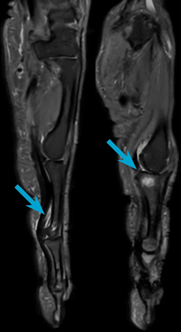
A 48-year-old female with multiple desmoids who underwent a biopsy on her left lower leg. There was an increase in the size and inhomogeneous mass of the caput laterale of the gastrocnemius muscle (arrows). AIR™ Recon DL, the 16-channel AIR™ AA Coil and the 24-channel PA Coil were used for this exam. (A) Coronal STIR FSE, 1.0 x 1.0 x 4.0 mm, 25 slices, 3:04 min.; (B) sagittal STIR FSE, 1.0 x 1.0 x 4.0 mm, 26 slices, 2:47 min.; (C) axial PD FS FSE, 0.9 x 0.9 x 5.0 mm, 76 slices, 4:18 min.; (D) axial DWI, (top, ADC; middle, synthetic b1400; bottom, b800), 3.0 x 2.4 x 6.0 mm, 60 slices, 4:27 min.; (E) axial 3D LAVA Flex +C, 1.0 x 1.0 x 5.0 mm, 80 slices, 1:34 min.; and (E) sagittal 3D LAVA Flex +C, 1.3 x 1.3 x 2.0 mm, 60 slices, 2:53 min.
B
Figure 2.
A 48-year-old female with multiple desmoids who underwent a biopsy on her left lower leg. There was an increase in the size and inhomogeneous mass of the caput laterale of the gastrocnemius muscle (arrows). AIR™ Recon DL, the 16-channel AIR™ AA Coil and the 24-channel PA Coil were used for this exam. (A) Coronal STIR FSE, 1.0 x 1.0 x 4.0 mm, 25 slices, 3:04 min.; (B) sagittal STIR FSE, 1.0 x 1.0 x 4.0 mm, 26 slices, 2:47 min.; (C) axial PD FS FSE, 0.9 x 0.9 x 5.0 mm, 76 slices, 4:18 min.; (D) axial DWI, (top, ADC; middle, synthetic b1400; bottom, b800), 3.0 x 2.4 x 6.0 mm, 60 slices, 4:27 min.; (E) axial 3D LAVA Flex +C, 1.0 x 1.0 x 5.0 mm, 80 slices, 1:34 min.; and (E) sagittal 3D LAVA Flex +C, 1.3 x 1.3 x 2.0 mm, 60 slices, 2:53 min.
C
Figure 2.
A 48-year-old female with multiple desmoids who underwent a biopsy on her left lower leg. There was an increase in the size and inhomogeneous mass of the caput laterale of the gastrocnemius muscle (arrows). AIR™ Recon DL, the 16-channel AIR™ AA Coil and the 24-channel PA Coil were used for this exam. (A) Coronal STIR FSE, 1.0 x 1.0 x 4.0 mm, 25 slices, 3:04 min.; (B) sagittal STIR FSE, 1.0 x 1.0 x 4.0 mm, 26 slices, 2:47 min.; (C) axial PD FS FSE, 0.9 x 0.9 x 5.0 mm, 76 slices, 4:18 min.; (D) axial DWI, (top, ADC; middle, synthetic b1400; bottom, b800), 3.0 x 2.4 x 6.0 mm, 60 slices, 4:27 min.; (E) axial 3D LAVA Flex +C, 1.0 x 1.0 x 5.0 mm, 80 slices, 1:34 min.; and (E) sagittal 3D LAVA Flex +C, 1.3 x 1.3 x 2.0 mm, 60 slices, 2:53 min.
D
Figure 2.
A 48-year-old female with multiple desmoids who underwent a biopsy on her left lower leg. There was an increase in the size and inhomogeneous mass of the caput laterale of the gastrocnemius muscle (arrows). AIR™ Recon DL, the 16-channel AIR™ AA Coil and the 24-channel PA Coil were used for this exam. (A) Coronal STIR FSE, 1.0 x 1.0 x 4.0 mm, 25 slices, 3:04 min.; (B) sagittal STIR FSE, 1.0 x 1.0 x 4.0 mm, 26 slices, 2:47 min.; (C) axial PD FS FSE, 0.9 x 0.9 x 5.0 mm, 76 slices, 4:18 min.; (D) axial DWI, (top, ADC; middle, synthetic b1400; bottom, b800), 3.0 x 2.4 x 6.0 mm, 60 slices, 4:27 min.; (E) axial 3D LAVA Flex +C, 1.0 x 1.0 x 5.0 mm, 80 slices, 1:34 min.; and (E) sagittal 3D LAVA Flex +C, 1.3 x 1.3 x 2.0 mm, 60 slices, 2:53 min.
E
Figure 2.
A 48-year-old female with multiple desmoids who underwent a biopsy on her left lower leg. There was an increase in the size and inhomogeneous mass of the caput laterale of the gastrocnemius muscle (arrows). AIR™ Recon DL, the 16-channel AIR™ AA Coil and the 24-channel PA Coil were used for this exam. (A) Coronal STIR FSE, 1.0 x 1.0 x 4.0 mm, 25 slices, 3:04 min.; (B) sagittal STIR FSE, 1.0 x 1.0 x 4.0 mm, 26 slices, 2:47 min.; (C) axial PD FS FSE, 0.9 x 0.9 x 5.0 mm, 76 slices, 4:18 min.; (D) axial DWI, (top, ADC; middle, synthetic b1400; bottom, b800), 3.0 x 2.4 x 6.0 mm, 60 slices, 4:27 min.; (E) axial 3D LAVA Flex +C, 1.0 x 1.0 x 5.0 mm, 80 slices, 1:34 min.; and (E) sagittal 3D LAVA Flex +C, 1.3 x 1.3 x 2.0 mm, 60 slices, 2:53 min.
F
Figure 2.
A 48-year-old female with multiple desmoids who underwent a biopsy on her left lower leg. There was an increase in the size and inhomogeneous mass of the caput laterale of the gastrocnemius muscle (arrows). AIR™ Recon DL, the 16-channel AIR™ AA Coil and the 24-channel PA Coil were used for this exam. (A) Coronal STIR FSE, 1.0 x 1.0 x 4.0 mm, 25 slices, 3:04 min.; (B) sagittal STIR FSE, 1.0 x 1.0 x 4.0 mm, 26 slices, 2:47 min.; (C) axial PD FS FSE, 0.9 x 0.9 x 5.0 mm, 76 slices, 4:18 min.; (D) axial DWI, (top, ADC; middle, synthetic b1400; bottom, b800), 3.0 x 2.4 x 6.0 mm, 60 slices, 4:27 min.; (E) axial 3D LAVA Flex +C, 1.0 x 1.0 x 5.0 mm, 80 slices, 1:34 min.; and (E) sagittal 3D LAVA Flex +C, 1.3 x 1.3 x 2.0 mm, 60 slices, 2:53 min.
result


PREVIOUS
${prev-page}
NEXT
${next-page}


Subscribe Now
Manage Subscription
FOLLOW US
Contact Us • Cookie Preferences • Privacy Policy • California Privacy PolicyDo Not Sell or Share My Personal Information • Terms & Conditions • Security
© 2024 GE HealthCare. GE is a trademark of General Electric Company. Used under trademark license.
In Practice
Upgrading MR systems for enhanced efficiency, quality and eco-friendliness
Upgrading MR systems for enhanced efficiency, quality and eco-friendliness
by Marc Ewig, MD, radiologist, and Timo Borberg, MD, radiologist, Die Radiologie am Raschplatz, Hannover, Germany
Centrally located in Hannover, Die Radiologie am Raschplatz (Radiology at Raschplatz) has long been a pioneer in medical imaging. In 1977, our practice installed one of the first CT systems in the country and, in 1984, the first MR system in northern Germany. In addition to our current fleet of four MR systems and two multi-slice CT systems, we also offer general X-ray, mammography and ultrasound imaging services.
Over the last two decades, we’ve worked with GE HealthCare to upgrade our MR magnets to the latest hardware and software, ensuring that we provide the most advanced technology to our patients and referring clinicians. In March 2017, we acquired a SIGNA™ Pioneer 3.0T to complement our existing Discovery™ MR750 3.0T. Recently, through the SIGNA™ Continuum, we upgraded a magnet originally installed in 2004 to SIGNA™ Victor and are in the process of upgrading a second magnet to SIGNA™ Artist. These upgrades are made possible due to the excellent homogeneity of the magnets.
The decision to upgrade our magnets – versus buying and installing new – benefits our practice in several ways. Our practice is on the sixth floor of a building at the main train station in Hannover. Replacing a magnet would incur significant construction costs and disruption to the local traffic and marketplace, as cranes and road closures would be required. By upgrading the magnet, the gradients, examination tables and other attachments can all be exchanged using the building elevator. Further, replacing a magnet requires additional installation time and more system downtime compared to an upgrade, which would disrupt our operations and reduce the number of patients we can accommodate.
Higher efficiency and image quality with shorter scan times
With the 1.5T system upgrades and concurrent software upgrades, we now have AIR™ Recon DL, GE HealthCare’s deep-learning-based reconstruction technology that delivers improved SNR and sharpens images by up to 60%, and also optimizes scan times up to 50% faster for improved productivity and patient experience. AIR™ Recon DL removes noise and ringing from the raw images for consistently crisp and detailed scans. The new technology with AIR™ Recon DL allows us to have excellent image quality on our 1.5T systems that we previously could not achieve.
The software upgrade in our practice was a significant leap, going from 25.2 to 29.1 or 30.0, depending on the system. We are using AIR™ Recon as a first step to keep the image quality robust and artifact-free. AIR™ Recon is a reconstruction algorithm and precursor to AIR™ Recon DL that reduces background noise and out of field-of-view artifacts for improved SNR and shorter scan times.
With the addition of AIR™ Recon DL, we can now consciously remove noise. In abdominal examinations, where breathing can lead to motion artifacts, we are utilizing AIR™ Recon DL to greatly reduce the breath-holding phases in order to increase patient comfort and increase productivity in such complex examinations. For MSK and spine examinations, we utilize AIR™ Recon DL to achieve both better resolution and shorter scan times.
Figure 1.
A 67-year-old female with a fatty mass in the distal thigh (A, arrow) between the femur and the quadriceps femoris muscle (B-D, arrows). MR exam showed signs of lipoma (F, arrow). AIR™ Recon DL, the 16-channel AIR™ AA Coil and the 24-channel PA Coil were used for this exam. (A) Coronal STIR FSE, 1.0 x 1.0 x 5.0 mm, 36 slices, 4:25 min.; (B) sagittal STIR FSE, 1.0 x 1.0 x 5.0 mm, 36 slices, 3:52 min.; (C) sagittal T1 FSE, 0.9 x 0.9 x 5.0 mm, 36 slices, 3:58 min.; (D) axial PD FS FSE, 1.0 x 1.0 x 5.0 mm, 42 slices, 1:22 min.; (E) coronal 3D LAVA Flex +C, 1.4 x 1.4 x 3.0 mm, 66 slices, 3:33 min.; and (F) sagittal 3D LAVA Flex +C, 1.0 x 1.0 x 2.0 mm, 100 slices, 3:08 min.
From the beginning, our goal was to achieve better image quality in a shorter examination time. Furthermore, it was important to obtain diagnostic-quality images through shortened examination times, even with difficult anatomy and restless, sick patients who are in pain. Currently, we average 30% shorter scan times while some anatomical areas have up to 50% shorter scan times.
In our practice, AIR™ Recon DL has the greatest impact in joint (MSK) examinations. We have significantly better exam quality, and due to the shorter scan times, we can accommodate more patients. Now that we have AIR™ Recon DL compatible with 3D and PROPELLER acquisitions, we are validating the use of both techniques. However, our initial impression is that we are achieving better image quality with acceleration using AIR™ Recon DL 3D. With the 2D PROPELLER and AIR™ Recon DL, we have a much more robust technique, especially when used in combination with the AIR™ Coil.
Due to our ability to shorten scan times and achieve exceptional images on our 1.5T systems, we began to rethink the luxury of having two 3.0T MR systems. A 3.0T MR system has higher energy requirements than a 1.5T system. Although certain examinations, such as breast and prostate MR, are best on a 3.0T system, we’ve successfully migrated other exams to our 1.5T systems with excellent results by utilizing AIR™ Recon DL. Therefore, we are decommissioning the Discovery™ MR750, which is still a great machine, to reduce our energy consumption and associated costs.
Embracing AIR Coils for all MR examinations
With the AIR™ Coils, GE HealthCare has introduced a new standard in MR coil technology that has become an integral part of our practice since it was first introduced on our 3.0T MR. In our experience, we have noticed the AIR™ Coils can reduce total examination time, reduce movement artifacts and help avoid examination terminations. Plus, because they are not rigid, they can be stored in a convenient position/location near the system.
When you hold the AIR™ Coils for the first time, there is a “wow” effect due to the impressively light weight and flexibility of the coil compared to conventional coils. Very quickly it is apparent that these two features translate to easier/simpler positioning on the patient, including any repositioning. The versatility of the AIR™ Coils means we can use them on different body regions, as they adapt to the patient and their specific body shape/habitus and the patient no longer has to adapt to the coil. In fact, the AIR™ Coils are also very pleasant for the patient due to the light weight and the patient is no longer constrained in a rigid coil. Additionally, the relatively flat and non-rigid AIR™ Coils provide significantly more space inside the MR bore, which certainly benefits larger-sized patients.
Figure 2.
A 48-year-old female with multiple desmoids who underwent a biopsy on her left lower leg. There was an increase in the size and inhomogeneous mass of the caput laterale of the gastrocnemius muscle (arrows). AIR™ Recon DL, the 16-channel AIR™ AA Coil and the 24-channel PA Coil were used for this exam. (A) Coronal STIR FSE, 1.0 x 1.0 x 4.0 mm, 25 slices, 3:04 min.; (B) sagittal STIR FSE, 1.0 x 1.0 x 4.0 mm, 26 slices, 2:47 min.; (C) axial PD FS FSE, 0.9 x 0.9 x 5.0 mm, 76 slices, 4:18 min.; (D) axial DWI, (top, ADC; middle, synthetic b1400; bottom, b800), 3.0 x 2.4 x 6.0 mm, 60 slices, 4:27 min.; (E) axial 3D LAVA Flex +C, 1.0 x 1.0 x 5.0 mm, 80 slices, 1:34 min.; and (E) sagittal 3D LAVA Flex +C, 1.3 x 1.3 x 2.0 mm, 60 slices, 2:53 min.
With AIR™ Touch Workflow, there is very good coil recognition with different field-of-views, enabling high-quality exams using the same coil on different body regions. This also reduces the number of coils required, further streamlining patient set-up. And, when the examination is shortened by using AIR™ Recon DL, there is a high level of patient satisfaction and the referrer is happy with the good image quality and less blurred images, which can be due to an uncomfortable patient moving. We have now migrated to using only AIR™ Coils for all of our MR examinations, including the 48-channel head coil that is designed to fit 99.9% of the patient population.
Last, the new high element PA coil that is embedded in the table makes it very easy to position the patient and provides very good coil detection that leads to improved image quality. We have experienced fewer artifacts and more homogenous images with the PA coil, especially in the body and spine. As a result, SIGNA™ Victor is often our preferred system for spine exams due to this new coil.
Upgrading a regret-free choice
Because AIR™ Recon DL enables a rapid reduction in examination times with the same or better image quality, and the use of AIR™ Coils further streamlines patient set-up and positioning, we have decreased our scheduled examination times to reflect our increased efficiency. We can now schedule the same number of patients across our three remaining MR systems as we had when utilizing all four MR systems.
Throughout our years working with GE HealthCare, the service and support have never let us down. This reassured us that the SIGNA™ Continuum was not only the best option for our practice logistically and financially, but that the upgrade would further elevate the quality and performance of our MR systems, enabling the continuation of high-quality MR imaging services even with shorter examination times. Given the opportunity to do this again, we would not change anything.
Figure 3.
A 74-year-old male with known primary carcinoma, multiple myeloma. Post-treatment the patient was able to walk, however, he experienced pain in the lower spine. MR exam depicted (D, E) pronounced osteolytic lesions and (C, F) bone metastasis. AIR™ Recon DL, oZTEo and the 24-channel PA Coil were used for this exam. (A) Sagittal T2 FSE, 0.6 x 0.7 x 4.0 mm, 14 slices, 2:05 min.; (B) sagittal T1 FSE, 0.7 x 0.8 x 4.0 mm, 14 slices, 1:45 min.; (C) coronal 3D STIR with HyperCube, 1.6 x 1.6 x 1.0 mm, 150 slices, 2:31 min.; (D) coronal T1 FSE, 0.9 x 0.9 x 3.0 mm, 28 slices, 2:18 min.; (E) coronal STIR FSE, 1.0 x 1.0 x 3.0 mm, 28 slices, 2:42 min.; and (F) coronal 3D oZTEo, 1.4 x 1.4 x 1.4 mm, 140 slices, 4:57 min.
Figure 4.
A 57-year-old female with chronic pain in the left metacarpophalangeal joint. MR exam depicted a defect zone of the radial extension hood at the level of the basal phalanx D II with accompanying stress reaction in the joint due to effusion formation. Rhizarthrosis in the thumb saddle joint with significant proximal malposition of the metacarpal D I was also depicted. AIR™ Recon DL and the 20-channel AIR™ MP Coil were used for this exam. The entire exam was 11:48 min., including contrast media administration. (A) Coronal T1 FSE, 0.8 x 0.8 x 2.0 mm, 18 slices, 1:11 min.; (B) coronal STIR FSE, 0.6 x 0.6 x 2.0 mm, 18 slices, 1:28 min.; (C) sagittal PD FS FSE, 0.8 x 0.8 x 2.0 mm, 42 slices, 2:32 min.; (D) axial PD FS FSE, 0.6 x 0.6 x 3.0 mm, 54 slices, 2:13 min.; (E) coronal T1 FS FSE +C, 0.8 x 0.8 x 2.0 mm, 18 slices, 1:33 min.; (F) axial T1 FS FSE +C, 0.5 x 0.5 x 3.0 mm, 54 slices, 2:51 min,; and (G) the 20-channel AIR™ MP Coil is wrapped around the patient’s hand, providing easy and comfortable patient positioning with very good SI coverage and homogeneous FatSat across the entire FOV.
DOWNLOAD ARTICLE HERE


