Figure 1.
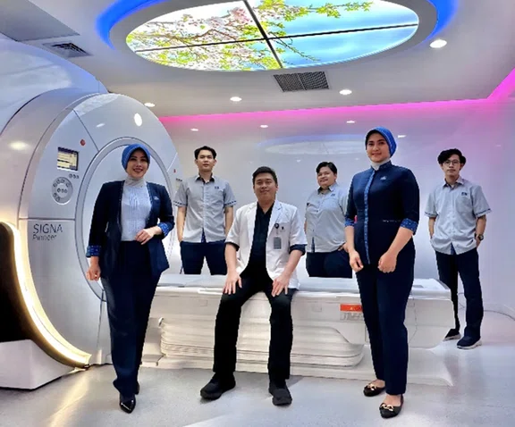
Radiologist and MR technologist team at Premier Bintaro Hospital in Tangerang, Indonesia. From left to right: Erni R. Rusmana, Ongki Imam Malik, Dr. Raditya Utomo, Rahmansyah Ardi Rahmanto, Wira Widya Putri and Fahan Muhandis.
A
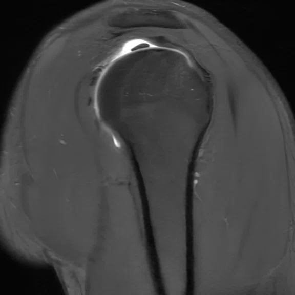
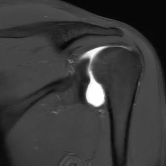
Figure 2.
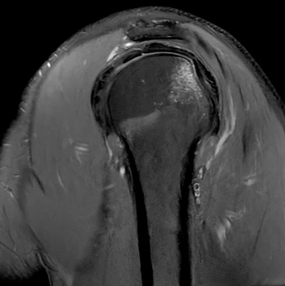
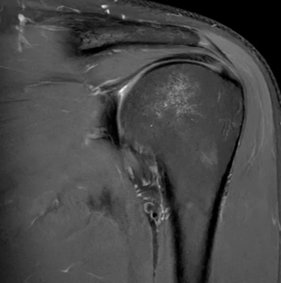
(A) Sagittal PD FatSat, 0.3 x 0.5 x 3 mm and (B) coronal PD FatSat, 0.3 x 0.5 x 3 mm, both acquired with AIR™ Recon DL High.
B
Figure 2.
(A) Sagittal PD FatSat, 0.3 x 0.5 x 3 mm and (B) coronal PD FatSat, 0.3 x 0.5 x 3 mm, both acquired with AIR™ Recon DL High.
1. Westbrook C. Handbook of MRI Technique (4th edition). John Wiley & Sons, Ltd. 2014.
2. Yu D, Turmezei TD, Kerslake RW. FIESTA: an MR arthrography celebration of shoulder joint anatomy, variants, and their mimics. Clin Anat. 2013;26(2):213-227.
A
Figure 3.
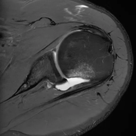
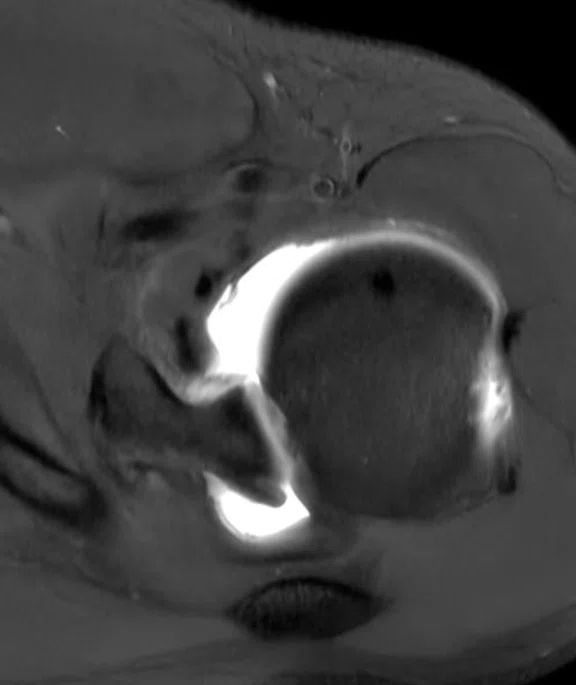
(A) Axial PD FatSat, 0.3 x 0.6 x 3 mm, (B) sagittal T1 FatSat, 0.4 x 0.8 x 3 mm, and (C) coronal T1 FatSat, 0.4 x 0.8 x 3 mm.
B
Figure 3.
(A) Axial PD FatSat, 0.3 x 0.6 x 3 mm, (B) sagittal T1 FatSat, 0.4 x 0.8 x 3 mm, and (C) coronal T1 FatSat, 0.4 x 0.8 x 3 mm.
C
Figure 3.
(A) Axial PD FatSat, 0.3 x 0.6 x 3 mm, (B) sagittal T1 FatSat, 0.4 x 0.8 x 3 mm, and (C) coronal T1 FatSat, 0.4 x 0.8 x 3 mm.
A
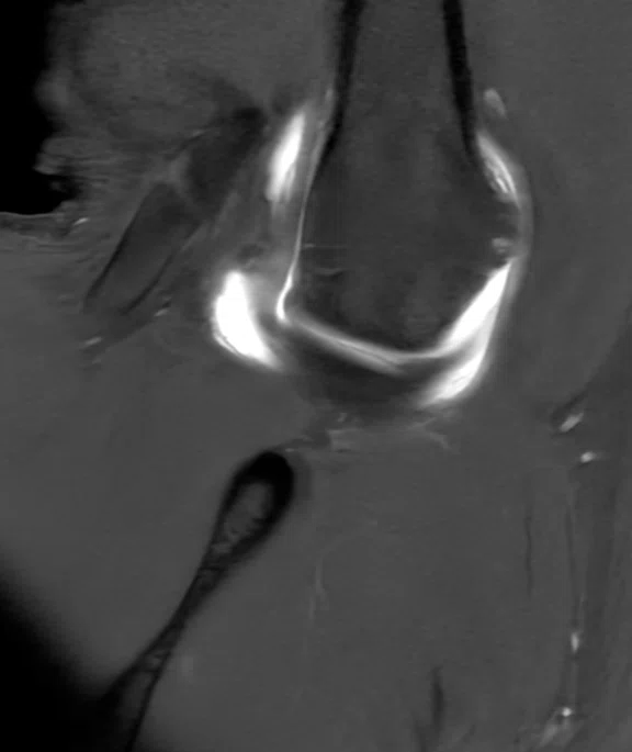
Figure 4.
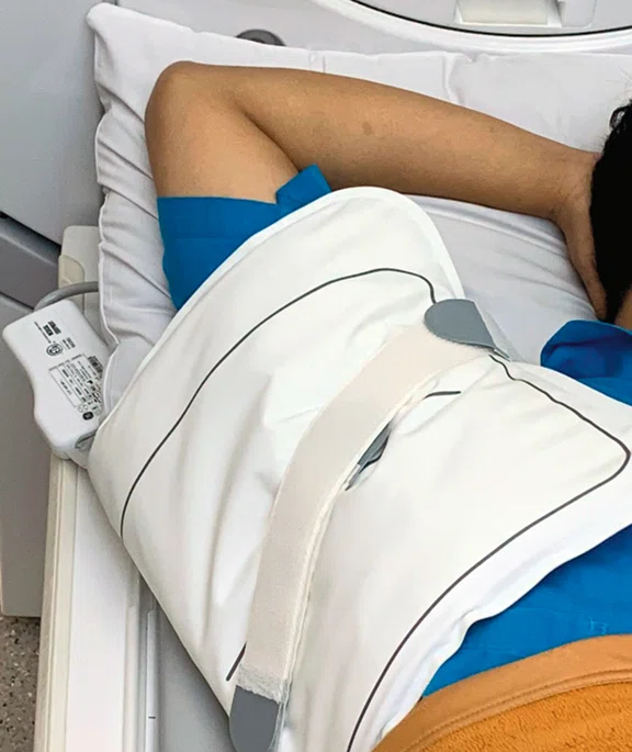
(A, B) ABER position with the 21ch (medium) AIR™ Multi-Purpose Coil, 0.4 x 0.8 x 3 mm, and (C) patient in the ABER position with the AIR™ Coil.
B
Figure 4.
(A, B) ABER position with the 21ch (medium) AIR™ Multi-Purpose Coil, 0.4 x 0.8 x 3 mm, and (C) patient in the ABER position with the AIR™ Coil.
C
Figure 4.
(A, B) ABER position with the 21ch (medium) AIR™ Multi-Purpose Coil, 0.4 x 0.8 x 3 mm, and (C) patient in the ABER position with the AIR™ Coil.
1. Westbrook C. Handbook of MRI Technique (4th edition). John Wiley & Sons, Ltd. 2014.
result


PREVIOUS
${prev-page}
NEXT
${next-page}
Subscribe Now
Manage Subscription
FOLLOW US
Contact Us • Cookie Preferences • Privacy Policy • California Privacy PolicyDo Not Sell or Share My Personal Information • Terms & Conditions • Security
© 2024 GE HealthCare. GE is a trademark of General Electric Company. Used under trademark license.
In Practice
A technical study of MR shoulder arthrography in Bankart lesion cases with AIR Recon DL image quality evaluation
A technical study of MR shoulder arthrography in Bankart lesion cases with AIR Recon DL image quality evaluation
By Raditya Utomo, MD, Radiology Consultant, and Wira Widya Putri, RT(R)(MR), MR technologist, Premier Bintaro Hospital, Tangerang, Indonesia
Premier Bintaro Hospital in Tangerang, Indonesia, has six Centers of Excellence, including the Ortho Sport and Wellness Center, which is focused on treating sports injuries such as those to the shoulder. For the center, MR is the most informative modality in the noninvasive evaluation of the musculoskeletal (MSK) system due to its multiplanar imaging capability and excellent soft-tissue contrast, enabling the assessment of anatomy and pathology from ligament injuries and articular cartilage lesions. One of the diagnostic examination techniques that can reveal partial tears of the articular surface of the rotator cuff is MR arthrography.
MR examinations with higher signal-to-noise ratio (SNR) and contrast-to-noise ratio (CNR) and spatial resolution values are required to depict good anatomical details. At the same time, shoulder MR examinations are expected to be carried out with fast scanning times aimed at improving patient comfort without compromising image quality and diagnostic information. The technological development that allows us to address both requirements in the same acquisition is AIR™ Recon DL. AIR™ Recon DL is an innovative deep-learning reconstruction technology from GE HealthCare that offers a fundamental change in the balance between image quality and scan time, allowing for short scan times that also generate highly detailed MR images with improved image reconstitution. Shoulder MR examinations requiring specific positioning, such as ABER (ABduction and External Rotation), may not be possible with conventional hard shell coils. However, imaging with AIR™ Coils makes this possible while maintaining image quality.
MR examinations with higher signal-to-noise ratio (SNR) and contrast-to-noise ratio (CNR) and spatial resolution values are required to depict good anatomical details. At the same time, shoulder MR examinations are expected to be carried out with fast scanning times aimed at improving patient comfort without compromising image quality and diagnostic information. The technological development that allows us to address both requirements in the same acquisition is AIR™ Recon DL. AIR™ Recon DL is an innovative deep-learning reconstruction technology from GE HealthCare that offers a fundamental change in the balance between image quality and scan time, allowing for short scan times that also generate highly detailed MR images with improved image reconstitution. Shoulder MR examinations requiring specific positioning, such as ABER (ABduction and External Rotation), may not be possible with conventional hard shell coils. However, imaging with AIR™ Coils makes this possible while maintaining image quality.
Procedure
MR arthrography of the shoulder in the radiology department at Premier Bintaro Hospital is performed on patients with indications of shoulder pain due to rotator cuff injuries, adhesive capsulitis, recurrent dislocations, impingement syndrome, Hill-Sachs lesions, Bankart lesions and labrum lesions.
The MR arthrography shoulder procedure consists of:
- Axial proton density (PD) sequences with and without FatSat, sagittal T1 and PD FatSat, and coronal T1 and PD Fat Sat, using a 16-channel dedicated shoulder coil.
- Axial T1 FatSat and PD, coronal T1 FatSat and sagittal T1 FatSat sequences. In the T1 FatSat sequences, the patient is in the ABER position with the 21-channel AIR™ Multi-Purpose Coil.
Discussion
In the MR arthrography examination, we increased the contrast of the image so that the anatomical boundaries of the shoulder joint are clearly visible, allowing the clinician to assess if there is a labrum tear or lesion on the joint membrane.
In the shoulder arthrography MR case presented here, we were able to visualize the labrum with an anterior inferior labral defect (Bankart lesion) and superior anterior labrum detachment (SLAP tear), which has a continuous impression of a Type V SLAP with a flattening of the humeral head in the posterior superior (Hill-Sachs lesion). After evaluating each part of the acromion with a flat inferior surface, there is no low-set or downsloping and no degenerative arthropathy, and no subacromial spur or sprain signs on the acromioclavicular joint. On the rotator cuff of the supraspinatus, subscapularis, infraspinatus and teres minor tendons, there were no signs of tendinosis or tears. On the bursa, both in the subacromial-subdeltoid and subscapularis, there was no effusion or signs of bursitis detected. In the long head biceps tendon, again there was no sign of tendinosis, tears or tenosynovitis. In the glenohumeral joint, no joint effusion was depicted and in the glenoid there was no visible deformity or fracture.
Due to the acquisition of very good detailed images using AIR™ Recon DL, it is possible to evaluate other structures such as the muscule tissue, vascular area and the nerves in the shoulder. The shoulder arthrography MR acquisition that we use to diagnose Bankart lesion abnormalities in the shoulder joint is based on Yu et al, 2013 and Westbrook 2014.1,2
AIR™ Recon DL has a significant impact on the MR arthrography examinations that we routinely perform. Specifically, the scan times with AIR™ Recon DL are significantly shorther than our conventional acquisitions without sacrificing the high resolution and image quality results in our MR arthrography examinations.
As shown in Table 1, the average time reduction in MR arthrography using AIR™ Recon DL is 61.3% compared to our standard, conventional acquisition. In addition to the faster exam, we have not experienced a reduction in image quality. In fact, we have found that the SNR and spatial resolution are greatly improved.
The MR shoulder arthrography examination at Premier Bintaro Hospital is performed on our SIGNA™ Pioneer 3.0T system equipped with AIR™ Recon DL and using the AIR™ Coil, which is very flexible and ideal for special patient positioning requirements such as ABER. As a result of using these innovative technologies, and in particular the use of AIR™ Recon DL, we could achieve sharper image quality with very good detail.
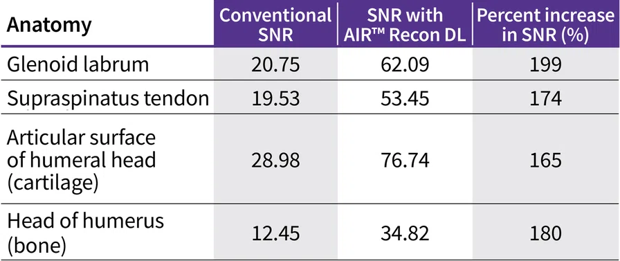
We also quantified the SNR improvement in the MR shoulder arthrography sequence with AIR™ Recon DL (Table 2).
CNR values obtained from several anatomies were also compared (Table 3). CNR is controlled by factors that affect SNR. The visualization of the lesion increases if the CNR between the pathology and the surrounding anatomy is high.1
Conclusion
Our results suggest that MR shoulder arthrography examinations with AIR™ Recon DL have greater diagnostic utility than conventional MR shoulder arthrography for assessing shoulder joint abnormalities with indications of a Bankart lesion.
By implementing AIR™ Recon DL in our MR shoulder arthrography examinations, we can achieve higher image quality with good SNR and CNR values with an average 61.3% faster scan time. Additionally, the AIR™ Coil design in either a medium or large size can fit nearly any shoulder, enhancing patient comfort and maximizing image quality particularly when special patient positioning such as ABER is required.
DOWNLOAD ARTICLE HERE


