‡Not licensed in accordance with Canadian law. Not available for sale in Canada. Not CE marked. Not available for sale in all regions.
Figure 2.
AIR Technology™ AA and PA Coils for abdominal imaging deliver better than expected coverage, signal homogeneity, high spatial resolution and homogeneous fat suppression. A thickening of the terminal ileum (red arrows) is consistent with Crohn’s disease. (A) Axial T2 SSFSE; (B) Axial DWI b1000; (C) Coronal T2 SSFSE; (D) Axial T2 SSFSE FatSat; and (E) Axial T1 LAVA ASPIR.
A
Figure 2.
AIR Technology™ AA and PA Coils for abdominal imaging deliver better than expected coverage, signal homogeneity, high spatial resolution and homogeneous fat suppression. A thickening of the terminal ileum (red arrows) is consistent with Crohn’s disease. (A) Axial T2 SSFSE; (B) Axial DWI b1000; (C) Coronal T2 SSFSE; (D) Axial T2 SSFSE FatSat; and (E) Axial T1 LAVA ASPIR.
C
Figure 2.
AIR Technology™ AA and PA Coils for abdominal imaging deliver better than expected coverage, signal homogeneity, high spatial resolution and homogeneous fat suppression. A thickening of the terminal ileum (red arrows) is consistent with Crohn’s disease. (A) Axial T2 SSFSE; (B) Axial DWI b1000; (C) Coronal T2 SSFSE; (D) Axial T2 SSFSE FatSat; and (E) Axial T1 LAVA ASPIR.
D
Figure 2.
AIR Technology™ AA and PA Coils for abdominal imaging deliver better than expected coverage, signal homogeneity, high spatial resolution and homogeneous fat suppression. A thickening of the terminal ileum (red arrows) is consistent with Crohn’s disease. (A) Axial T2 SSFSE; (B) Axial DWI b1000; (C) Coronal T2 SSFSE; (D) Axial T2 SSFSE FatSat; and (E) Axial T1 LAVA ASPIR.
B
Figure 2.
AIR Technology™ AA and PA Coils for abdominal imaging deliver better than expected coverage, signal homogeneity, high spatial resolution and homogeneous fat suppression. A thickening of the terminal ileum (red arrows) is consistent with Crohn’s disease. (A) Axial T2 SSFSE; (B) Axial DWI b1000; (C) Coronal T2 SSFSE; (D) Axial T2 SSFSE FatSat; and (E) Axial T1 LAVA ASPIR.
E
Figure 2.
AIR Technology™ AA and PA Coils for abdominal imaging deliver better than expected coverage, signal homogeneity, high spatial resolution and homogeneous fat suppression. A thickening of the terminal ileum (red arrows) is consistent with Crohn’s disease. (A) Axial T2 SSFSE; (B) Axial DWI b1000; (C) Coronal T2 SSFSE; (D) Axial T2 SSFSE FatSat; and (E) Axial T1 LAVA ASPIR.
A
Figure 3.
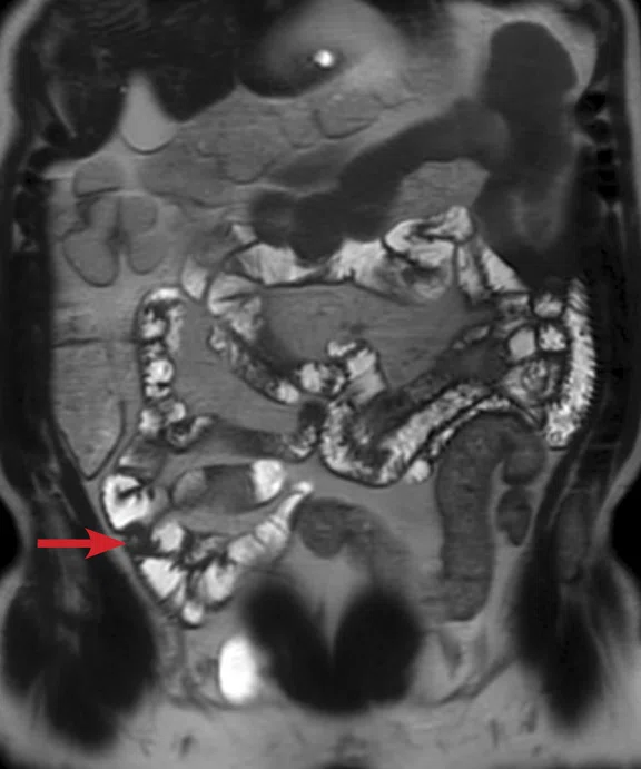
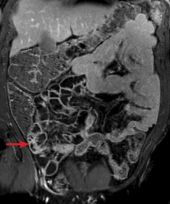
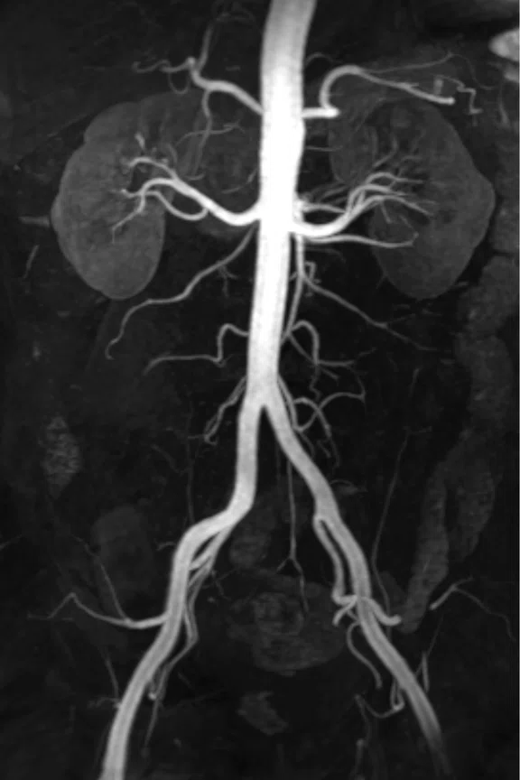
A thickening of the terminal ileum (red arrows) is consistent with Crohn’s disease. (A) Coronal T2 SSFSE; (B) Coronal T1 LAVA ASPIR; and (C) MIP from DISCO, arterial phase.
B
Figure 3.
A thickening of the terminal ileum (red arrows) is consistent with Crohn’s disease. (A) Coronal T2 SSFSE; (B) Coronal T1 LAVA ASPIR; and (C) MIP from DISCO, arterial phase.
C
Figure 3.
A thickening of the terminal ileum (red arrows) is consistent with Crohn’s disease. (A) Coronal T2 SSFSE; (B) Coronal T1 LAVA ASPIR; and (C) MIP from DISCO, arterial phase.
A
Figure 4.
Same patient as Figure 3. (A) Coronal T2 FIESTA and (B) Axial T1 LAVA ASPIR.
B
Figure 4.
Same patient as Figure 3. (A) Coronal T2 FIESTA and (B) Axial T1 LAVA ASPIR.
A
Figure 6.
There is no enhancement in the dynamic sequence and restricted diff usion is consistent with a prostate abscess. (A) Axial DISCO LAVA; (B) enhancement integral map; (C) Axial DWI FOCUS b800; (D) ADC map; and (E) MAGiC DWI b1500.
B
Figure 6.
There is no enhancement in the dynamic sequence and restricted diff usion is consistent with a prostate abscess. (A) Axial DISCO LAVA; (B) enhancement integral map; (C) Axial DWI FOCUS b800; (D) ADC map; and (E) MAGiC DWI b1500.
C
Figure 6.
There is no enhancement in the dynamic sequence and restricted diff usion is consistent with a prostate abscess. (A) Axial DISCO LAVA; (B) enhancement integral map; (C) Axial DWI FOCUS b800; (D) ADC map; and (E) MAGiC DWI b1500.
D
Figure 6.
There is no enhancement in the dynamic sequence and restricted diff usion is consistent with a prostate abscess. (A) Axial DISCO LAVA; (B) enhancement integral map; (C) Axial DWI FOCUS b800; (D) ADC map; and (E) MAGiC DWI b1500.
E
Figure 6.
There is no enhancement in the dynamic sequence and restricted diff usion is consistent with a prostate abscess. (A) Axial DISCO LAVA; (B) enhancement integral map; (C) Axial DWI FOCUS b800; (D) ADC map; and (E) MAGiC DWI b1500.
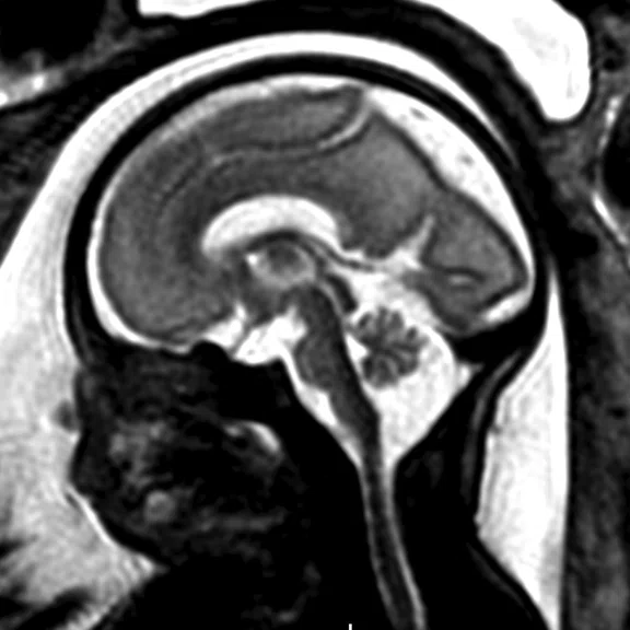
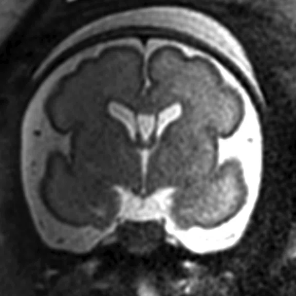
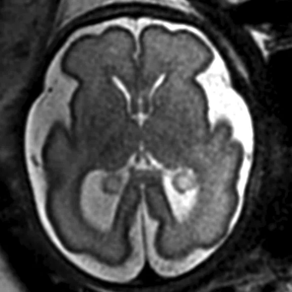
Figure 1.
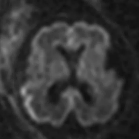
AIR Technology™ AA provides excellent signal penetration for high-quality images. (A) Sagittal T2 SSFSE; (B) Coronal T2 SSFSE; (C) Axial T2 SSFSE; and (D) Axial DWI b800.
A
Figure 1.
AIR Technology™ AA provides excellent signal penetration for high-quality images. (A) Sagittal T2 SSFSE; (B) Coronal T2 SSFSE; (C) Axial T2 SSFSE; and (D) Axial DWI b800.
B
Figure 1.
AIR Technology™ AA provides excellent signal penetration for high-quality images. (A) Sagittal T2 SSFSE; (B) Coronal T2 SSFSE; (C) Axial T2 SSFSE; and (D) Axial DWI b800.
C
Figure 1.
AIR Technology™ AA provides excellent signal penetration for high-quality images. (A) Sagittal T2 SSFSE; (B) Coronal T2 SSFSE; (C) Axial T2 SSFSE; and (D) Axial DWI b800.
D
Figure 1.
AIR Technology™ AA provides excellent signal penetration for high-quality images. (A) Sagittal T2 SSFSE; (B) Coronal T2 SSFSE; (C) Axial T2 SSFSE; and (D) Axial DWI b800.
‡Not licensed in accordance with Canadian law. Not available for sale in Canada. Not CE marked. Not available for sale in all regions.
A
Figure 2.
AIR Technology™ AA and PA Coils for abdominal imaging deliver better than expected coverage, signal homogeneity, high spatial resolution and homogeneous fat suppression. A thickening of the terminal ileum (red arrows) is consistent with Crohn’s disease. (A) Axial T2 SSFSE; (B) Axial DWI b1000; (C) Coronal T2 SSFSE; (D) Axial T2 SSFSE FatSat; and (E) Axial T1 LAVA ASPIR.
C
Figure 2.
AIR Technology™ AA and PA Coils for abdominal imaging deliver better than expected coverage, signal homogeneity, high spatial resolution and homogeneous fat suppression. A thickening of the terminal ileum (red arrows) is consistent with Crohn’s disease. (A) Axial T2 SSFSE; (B) Axial DWI b1000; (C) Coronal T2 SSFSE; (D) Axial T2 SSFSE FatSat; and (E) Axial T1 LAVA ASPIR.
D
Figure 2.
AIR Technology™ AA and PA Coils for abdominal imaging deliver better than expected coverage, signal homogeneity, high spatial resolution and homogeneous fat suppression. A thickening of the terminal ileum (red arrows) is consistent with Crohn’s disease. (A) Axial T2 SSFSE; (B) Axial DWI b1000; (C) Coronal T2 SSFSE; (D) Axial T2 SSFSE FatSat; and (E) Axial T1 LAVA ASPIR.
B
Figure 2.
AIR Technology™ AA and PA Coils for abdominal imaging deliver better than expected coverage, signal homogeneity, high spatial resolution and homogeneous fat suppression. A thickening of the terminal ileum (red arrows) is consistent with Crohn’s disease. (A) Axial T2 SSFSE; (B) Axial DWI b1000; (C) Coronal T2 SSFSE; (D) Axial T2 SSFSE FatSat; and (E) Axial T1 LAVA ASPIR.
E
Figure 2.
AIR Technology™ AA and PA Coils for abdominal imaging deliver better than expected coverage, signal homogeneity, high spatial resolution and homogeneous fat suppression. A thickening of the terminal ileum (red arrows) is consistent with Crohn’s disease. (A) Axial T2 SSFSE; (B) Axial DWI b1000; (C) Coronal T2 SSFSE; (D) Axial T2 SSFSE FatSat; and (E) Axial T1 LAVA ASPIR.
A
Figure 3.
A thickening of the terminal ileum (red arrows) is consistent with Crohn’s disease. (A) Coronal T2 SSFSE; (B) Coronal T1 LAVA ASPIR; and (C) MIP from DISCO, arterial phase.
B
Figure 3.
A thickening of the terminal ileum (red arrows) is consistent with Crohn’s disease. (A) Coronal T2 SSFSE; (B) Coronal T1 LAVA ASPIR; and (C) MIP from DISCO, arterial phase.
C
Figure 3.
A thickening of the terminal ileum (red arrows) is consistent with Crohn’s disease. (A) Coronal T2 SSFSE; (B) Coronal T1 LAVA ASPIR; and (C) MIP from DISCO, arterial phase.
A
Figure 4.
Same patient as Figure 3. (A) Coronal T2 FIESTA and (B) Axial T1 LAVA ASPIR.
B
Figure 4.
Same patient as Figure 3. (A) Coronal T2 FIESTA and (B) Axial T1 LAVA ASPIR.
Figure 5.
AIR Technology™ AA and PA provide high spatial resolution in T2 sequences, which improves visualization of the cancerous lesion for staging. Note the lesion with a high signal in the central prostate. (A) Axial T2 and (B) Axial T2 Cube.
A
Figure 5.
AIR Technology™ AA and PA provide high spatial resolution in T2 sequences, which improves visualization of the cancerous lesion for staging. Note the lesion with a high signal in the central prostate. (A) Axial T2 and (B) Axial T2 Cube.
B
Figure 5.
AIR Technology™ AA and PA provide high spatial resolution in T2 sequences, which improves visualization of the cancerous lesion for staging. Note the lesion with a high signal in the central prostate. (A) Axial T2 and (B) Axial T2 Cube.
A
Figure 6.
There is no enhancement in the dynamic sequence and restricted diff usion is consistent with a prostate abscess. (A) Axial DISCO LAVA; (B) enhancement integral map; (C) Axial DWI FOCUS b800; (D) ADC map; and (E) MAGiC DWI b1500.
B
Figure 6.
There is no enhancement in the dynamic sequence and restricted diff usion is consistent with a prostate abscess. (A) Axial DISCO LAVA; (B) enhancement integral map; (C) Axial DWI FOCUS b800; (D) ADC map; and (E) MAGiC DWI b1500.
C
Figure 6.
There is no enhancement in the dynamic sequence and restricted diff usion is consistent with a prostate abscess. (A) Axial DISCO LAVA; (B) enhancement integral map; (C) Axial DWI FOCUS b800; (D) ADC map; and (E) MAGiC DWI b1500.
D
Figure 6.
There is no enhancement in the dynamic sequence and restricted diff usion is consistent with a prostate abscess. (A) Axial DISCO LAVA; (B) enhancement integral map; (C) Axial DWI FOCUS b800; (D) ADC map; and (E) MAGiC DWI b1500.
E
Figure 6.
There is no enhancement in the dynamic sequence and restricted diff usion is consistent with a prostate abscess. (A) Axial DISCO LAVA; (B) enhancement integral map; (C) Axial DWI FOCUS b800; (D) ADC map; and (E) MAGiC DWI b1500.
Figure 5.
AIR Technology™ AA and PA provide high spatial resolution in T2 sequences, which improves visualization of the cancerous lesion for staging. Note the lesion with a high signal in the central prostate. (A) Axial T2 and (B) Axial T2 Cube.
A
Figure 5.
AIR Technology™ AA and PA provide high spatial resolution in T2 sequences, which improves visualization of the cancerous lesion for staging. Note the lesion with a high signal in the central prostate. (A) Axial T2 and (B) Axial T2 Cube.
B
Figure 5.
AIR Technology™ AA and PA provide high spatial resolution in T2 sequences, which improves visualization of the cancerous lesion for staging. Note the lesion with a high signal in the central prostate. (A) Axial T2 and (B) Axial T2 Cube.
A
Figure 1.
AIR Technology™ AA provides excellent signal penetration for high-quality images. (A) Sagittal T2 SSFSE; (B) Coronal T2 SSFSE; (C) Axial T2 SSFSE; and (D) Axial DWI b800.
B
Figure 1.
AIR Technology™ AA provides excellent signal penetration for high-quality images. (A) Sagittal T2 SSFSE; (B) Coronal T2 SSFSE; (C) Axial T2 SSFSE; and (D) Axial DWI b800.
C
Figure 1.
AIR Technology™ AA provides excellent signal penetration for high-quality images. (A) Sagittal T2 SSFSE; (B) Coronal T2 SSFSE; (C) Axial T2 SSFSE; and (D) Axial DWI b800.
D
Figure 1.
AIR Technology™ AA provides excellent signal penetration for high-quality images. (A) Sagittal T2 SSFSE; (B) Coronal T2 SSFSE; (C) Axial T2 SSFSE; and (D) Axial DWI b800.
Not licensed in accordance with Canadian law. Not available for sale in Canada. Not CE marked. Not available for sale in all regions.
result


PREVIOUS
${prev-page}
NEXT
${next-page}
Subscribe Now
Manage Subscription
FOLLOW US
Contact Us • Cookie Preferences • Privacy Policy • California Privacy PolicyDo Not Sell or Share My Personal Information • Terms & Conditions • Security
© 2024 GE HealthCare. GE is a trademark of General Electric Company. Used under trademark license.
CASE STUDIES
Body imaging with AIR Technology Anterior Array and Posterior Array
Body imaging with AIR Technology Anterior Array and Posterior Array
Submitted by Quirónsalud Madrid University Hospital
CASE STUDIES
Body imaging with AIR Technology Anterior Array and Posterior Array
Body imaging with AIR Technology Anterior Array and Posterior Array
Submitted by Quirónsalud Madrid University Hospital
Fetal imaging
By Manuel Recio Rodríguez, MD, Associate Chief of Diagnostic Imaging Department
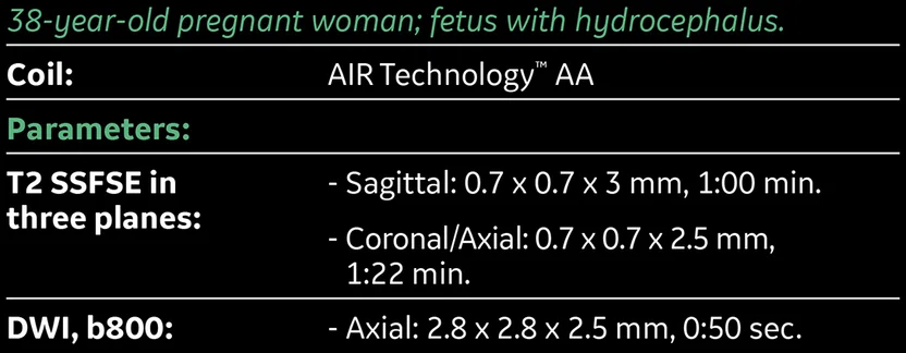
With the AIR Technology™‡ Anterior Array (AA), we can achieve good signal penetration for fetal imaging. This enables us to obtain high-quality images of the fetal brain with short acquisition times using T2 and DWI sequences.
Abdominal imaging
By Vicente Martinez de Vega, MD, Chief of Diagnostic Imaging Department
Abdominal imaging with the AIR Technology™ Coils is better than we expected, particularly the coverage, signal homogeneity, high spatial resolution and scanning speed. We were also able to achieve short acquisition times and homogenous fat suppression. In this particular case, a thickening of the terminal ileum can be noticed in a very short segment, which is consistent with Crohn’s disease.
Figure 2.
AIR Technology™ AA and PA Coils for abdominal imaging deliver better than expected coverage, signal homogeneity, high spatial resolution and homogeneous fat suppression. A thickening of the terminal ileum (red arrows) is consistent with Crohn’s disease. (A) Axial T2 SSFSE; (B) Axial DWI b1000; (C) Coronal T2 SSFSE; (D) Axial T2 SSFSE FatSat; and (E) Axial T1 LAVA ASPIR.
Abdominal imaging
By Vicente Martinez de Vega, MD, Chief of Diagnostic Imaging Department
Abdominal imaging with the AIR Technology™ Coils is better than we expected, particularly the coverage, signal homogeneity, high spatial resolution and scanning speed. We were also able to achieve short acquisition times and homogenous fat suppression. In this particular case, a thickening of the terminal ileum can be noticed in a very short segment, which is consistent with Crohn’s disease.
Prostate imaging
By Manuel Recio Rodríguez, MD, Associate Chief of Diagnostic Imaging Department
Prostate MR is growing in use and referrals in our institution. However, in order to clearly depict the cancer to determine the extent of disease, it is necessary to obtain T2 sequences with high spatial resolution. Also, T2 Cube images can be used to merge with ultrasound to assist in performing targeted biopsies. In this particular case, a lesion with high signal in the T2-weighted sequence in the central prostate can be seen. There is no enhancement in the dynamic sequence and restricted diffusion is consistent with a prostate abscess. Dynamic acquisition with DISCO LAVA provides high spatial and temporal resolution (4.5 seconds per phase) and the diffusion imaging is very high quality.
Fetal imaging
By Manuel Recio Rodríguez, MD, Associate Chief of Diagnostic Imaging Department
With the AIR Technology™‡ Anterior Array (AA), we can achieve good signal penetration for fetal imaging. This enables us to obtain high-quality images of the fetal brain with short acquisition times using T2 and DWI sequences.
Figure 2.
AIR Technology™ AA and PA Coils for abdominal imaging deliver better than expected coverage, signal homogeneity, high spatial resolution and homogeneous fat suppression. A thickening of the terminal ileum (red arrows) is consistent with Crohn’s disease. (A) Axial T2 SSFSE; (B) Axial DWI b1000; (C) Coronal T2 SSFSE; (D) Axial T2 SSFSE FatSat; and (E) Axial T1 LAVA ASPIR.
Prostate imaging
By Manuel Recio Rodríguez, MD, Associate Chief of Diagnostic Imaging Department
Prostate MR is growing in use and referrals in our institution. However, in order to clearly depict the cancer to determine the extent of disease, it is necessary to obtain T2 sequences with high spatial resolution. Also, T2 Cube images can be used to merge with ultrasound to assist in performing targeted biopsies. In this particular case, a lesion with high signal in the T2-weighted sequence in the central prostate can be seen. There is no enhancement in the dynamic sequence and restricted diffusion is consistent with a prostate abscess. Dynamic acquisition with DISCO LAVA provides high spatial and temporal resolution (4.5 seconds per phase) and the diffusion imaging is very high quality.


