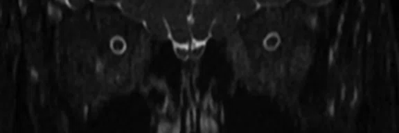
A
Figure 1.
Cervical spine and carotid CE-MRA. (A) Sagittal T2 FSE Flex, 0.9 x 0.9 x 3 mm, 3:45 min.; (B) Sagittal T1 optimized, 0.8 x 1 x 3 mm, 2:47 min.; (C) Coronal carotid CE-MRA, 0.9 x 0.9 x 1.2 mm, 0:40 sec.; and (D) Axial T2 HyperCube, 0.7 x 0.7 x 1.8 mm, 2:24 min.
B
Figure 1.
Cervical spine and carotid CE-MRA. (A) Sagittal T2 FSE Flex, 0.9 x 0.9 x 3 mm, 3:45 min.; (B) Sagittal T1 optimized, 0.8 x 1 x 3 mm, 2:47 min.; (C) Coronal carotid CE-MRA, 0.9 x 0.9 x 1.2 mm, 0:40 sec.; and (D) Axial T2 HyperCube, 0.7 x 0.7 x 1.8 mm, 2:24 min.
C
Figure 1.
Cervical spine and carotid CE-MRA. (A) Sagittal T2 FSE Flex, 0.9 x 0.9 x 3 mm, 3:45 min.; (B) Sagittal T1 optimized, 0.8 x 1 x 3 mm, 2:47 min.; (C) Coronal carotid CE-MRA, 0.9 x 0.9 x 1.2 mm, 0:40 sec.; and (D) Axial T2 HyperCube, 0.7 x 0.7 x 1.8 mm, 2:24 min.
D
Figure 1.
Cervical spine and carotid CE-MRA. (A) Sagittal T2 FSE Flex, 0.9 x 0.9 x 3 mm, 3:45 min.; (B) Sagittal T1 optimized, 0.8 x 1 x 3 mm, 2:47 min.; (C) Coronal carotid CE-MRA, 0.9 x 0.9 x 1.2 mm, 0:40 sec.; and (D) Axial T2 HyperCube, 0.7 x 0.7 x 1.8 mm, 2:24 min.
A
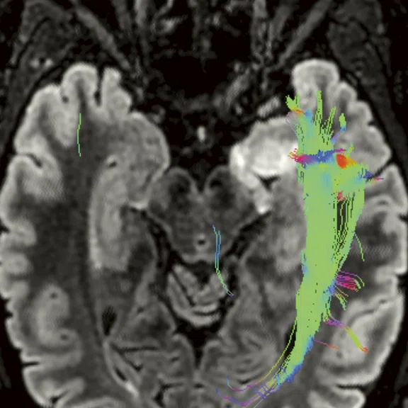
Figure 4.
Patient with low-grade glioma. (A, B) DTI HyperBand with 32 directions, 2.2 x 1.8 x 4 mm, 3:43 min.
B
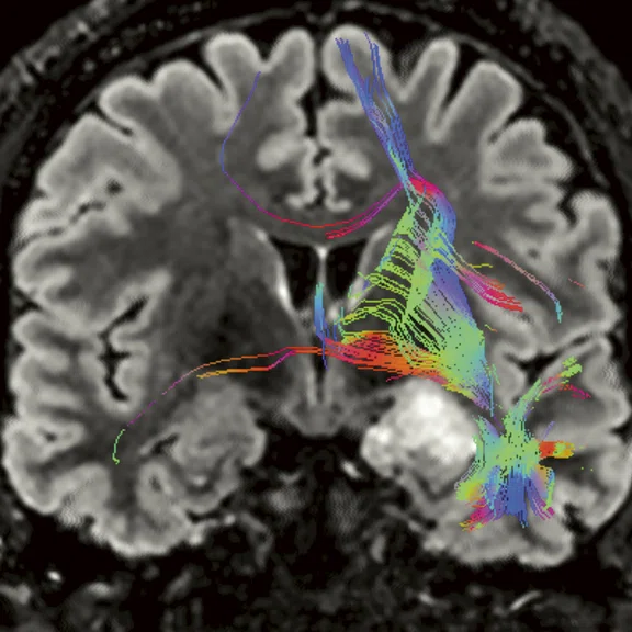
Figure 4.
Patient with low-grade glioma. (A, B) DTI HyperBand with 32 directions, 2.2 x 1.8 x 4 mm, 3:43 min.
A
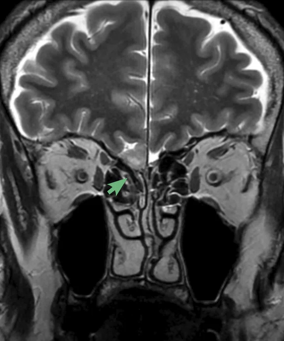
Figure 5.
Patient with meningioma scanned with the 48-channel Head Coil. (A) Coronal T2, 0.5 x 0.6 x 3 mm, 2:15 min.; (B) Axial LAVA ASPIR post contrast, 0.8 x 0.8 x 1 mm, 3:50 min. with (C) Coronal reformat.
B
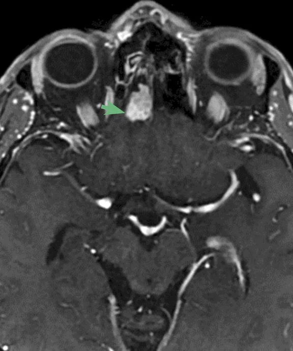
Figure 5.
Patient with meningioma scanned with the 48-channel Head Coil. (A) Coronal T2, 0.5 x 0.6 x 3 mm, 2:15 min.; (B) Axial LAVA ASPIR post contrast, 0.8 x 0.8 x 1 mm, 3:50 min. with (C) Coronal reformat.
C
Figure 5.
Patient with meningioma scanned with the 48-channel Head Coil. (A) Coronal T2, 0.5 x 0.6 x 3 mm, 2:15 min.; (B) Axial LAVA ASPIR post contrast, 0.8 x 0.8 x 1 mm, 3:50 min. with (C) Coronal reformat.
A
Figure 6.
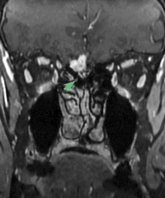
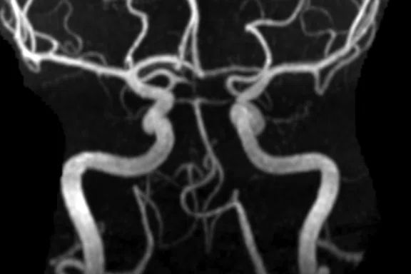
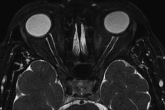
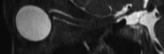
Standard brain and orbits protocols acquired with the 48-channel Head Coil. (A) TOF with HyperSense, 0.7 x 0.7 x 1 mm, 3:21 min.; with (C) Sagittal and (D) Coronal reformats; and (B) Axial T2 HyperCube with Flex and HyperSense, 3:53 min.
B
Figure 6.
Standard brain and orbits protocols acquired with the 48-channel Head Coil. (A) TOF with HyperSense, 0.7 x 0.7 x 1 mm, 3:21 min.; with (C) Sagittal and (D) Coronal reformats; and (B) Axial T2 HyperCube with Flex and HyperSense, 3:53 min.
C
Figure 6.
Standard brain and orbits protocols acquired with the 48-channel Head Coil. (A) TOF with HyperSense, 0.7 x 0.7 x 1 mm, 3:21 min.; with (C) Sagittal and (D) Coronal reformats; and (B) Axial T2 HyperCube with Flex and HyperSense, 3:53 min.
D
Figure 6.
Standard brain and orbits protocols acquired with the 48-channel Head Coil. (A) TOF with HyperSense, 0.7 x 0.7 x 1 mm, 3:21 min.; with (C) Sagittal and (D) Coronal reformats; and (B) Axial T2 HyperCube with Flex and HyperSense, 3:53 min.
A
Figure 1.
Cervical spine and carotid CE-MRA. (A) Sagittal T2 FSE Flex, 0.9 x 0.9 x 3 mm, 3:45 min.; (B) Sagittal T1 optimized, 0.8 x 1 x 3 mm, 2:47 min.; (C) Coronal carotid CE-MRA, 0.9 x 0.9 x 1.2 mm, 0:40 sec.; and (D) Axial T2 HyperCube, 0.7 x 0.7 x 1.8 mm, 2:24 min.
B
Figure 1.
Cervical spine and carotid CE-MRA. (A) Sagittal T2 FSE Flex, 0.9 x 0.9 x 3 mm, 3:45 min.; (B) Sagittal T1 optimized, 0.8 x 1 x 3 mm, 2:47 min.; (C) Coronal carotid CE-MRA, 0.9 x 0.9 x 1.2 mm, 0:40 sec.; and (D) Axial T2 HyperCube, 0.7 x 0.7 x 1.8 mm, 2:24 min.
C
Figure 1.
Cervical spine and carotid CE-MRA. (A) Sagittal T2 FSE Flex, 0.9 x 0.9 x 3 mm, 3:45 min.; (B) Sagittal T1 optimized, 0.8 x 1 x 3 mm, 2:47 min.; (C) Coronal carotid CE-MRA, 0.9 x 0.9 x 1.2 mm, 0:40 sec.; and (D) Axial T2 HyperCube, 0.7 x 0.7 x 1.8 mm, 2:24 min.
D
Figure 1.
Cervical spine and carotid CE-MRA. (A) Sagittal T2 FSE Flex, 0.9 x 0.9 x 3 mm, 3:45 min.; (B) Sagittal T1 optimized, 0.8 x 1 x 3 mm, 2:47 min.; (C) Coronal carotid CE-MRA, 0.9 x 0.9 x 1.2 mm, 0:40 sec.; and (D) Axial T2 HyperCube, 0.7 x 0.7 x 1.8 mm, 2:24 min.
A
Figure 2.
Patient with multiple sclerosis. Higher SNR with the 48-channel Head Coil enables better gray matter delineation (green arrows) and enhanced lesion depiction (red arrows). (A-C) 48-channel Head Coil, Sagittal Cube FLAIR HyperSense, 1 x 1.1 x 1.2 mm, 3:30 min.; and (D-F) conventional HNU, Sagittal Cube FLAIR HyperSense, 1 x 1.1 x 1.2 mm, 3:45 min.
B
Figure 2.
Patient with multiple sclerosis. Higher SNR with the 48-channel Head Coil enables better gray matter delineation (green arrows) and enhanced lesion depiction (red arrows). (A-C) 48-channel Head Coil, Sagittal Cube FLAIR HyperSense, 1 x 1.1 x 1.2 mm, 3:30 min.; and (D-F) conventional HNU, Sagittal Cube FLAIR HyperSense, 1 x 1.1 x 1.2 mm, 3:45 min.
C
Figure 2.
Patient with multiple sclerosis. Higher SNR with the 48-channel Head Coil enables better gray matter delineation (green arrows) and enhanced lesion depiction (red arrows). (A-C) 48-channel Head Coil, Sagittal Cube FLAIR HyperSense, 1 x 1.1 x 1.2 mm, 3:30 min.; and (D-F) conventional HNU, Sagittal Cube FLAIR HyperSense, 1 x 1.1 x 1.2 mm, 3:45 min.
D
Figure 2.
Patient with multiple sclerosis. Higher SNR with the 48-channel Head Coil enables better gray matter delineation (green arrows) and enhanced lesion depiction (red arrows). (A-C) 48-channel Head Coil, Sagittal Cube FLAIR HyperSense, 1 x 1.1 x 1.2 mm, 3:30 min.; and (D-F) conventional HNU, Sagittal Cube FLAIR HyperSense, 1 x 1.1 x 1.2 mm, 3:45 min.
E
Figure 2.
Patient with multiple sclerosis. Higher SNR with the 48-channel Head Coil enables better gray matter delineation (green arrows) and enhanced lesion depiction (red arrows). (A-C) 48-channel Head Coil, Sagittal Cube FLAIR HyperSense, 1 x 1.1 x 1.2 mm, 3:30 min.; and (D-F) conventional HNU, Sagittal Cube FLAIR HyperSense, 1 x 1.1 x 1.2 mm, 3:45 min.
F
Figure 2.
Patient with multiple sclerosis. Higher SNR with the 48-channel Head Coil enables better gray matter delineation (green arrows) and enhanced lesion depiction (red arrows). (A-C) 48-channel Head Coil, Sagittal Cube FLAIR HyperSense, 1 x 1.1 x 1.2 mm, 3:30 min.; and (D-F) conventional HNU, Sagittal Cube FLAIR HyperSense, 1 x 1.1 x 1.2 mm, 3:45 min.
A
Figure 3.
Patient with low-grade glioma. Higher SNR with the (A-C) 48-channel Head Coil enables better in-plane resolution (green arrow) and enhanced lesion depiction (red arrow) than (D-F) images acquired with conventional HNU. (A, D) Cube FLAIR with HyperSense, 1 x 1.1 x 1.2 mm, 3:38 min. with 48-channel Head Coil and 3:45 min. with conventional HNU; (B, C, E, F) Axial T2 PROPELLER, 0.5 x 0.5 x 3 mm, 2:10 min. with 48-channel Head Coil and 0.6 x 0.6 x 3 mm, 2:23 min. with conventional HNU.
B
Figure 3.
Patient with low-grade glioma. Higher SNR with the (A-C) 48-channel Head Coil enables better in-plane resolution (green arrow) and enhanced lesion depiction (red arrow) than (D-F) images acquired with conventional HNU. (A, D) Cube FLAIR with HyperSense, 1 x 1.1 x 1.2 mm, 3:38 min. with 48-channel Head Coil and 3:45 min. with conventional HNU; (B, C, E, F) Axial T2 PROPELLER, 0.5 x 0.5 x 3 mm, 2:10 min. with 48-channel Head Coil and 0.6 x 0.6 x 3 mm, 2:23 min. with conventional HNU.
C
Figure 3.
Patient with low-grade glioma. Higher SNR with the (A-C) 48-channel Head Coil enables better in-plane resolution (green arrow) and enhanced lesion depiction (red arrow) than (D-F) images acquired with conventional HNU. (A, D) Cube FLAIR with HyperSense, 1 x 1.1 x 1.2 mm, 3:38 min. with 48-channel Head Coil and 3:45 min. with conventional HNU; (B, C, E, F) Axial T2 PROPELLER, 0.5 x 0.5 x 3 mm, 2:10 min. with 48-channel Head Coil and 0.6 x 0.6 x 3 mm, 2:23 min. with conventional HNU.
D
Figure 3.
Patient with low-grade glioma. Higher SNR with the (A-C) 48-channel Head Coil enables better in-plane resolution (green arrow) and enhanced lesion depiction (red arrow) than (D-F) images acquired with conventional HNU. (A, D) Cube FLAIR with HyperSense, 1 x 1.1 x 1.2 mm, 3:38 min. with 48-channel Head Coil and 3:45 min. with conventional HNU; (B, C, E, F) Axial T2 PROPELLER, 0.5 x 0.5 x 3 mm, 2:10 min. with 48-channel Head Coil and 0.6 x 0.6 x 3 mm, 2:23 min. with conventional HNU.
E
Figure 3.
Patient with low-grade glioma. Higher SNR with the (A-C) 48-channel Head Coil enables better in-plane resolution (green arrow) and enhanced lesion depiction (red arrow) than (D-F) images acquired with conventional HNU. (A, D) Cube FLAIR with HyperSense, 1 x 1.1 x 1.2 mm, 3:38 min. with 48-channel Head Coil and 3:45 min. with conventional HNU; (B, C, E, F) Axial T2 PROPELLER, 0.5 x 0.5 x 3 mm, 2:10 min. with 48-channel Head Coil and 0.6 x 0.6 x 3 mm, 2:23 min. with conventional HNU.
F
Figure 3.
Patient with low-grade glioma. Higher SNR with the (A-C) 48-channel Head Coil enables better in-plane resolution (green arrow) and enhanced lesion depiction (red arrow) than (D-F) images acquired with conventional HNU. (A, D) Cube FLAIR with HyperSense, 1 x 1.1 x 1.2 mm, 3:38 min. with 48-channel Head Coil and 3:45 min. with conventional HNU; (B, C, E, F) Axial T2 PROPELLER, 0.5 x 0.5 x 3 mm, 2:10 min. with 48-channel Head Coil and 0.6 x 0.6 x 3 mm, 2:23 min. with conventional HNU.
A
Figure 2.
Patient with multiple sclerosis. Higher SNR with the 48-channel Head Coil enables better gray matter delineation (green arrows) and enhanced lesion depiction (red arrows). (A-C) 48-channel Head Coil, Sagittal Cube FLAIR HyperSense, 1 x 1.1 x 1.2 mm, 3:30 min.; and (D-F) conventional HNU, Sagittal Cube FLAIR HyperSense, 1 x 1.1 x 1.2 mm, 3:45 min.
B
Figure 2.
Patient with multiple sclerosis. Higher SNR with the 48-channel Head Coil enables better gray matter delineation (green arrows) and enhanced lesion depiction (red arrows). (A-C) 48-channel Head Coil, Sagittal Cube FLAIR HyperSense, 1 x 1.1 x 1.2 mm, 3:30 min.; and (D-F) conventional HNU, Sagittal Cube FLAIR HyperSense, 1 x 1.1 x 1.2 mm, 3:45 min.
C
Figure 2.
Patient with multiple sclerosis. Higher SNR with the 48-channel Head Coil enables better gray matter delineation (green arrows) and enhanced lesion depiction (red arrows). (A-C) 48-channel Head Coil, Sagittal Cube FLAIR HyperSense, 1 x 1.1 x 1.2 mm, 3:30 min.; and (D-F) conventional HNU, Sagittal Cube FLAIR HyperSense, 1 x 1.1 x 1.2 mm, 3:45 min.
D
Figure 2.
Patient with multiple sclerosis. Higher SNR with the 48-channel Head Coil enables better gray matter delineation (green arrows) and enhanced lesion depiction (red arrows). (A-C) 48-channel Head Coil, Sagittal Cube FLAIR HyperSense, 1 x 1.1 x 1.2 mm, 3:30 min.; and (D-F) conventional HNU, Sagittal Cube FLAIR HyperSense, 1 x 1.1 x 1.2 mm, 3:45 min.
E
Figure 2.
Patient with multiple sclerosis. Higher SNR with the 48-channel Head Coil enables better gray matter delineation (green arrows) and enhanced lesion depiction (red arrows). (A-C) 48-channel Head Coil, Sagittal Cube FLAIR HyperSense, 1 x 1.1 x 1.2 mm, 3:30 min.; and (D-F) conventional HNU, Sagittal Cube FLAIR HyperSense, 1 x 1.1 x 1.2 mm, 3:45 min.
F
Figure 2.
Patient with multiple sclerosis. Higher SNR with the 48-channel Head Coil enables better gray matter delineation (green arrows) and enhanced lesion depiction (red arrows). (A-C) 48-channel Head Coil, Sagittal Cube FLAIR HyperSense, 1 x 1.1 x 1.2 mm, 3:30 min.; and (D-F) conventional HNU, Sagittal Cube FLAIR HyperSense, 1 x 1.1 x 1.2 mm, 3:45 min.
A
Figure 3.
Patient with low-grade glioma. Higher SNR with the (A-C) 48-channel Head Coil enables better in-plane resolution (green arrow) and enhanced lesion depiction (red arrow) than (D-F) images acquired with conventional HNU. (A, D) Cube FLAIR with HyperSense, 1 x 1.1 x 1.2 mm, 3:38 min. with 48-channel Head Coil and 3:45 min. with conventional HNU; (B, C, E, F) Axial T2 PROPELLER, 0.5 x 0.5 x 3 mm, 2:10 min. with 48-channel Head Coil and 0.6 x 0.6 x 3 mm, 2:23 min. with conventional HNU.
B
Figure 3.
Patient with low-grade glioma. Higher SNR with the (A-C) 48-channel Head Coil enables better in-plane resolution (green arrow) and enhanced lesion depiction (red arrow) than (D-F) images acquired with conventional HNU. (A, D) Cube FLAIR with HyperSense, 1 x 1.1 x 1.2 mm, 3:38 min. with 48-channel Head Coil and 3:45 min. with conventional HNU; (B, C, E, F) Axial T2 PROPELLER, 0.5 x 0.5 x 3 mm, 2:10 min. with 48-channel Head Coil and 0.6 x 0.6 x 3 mm, 2:23 min. with conventional HNU.
C
Figure 3.
Patient with low-grade glioma. Higher SNR with the (A-C) 48-channel Head Coil enables better in-plane resolution (green arrow) and enhanced lesion depiction (red arrow) than (D-F) images acquired with conventional HNU. (A, D) Cube FLAIR with HyperSense, 1 x 1.1 x 1.2 mm, 3:38 min. with 48-channel Head Coil and 3:45 min. with conventional HNU; (B, C, E, F) Axial T2 PROPELLER, 0.5 x 0.5 x 3 mm, 2:10 min. with 48-channel Head Coil and 0.6 x 0.6 x 3 mm, 2:23 min. with conventional HNU.
D
Figure 3.
Patient with low-grade glioma. Higher SNR with the (A-C) 48-channel Head Coil enables better in-plane resolution (green arrow) and enhanced lesion depiction (red arrow) than (D-F) images acquired with conventional HNU. (A, D) Cube FLAIR with HyperSense, 1 x 1.1 x 1.2 mm, 3:38 min. with 48-channel Head Coil and 3:45 min. with conventional HNU; (B, C, E, F) Axial T2 PROPELLER, 0.5 x 0.5 x 3 mm, 2:10 min. with 48-channel Head Coil and 0.6 x 0.6 x 3 mm, 2:23 min. with conventional HNU.
E
Figure 3.
Patient with low-grade glioma. Higher SNR with the (A-C) 48-channel Head Coil enables better in-plane resolution (green arrow) and enhanced lesion depiction (red arrow) than (D-F) images acquired with conventional HNU. (A, D) Cube FLAIR with HyperSense, 1 x 1.1 x 1.2 mm, 3:38 min. with 48-channel Head Coil and 3:45 min. with conventional HNU; (B, C, E, F) Axial T2 PROPELLER, 0.5 x 0.5 x 3 mm, 2:10 min. with 48-channel Head Coil and 0.6 x 0.6 x 3 mm, 2:23 min. with conventional HNU.
F
Figure 3.
Patient with low-grade glioma. Higher SNR with the (A-C) 48-channel Head Coil enables better in-plane resolution (green arrow) and enhanced lesion depiction (red arrow) than (D-F) images acquired with conventional HNU. (A, D) Cube FLAIR with HyperSense, 1 x 1.1 x 1.2 mm, 3:38 min. with 48-channel Head Coil and 3:45 min. with conventional HNU; (B, C, E, F) Axial T2 PROPELLER, 0.5 x 0.5 x 3 mm, 2:10 min. with 48-channel Head Coil and 0.6 x 0.6 x 3 mm, 2:23 min. with conventional HNU.
A
Figure 6.
Standard brain and orbits protocols acquired with the 48-channel Head Coil. (A) TOF with HyperSense, 0.7 x 0.7 x 1 mm, 3:21 min.; with (C) Sagittal and (D) Coronal reformats; and (B) Axial T2 HyperCube with Flex and HyperSense, 3:53 min.
B
Figure 6.
Standard brain and orbits protocols acquired with the 48-channel Head Coil. (A) TOF with HyperSense, 0.7 x 0.7 x 1 mm, 3:21 min.; with (C) Sagittal and (D) Coronal reformats; and (B) Axial T2 HyperCube with Flex and HyperSense, 3:53 min.
C
Figure 6.
Standard brain and orbits protocols acquired with the 48-channel Head Coil. (A) TOF with HyperSense, 0.7 x 0.7 x 1 mm, 3:21 min.; with (C) Sagittal and (D) Coronal reformats; and (B) Axial T2 HyperCube with Flex and HyperSense, 3:53 min.
D
Figure 6.
Standard brain and orbits protocols acquired with the 48-channel Head Coil. (A) TOF with HyperSense, 0.7 x 0.7 x 1 mm, 3:21 min.; with (C) Sagittal and (D) Coronal reformats; and (B) Axial T2 HyperCube with Flex and HyperSense, 3:53 min.
result


PREVIOUS
${prev-page}
NEXT
${next-page}
Subscribe Now
Manage Subscription
FOLLOW US
Contact Us • Cookie Preferences • Privacy Policy • California Privacy PolicyDo Not Sell or Share My Personal Information • Terms & Conditions • Security
© 2024 GE HealthCare. GE is a trademark of General Electric Company. Used under trademark license.
CASE STUDIES
Neuro imaging with 48-channel Head Coil
Neuro imaging with 48-channel Head Coil
By Krisztina Baráth, MD, neuroradiologist, and Brigitte Trudel, RT(R)(MR), MRI Chief Technologist, RNR Institute of Radiology and Neuroradiology at Glattzentrum
When scanning with the new 48-channel Head Coil on the SIGNA™ Pioneer 3.0T MR system, routine neuro acquisitions show significantly higher signal-to-noise ratio (SNR) compared to prior acquisitions with the conventional Head Neck Unit (HNU) coil.
With the embedded AIR Technology™ element design, we can observe a very homogenous signal distribution over the whole field-of-view without any signal drop in the center of the brain. In our experience, we know this is not the case for every dedicated neuro coil available on the market.
Additionally, the coverage of the coil in the z-direction gives us the versatility to easily scan the brain and cervical spine for multiple sclerosis as well as carotid MRA studies.
The 48-channel Head Coil has an adaptable design with an additional 3 cm expansion to gain more room for very large-sized heads and necks. It also helps reduce the patient feeling confined or having their nose in contact with the front of the coil. The coil is compatible with the comfort tilt device, which is very important when scanning elderly patients suffering from kyphosis because it helps them lie comfortably on the table. It is essential for our dementia protocols that the patient not move during scanning due to discomfort.
By combining the advantage of extra SNR with high ARC factors and new acceleration techniques, such as HyperSense and HyperBand, we are able to decrease significantly our total examination time for neuro protocols by 25% while maintaining or even increasing image quality and spatial resolution.
The 48-channel Head Coil is a real asset for us as a neuroradiological institute and it further extends the clinical benefits of a powerful 3.0T MR system such as the SIGNA™ Pioneer.
Figure 2.
Patient with multiple sclerosis. Higher SNR with the 48-channel Head Coil enables better gray matter delineation (green arrows) and enhanced lesion depiction (red arrows). (A-C) 48-channel Head Coil, Sagittal Cube FLAIR HyperSense, 1 x 1.1 x 1.2 mm, 3:30 min.; and (D-F) conventional HNU, Sagittal Cube FLAIR HyperSense, 1 x 1.1 x 1.2 mm, 3:45 min.
Figure 3.
Patient with low-grade glioma. Higher SNR with the (A-C) 48-channel Head Coil enables better in-plane resolution (green arrow) and enhanced lesion depiction (red arrow) than (D-F) images acquired with conventional HNU. (A, D) Cube FLAIR with HyperSense, 1 x 1.1 x 1.2 mm, 3:38 min. with 48-channel Head Coil and 3:45 min. with conventional HNU; (B, C, E, F) Axial T2 PROPELLER, 0.5 x 0.5 x 3 mm, 2:10 min. with 48-channel Head Coil and 0.6 x 0.6 x 3 mm, 2:23 min. with conventional HNU.
CASE STUDIES
Neuro imaging with 48-channel Head Coil
Neuro imaging with 48-channel Head Coil
By Krisztina Baráth, MD, neuroradiologist, and Brigitte Trudel, RT(R)(MR), MRI Chief Technologist, RNR Institute of Radiology and Neuroradiology at Glattzentrum
Figure 2.
Patient with multiple sclerosis. Higher SNR with the 48-channel Head Coil enables better gray matter delineation (green arrows) and enhanced lesion depiction (red arrows). (A-C) 48-channel Head Coil, Sagittal Cube FLAIR HyperSense, 1 x 1.1 x 1.2 mm, 3:30 min.; and (D-F) conventional HNU, Sagittal Cube FLAIR HyperSense, 1 x 1.1 x 1.2 mm, 3:45 min.
Figure 3.
Patient with low-grade glioma. Higher SNR with the (A-C) 48-channel Head Coil enables better in-plane resolution (green arrow) and enhanced lesion depiction (red arrow) than (D-F) images acquired with conventional HNU. (A, D) Cube FLAIR with HyperSense, 1 x 1.1 x 1.2 mm, 3:38 min. with 48-channel Head Coil and 3:45 min. with conventional HNU; (B, C, E, F) Axial T2 PROPELLER, 0.5 x 0.5 x 3 mm, 2:10 min. with 48-channel Head Coil and 0.6 x 0.6 x 3 mm, 2:23 min. with conventional HNU.

When scanning with the new 48-channel Head Coil on the SIGNA™ Pioneer 3.0T MR system, routine neuro acquisitions show significantly higher signal-to-noise ratio (SNR) compared to prior acquisitions with the conventional Head Neck Unit (HNU) coil.
With the embedded AIR Technology™ element design, we can observe a very homogenous signal distribution over the whole field-of-view without any signal drop in the center of the brain. In our experience, we know this is not the case for every dedicated neuro coil available on the market.
Additionally, the coverage of the coil in the z-direction gives us the versatility to easily scan the brain and cervical spine for multiple sclerosis as well as carotid MRA studies.
The 48-channel Head Coil has an adaptable design with an additional 3 cm expansion to gain more room for very large-sized heads and necks. It also helps reduce the patient feeling confined or having their nose in contact with the front of the coil. The coil is compatible with the comfort tilt device, which is very important when scanning elderly patients suffering from kyphosis because it helps them lie comfortably on the table. It is essential for our dementia protocols that the patient not move during scanning due to discomfort.
By combining the advantage of extra SNR with high ARC factors and new acceleration techniques, such as HyperSense and HyperBand, we are able to decrease significantly our total examination time for neuro protocols by 25% while maintaining or even increasing image quality and spatial resolution.
The 48-channel Head Coil is a real asset for us as a neuroradiological institute and it further extends the clinical benefits of a powerful 3.0T MR system such as the SIGNA™ Pioneer.


