A
Figure 1.
AIR™ Recon DL has made a significant difference for orthopedic imaging at Benefis Health System, including the ability to capture high-resolution images of the extremities, as shown in this finger exam. (A) Axial PD FRFSE FatSat, 0.4 x 0.5 x 3 mm, 60 slices in 3:02 min. and (B) coronal PD STIR, 0.6 x 0.8 x 3 mm, 18 slices in 1:54 min.
B
Figure 1.
AIR™ Recon DL has made a significant difference for orthopedic imaging at Benefis Health System, including the ability to capture high-resolution images of the extremities, as shown in this finger exam. (A) Axial PD FRFSE FatSat, 0.4 x 0.5 x 3 mm, 60 slices in 3:02 min. and (B) coronal PD STIR, 0.6 x 0.8 x 3 mm, 18 slices in 1:54 min.
A
Figure 6.
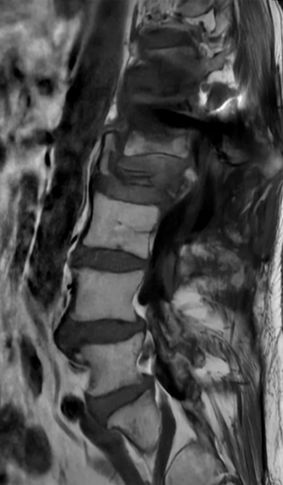
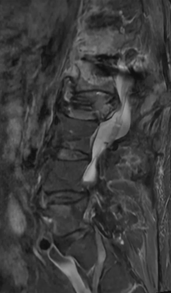
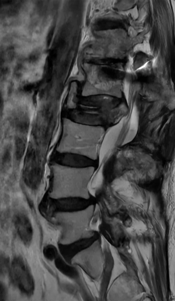
Even in the presence of metal, SIGNA™ Voyager with AIR™ Recon DL delivers excellent image quality, as shown in this lumbar spine exam. (A) Sagittal T2 FRFSE, 0.6 x 0.9 x 4 mm, 20 slices in 1:43 min., (B) sagittal PD STIR, 1 x 1.3 x 4 mm, 20 slices in 2:05 min. and (C) sagittal T1 FSE, 0.7 x 1 x 4 mm, 20 slices in 2:12 min.
B
Figure 6.
Even in the presence of metal, SIGNA™ Voyager with AIR™ Recon DL delivers excellent image quality, as shown in this lumbar spine exam. (A) Sagittal T2 FRFSE, 0.6 x 0.9 x 4 mm, 20 slices in 1:43 min., (B) sagittal PD STIR, 1 x 1.3 x 4 mm, 20 slices in 2:05 min. and (C) sagittal T1 FSE, 0.7 x 1 x 4 mm, 20 slices in 2:12 min.
C
Figure 6.
Even in the presence of metal, SIGNA™ Voyager with AIR™ Recon DL delivers excellent image quality, as shown in this lumbar spine exam. (A) Sagittal T2 FRFSE, 0.6 x 0.9 x 4 mm, 20 slices in 1:43 min., (B) sagittal PD STIR, 1 x 1.3 x 4 mm, 20 slices in 2:05 min. and (C) sagittal T1 FSE, 0.7 x 1 x 4 mm, 20 slices in 2:12 min.
A
Figure 2.
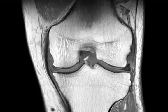
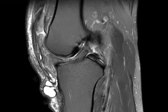
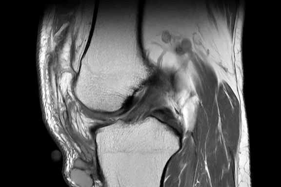
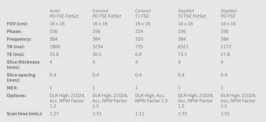
With SIGNA™ Voyager and AIR™ Recon DL, Benefis is now routinely performing MR knee exams in less than 10 minutes. In this case, the total exam time was 6:46 min. (A) Axial PD FSE FatSat, (B) coronal PD FSE FatSat, (C) coronal T1 FSE, (D) sagittal T2 FSE FatSat and (E) sagittal PD FSE, all acquired with AIR™ Recon DL.
B
Figure 2.
With SIGNA™ Voyager and AIR™ Recon DL, Benefis is now routinely performing MR knee exams in less than 10 minutes. In this case, the total exam time was 6:46 min. (A) Axial PD FSE FatSat, (B) coronal PD FSE FatSat, (C) coronal T1 FSE, (D) sagittal T2 FSE FatSat and (E) sagittal PD FSE, all acquired with AIR™ Recon DL.
C
Figure 2.
With SIGNA™ Voyager and AIR™ Recon DL, Benefis is now routinely performing MR knee exams in less than 10 minutes. In this case, the total exam time was 6:46 min. (A) Axial PD FSE FatSat, (B) coronal PD FSE FatSat, (C) coronal T1 FSE, (D) sagittal T2 FSE FatSat and (E) sagittal PD FSE, all acquired with AIR™ Recon DL.
D
Figure 2.
With SIGNA™ Voyager and AIR™ Recon DL, Benefis is now routinely performing MR knee exams in less than 10 minutes. In this case, the total exam time was 6:46 min. (A) Axial PD FSE FatSat, (B) coronal PD FSE FatSat, (C) coronal T1 FSE, (D) sagittal T2 FSE FatSat and (E) sagittal PD FSE, all acquired with AIR™ Recon DL.
E
Figure 2.
With SIGNA™ Voyager and AIR™ Recon DL, Benefis is now routinely performing MR knee exams in less than 10 minutes. In this case, the total exam time was 6:46 min. (A) Axial PD FSE FatSat, (B) coronal PD FSE FatSat, (C) coronal T1 FSE, (D) sagittal T2 FSE FatSat and (E) sagittal PD FSE, all acquired with AIR™ Recon DL.
A
Figure 3.
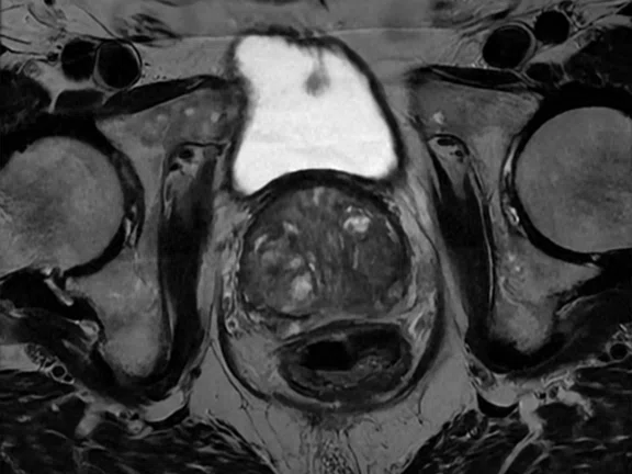
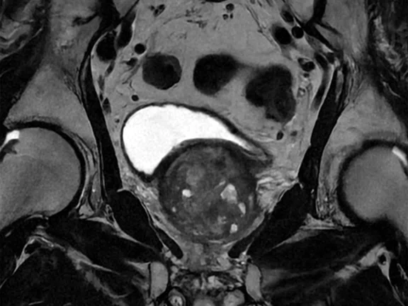
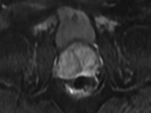
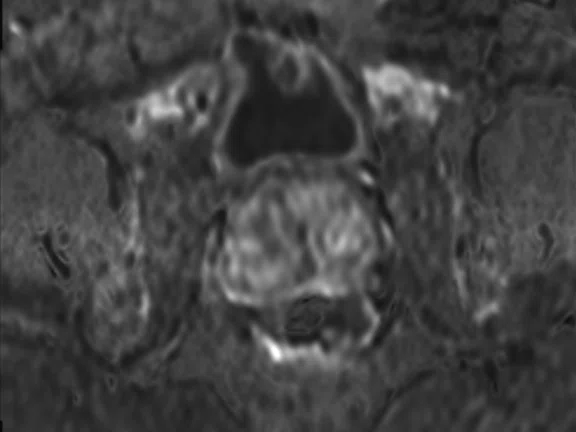
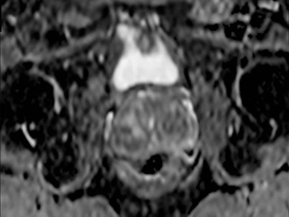
All prostate exams are now performed with the 16ch AIR™ AA coil. (A) Axial T2 FRFSE, 0.6 x 0.8 x 3 mm, 50 slices in 3:42 min., (B) coronal T2 FRFSE, 0.6 x 0.6 x 3 mm, 36 slices in 3:15 min. and (C-E) axial MAGiC DWI, 2.2 x 4.3 x 3 mm, 50 slices in 9:58 min. with (C) b800, (D) MAGiC (synthetic) b1400 and (E) ADC map.
B
Figure 3.
All prostate exams are now performed with the 16ch AIR™ AA coil. (A) Axial T2 FRFSE, 0.6 x 0.8 x 3 mm, 50 slices in 3:42 min., (B) coronal T2 FRFSE, 0.6 x 0.6 x 3 mm, 36 slices in 3:15 min. and (C-E) axial MAGiC DWI, 2.2 x 4.3 x 3 mm, 50 slices in 9:58 min. with (C) b800, (D) MAGiC (synthetic) b1400 and (E) ADC map.
C
Figure 3.
All prostate exams are now performed with the 16ch AIR™ AA coil. (A) Axial T2 FRFSE, 0.6 x 0.8 x 3 mm, 50 slices in 3:42 min., (B) coronal T2 FRFSE, 0.6 x 0.6 x 3 mm, 36 slices in 3:15 min. and (C-E) axial MAGiC DWI, 2.2 x 4.3 x 3 mm, 50 slices in 9:58 min. with (C) b800, (D) MAGiC (synthetic) b1400 and (E) ADC map.
D
Figure 3.
All prostate exams are now performed with the 16ch AIR™ AA coil. (A) Axial T2 FRFSE, 0.6 x 0.8 x 3 mm, 50 slices in 3:42 min., (B) coronal T2 FRFSE, 0.6 x 0.6 x 3 mm, 36 slices in 3:15 min. and (C-E) axial MAGiC DWI, 2.2 x 4.3 x 3 mm, 50 slices in 9:58 min. with (C) b800, (D) MAGiC (synthetic) b1400 and (E) ADC map.
E
Figure 3.
All prostate exams are now performed with the 16ch AIR™ AA coil. (A) Axial T2 FRFSE, 0.6 x 0.8 x 3 mm, 50 slices in 3:42 min., (B) coronal T2 FRFSE, 0.6 x 0.6 x 3 mm, 36 slices in 3:15 min. and (C-E) axial MAGiC DWI, 2.2 x 4.3 x 3 mm, 50 slices in 9:58 min. with (C) b800, (D) MAGiC (synthetic) b1400 and (E) ADC map.
A
Figure 4.
The combination of AIR™ AA Coil and AIR™ Recon DL has led to higher SNR, shorter scan times and greater patient comfort at Benefis. The AIR™ AA Coil facilitates the acquisition of large field-of-views, as shown in this long leg bone exam. (A) Coronal T1 FSE, 0.6 x 0.8 x 4 mm, 28 slices in 1:02 min., (B) coronal PD STIR, 1 x 1.2 x 4 mm, 28 slices in 1:38 min., (C) axial T1 FSE, 0.6 x 0.6 x 4 mm, 89 slices in 3:08 min. and (D) axial PD STIR, 0.7 x 0.9 x 5 mm, 89 slices in 4:00 min.
B
Figure 4.
The combination of AIR™ AA Coil and AIR™ Recon DL has led to higher SNR, shorter scan times and greater patient comfort at Benefis. The AIR™ AA Coil facilitates the acquisition of large field-of-views, as shown in this long leg bone exam. (A) Coronal T1 FSE, 0.6 x 0.8 x 4 mm, 28 slices in 1:02 min., (B) coronal PD STIR, 1 x 1.2 x 4 mm, 28 slices in 1:38 min., (C) axial T1 FSE, 0.6 x 0.6 x 4 mm, 89 slices in 3:08 min. and (D) axial PD STIR, 0.7 x 0.9 x 5 mm, 89 slices in 4:00 min.
C
Figure 4.
The combination of AIR™ AA Coil and AIR™ Recon DL has led to higher SNR, shorter scan times and greater patient comfort at Benefis. The AIR™ AA Coil facilitates the acquisition of large field-of-views, as shown in this long leg bone exam. (A) Coronal T1 FSE, 0.6 x 0.8 x 4 mm, 28 slices in 1:02 min., (B) coronal PD STIR, 1 x 1.2 x 4 mm, 28 slices in 1:38 min., (C) axial T1 FSE, 0.6 x 0.6 x 4 mm, 89 slices in 3:08 min. and (D) axial PD STIR, 0.7 x 0.9 x 5 mm, 89 slices in 4:00 min.
D
Figure 4.
The combination of AIR™ AA Coil and AIR™ Recon DL has led to higher SNR, shorter scan times and greater patient comfort at Benefis. The AIR™ AA Coil facilitates the acquisition of large field-of-views, as shown in this long leg bone exam. (A) Coronal T1 FSE, 0.6 x 0.8 x 4 mm, 28 slices in 1:02 min., (B) coronal PD STIR, 1 x 1.2 x 4 mm, 28 slices in 1:38 min., (C) axial T1 FSE, 0.6 x 0.6 x 4 mm, 89 slices in 3:08 min. and (D) axial PD STIR, 0.7 x 0.9 x 5 mm, 89 slices in 4:00 min.
A
Figure 5.
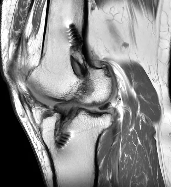
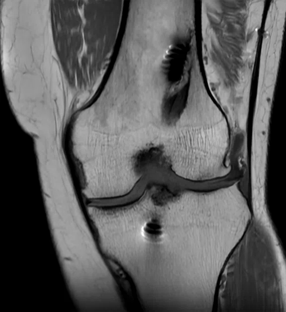
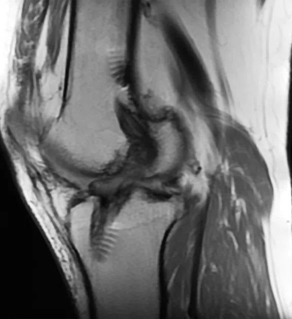
Even in the presence of metal, SIGNA™ Voyager with AIR™ Recon DL and MAVRIC SL delivers excellent image quality, as shown in this knee exam. (A) Sagittal PD FSE, 0.4 x 0.6 x 4 mm, 28 slices in 1:01 min. with AIR™ Recon DL, (B) coronal T1 FSE, 0.5 x 0.7 x 4 mm, 30 slices in 1:06 min. with AIR™ Recon DL and (C) sagittal T2 MAVRIC SL, 0.7 x 0.9 x 5 mm, 24 slices in 5:27min.
B
Figure 5.
Even in the presence of metal, SIGNA™ Voyager with AIR™ Recon DL and MAVRIC SL delivers excellent image quality, as shown in this knee exam. (A) Sagittal PD FSE, 0.4 x 0.6 x 4 mm, 28 slices in 1:01 min. with AIR™ Recon DL, (B) coronal T1 FSE, 0.5 x 0.7 x 4 mm, 30 slices in 1:06 min. with AIR™ Recon DL and (C) sagittal T2 MAVRIC SL, 0.7 x 0.9 x 5 mm, 24 slices in 5:27min.
C
Figure 5.
Even in the presence of metal, SIGNA™ Voyager with AIR™ Recon DL and MAVRIC SL delivers excellent image quality, as shown in this knee exam. (A) Sagittal PD FSE, 0.4 x 0.6 x 4 mm, 28 slices in 1:01 min. with AIR™ Recon DL, (B) coronal T1 FSE, 0.5 x 0.7 x 4 mm, 30 slices in 1:06 min. with AIR™ Recon DL and (C) sagittal T2 MAVRIC SL, 0.7 x 0.9 x 5 mm, 24 slices in 5:27min.
result


PREVIOUS
${prev-page}
NEXT
${next-page}
Subscribe Now
Manage Subscription
FOLLOW US
Contact Us • Cookie Preferences • Privacy Policy • California Privacy PolicyDo Not Sell or Share My Personal Information • Terms & Conditions • Security
© 2024 GE HealthCare. GE is a trademark of General Electric Company. Used under trademark license.
IN PRACTICE
Community hospital expands imaging capabilities and capacity by investing in advanced MR technology
Community hospital expands imaging capabilities and capacity by investing in advanced MR technology
Benefis Health System in Great Falls, Montana, serves approximately 230,000 residents in a vast, 14-county region that covers more than 40,000 square miles. Despite this geographic reach, approximately one-third of patients come from outside their primary service area to be cared for in Great Falls. The health system recognized the need to expand into other markets and bring services closer to their patients and embarked on a strategic growth plan that included investing in new advanced MR systems.
Benefis Health System in Great Falls, Montana, serves approximately 230,000 residents in a vast, 14-county region that covers more than 40,000 square miles. Despite this geographic reach, approximately one-third of patients come from outside their primary service area to be cared for in Great Falls. The health system recognized the need to expand into other markets and bring services closer to their patients and embarked on a strategic growth plan that included investing in new advanced MR systems.
Forrest Ehlinger, Chief Resources Officer and Executive Vice President at Benefis, says advanced imaging is essential to caring for their geographically dispersed community. “Imaging is a core function for our patient population. It allows us to move forward with all of our other service lines, such as cardiac, orthopedics and general surgery, because they all rely heavily on that function,” he explains.
Strategically growing advanced imaging
Since 2020, Benefis has expanded throughout the region, acquiring two standalone imaging centers 90 minutes south in Helena and upgrading much of its existing imaging equipment to meet the increased demand for high-quality imaging in the region.
“As we’re expanding into other markets, we’re trying to bring those services closer to our patients to make it as convenient as possible for them to get the care that they need.”
Forrest Ehlinger
These upgrades included three SIGNA™ Voyager AIR™ IQ Edition 1.5T MR systems, the first installed at the main hospital in Great Falls in Autumn 2021. That first system was so well-received by physicians and patients that the health system quickly installed two more: one in its nearby orthopedic outpatient clinic and one to replace a 25-year-old MR system at an imaging center in Helena.
“We determined it was best for everybody to continue with the same functionality and systems so that we had overlap between Great Falls and Helena. We’ve been really happy with the results in Great Falls, and we’re excited about how it will improve both care for our patients and the functionality for our providers in the Helena market,” says Ehlinger.
SIGNA™ Voyager AIR™ IQ Edition is a high-performance 1.5T, 70 cm wide bore MR system that provides high-quality imaging and increased throughput at a lower operating cost. It’s designed to maximize productivity and workflow while delivering extraordinary clinical potential and exceptional patient comfort. And it has one of the smallest footprints and lowest power consumptions in the industry for a 1.5T wide bore system. It includes the AIR™ family of products – including AIR™ Recon DL and AIR™ Coils – to deliver clinical versatility and comfort, intelligent productivity improvements and consistently superior image quality.
Figure 1.
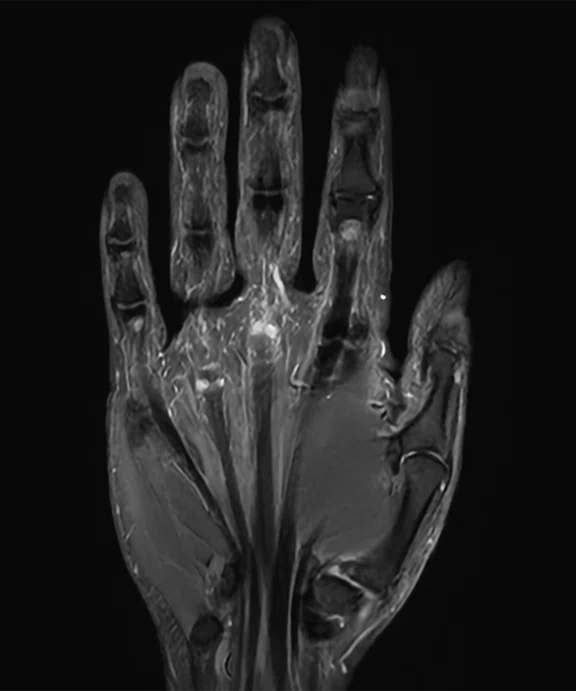
AIR™ Recon DL has made a significant difference for orthopedic imaging at Benefis Health System, including the ability to capture high-resolution images of the extremities, as shown in this finger exam. (A) Axial PD FRFSE FatSat, 0.4 x 0.5 x 3 mm, 60 slices in 3:02 min. and (B) coronal PD STIR, 0.6 x 0.8 x 3 mm, 18 slices in 1:54 min.
Figure 2.
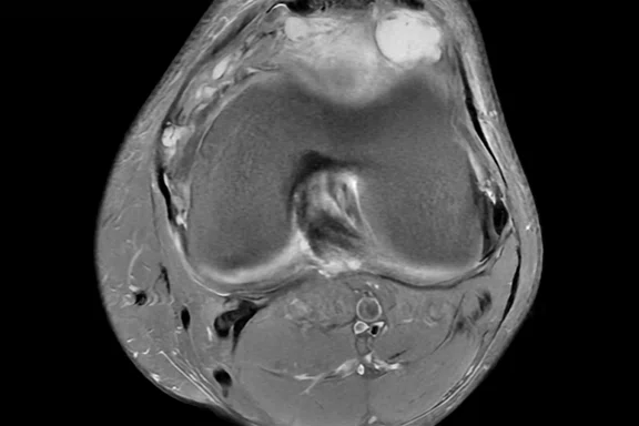
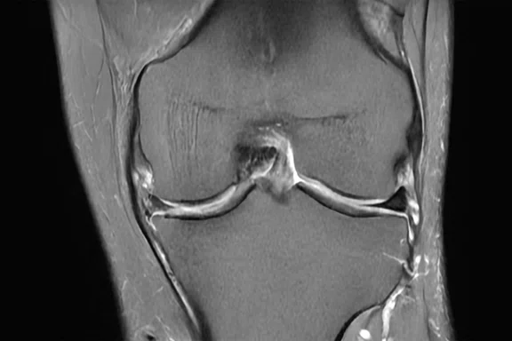
With SIGNA™ Voyager and AIR™ Recon DL, Benefis is now routinely performing MR knee exams in less than 10 minutes. In this case, the total exam time was 6:46 min. (A) Axial PD FSE FatSat, (B) coronal PD FSE FatSat, (C) coronal T1 FSE, (D) sagittal T2 FSE FatSat and (E) sagittal PD FSE, all acquired with AIR™ Recon DL.
Paul Fulbright, director of imaging at Benefis, says the health system chose SIGNA™ Voyager AIR™ IQ Edition because it has the latest technology with AIR™ Recon DL. “When they showed me the first time, it was pretty impressive. When I showed the orthopedic doctors the images and how short the exam time was, they were just blown away by it. That’s the technology that reeled me in.”
The three SIGNA™ Voyager systems, plus an existing non-GE HealthCare MR system, scan more than 50 patients per day across the three locations. Fulbright has seen significant improvements across all exams, including body, orthopedic, neuro and MSK imaging. “The advantage definitely is speed and quality. Orthopedic images are amazing on these new systems,” he says.
Figure 3.
All prostate exams are now performed with the 16ch AIR™ AA coil. (A) Axial T2 FRFSE, 0.6 x 0.8 x 3 mm, 50 slices in 3:42 min., (B) coronal T2 FRFSE, 0.6 x 0.6 x 3 mm, 36 slices in 3:15 min. and (C-E) axial MAGiC DWI, 2.2 x 4.3 x 3 mm, 50 slices in 9:58 min. with (C) b800, (D) MAGiC (synthetic) b1400 and (E) ADC map.
Figure 4.
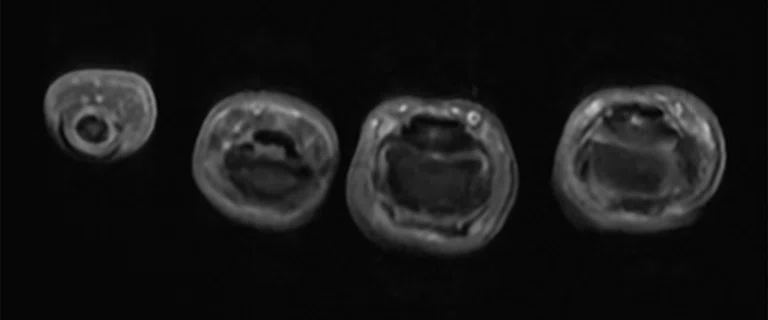
The combination of AIR™ AA Coil and AIR™ Recon DL has led to higher SNR, shorter scan times and greater patient comfort at Benefis. The AIR™ AA Coil facilitates the acquisition of large field-of-views, as shown in this long leg bone exam. (A) Coronal T1 FSE, 0.6 x 0.8 x 4 mm, 28 slices in 1:02 min., (B) coronal PD STIR, 1 x 1.2 x 4 mm, 28 slices in 1:38 min., (C) axial T1 FSE, 0.6 x 0.6 x 4 mm, 89 slices in 3:08 min. and (D) axial PD STIR, 0.7 x 0.9 x 5 mm, 89 slices in 4:00 min.
An increase in patient satisfaction
SIGNA™ Voyager AIR™ IQ Edition has helped Benefis improve patient throughput. Prior to installing these new systems, Benefis had a nearly month-long backlog of patients waiting for MR exams. Now they can schedule patients within just a few days. Fulbright estimates scanning timeslots are 33% shorter, with the majority of scanning time taking just 10-15 minutes. Previously, patients were scheduled in 45-minute slots and now the majority of scans are scheduled in 30-minute slots. At the outpatient imaging center in Helena, patients undergoing MR exams can now get in and out of the clinic within an hour.
“Our throughput has been amazing. We can get so many more patients done and have great images on top of it. It’s a huge satisfier. Patients are saying, ‘This is so fast.’ Doing a 10-minute shoulder MR or a 10-minute knee MR exam is just amazing.”
Paul Fulbright
Fulbright believes this could have clinical implications for patients, especially if they need multiple MR exams over time to monitor disease progression or treatment efficacy. “To be able to get more patients through means you can treat them quicker and get them on their treatment pathway sooner,” he says.
AIR™ Coils have further improved patient comfort and satisfaction during exams, particularly in the abdomen because they’re lightweight and less rigid. “We’re big fans of the AIR™ Coils. They’re light, they’re flexible and we wish we had every size,” he says. “It feels like a blanket and people are amazed, so that part is a customer satisfier. Patients comment, ‘that was so much better than last time and it’s so much faster.’”
Staff satisfaction on the rise
Benefis’ technologists and radiologists are equally impressed and satisfied with the SIGNA™ Voyager systems. They have been able to transfer many studies previously done on their existing 3.0T scanner to the SIGNA™ Voyager 1.5T systems, allowing them to balance out their workload. “We made MR more accessible. And now we can spread out that workload so it’s not siloed on one system,” says Fulbright.
The SIGNA™ Voyager systems have also allowed Benefis to expand imaging capabilities. The 70 cm wide bore has opened up opportunities for other exams, such as abdominal scans. “We didn’t ever put abdominal exams on the old 1.5T system. Now we do it all the time. With the wide bore, we don’t have to worry about somebody not fitting in the scanner anymore,” he says.
The new SIGNA™ Voyager systems have impacted image quality across all types of exams. “The biggest feedback I hear is that there’s so much definition now. In orthopedic work, there’s higher confidence because the clarity is amazing. It makes a big difference,” says Fulbright.
Figure 5.
Even in the presence of metal, SIGNA™ Voyager with AIR™ Recon DL and MAVRIC SL delivers excellent image quality, as shown in this knee exam. (A) Sagittal PD FSE, 0.4 x 0.6 x 4 mm, 28 slices in 1:01 min. with AIR™ Recon DL, (B) coronal T1 FSE, 0.5 x 0.7 x 4 mm, 30 slices in 1:06 min. with AIR™ Recon DL and (C) sagittal T2 MAVRIC SL, 0.7 x 0.9 x 5 mm, 24 slices in 5:27min.
Figure 6.
Even in the presence of metal, SIGNA™ Voyager with AIR™ Recon DL delivers excellent image quality, as shown in this lumbar spine exam. (A) Sagittal T2 FRFSE, 0.6 x 0.9 x 4 mm, 20 slices in 1:43 min., (B) sagittal PD STIR, 1 x 1.3 x 4 mm, 20 slices in 2:05 min. and (C) sagittal T1 FSE, 0.7 x 1 x 4 mm, 20 slices in 2:12 min.
Technologists and radiology staff are experiencing a spike in efficiency and satisfaction across locations. In the hospital setting, the SIGNA™ Voyager system has allowed staff to better accommodate inpatient scans in between outpatient scans. At the outpatient orthopedic center and standalone MR imaging center, they only need to schedule day shifts Monday through Friday, without a backlog of exams.
“Technologists are satisfied because they can get more done. On the hospital side, they need to squeeze in inpatient exams between outpatient exams, which is now easier because it only takes 10 to 12 minutes. They’re happy because they can get all their work done more efficiently, and they aren’t staying overtime.”
Paul Fulbright
All of this leads to staff accommodating a higher volume of patients, without more stress, and a better experience for referring physicians, an important source of revenue for Benefis. “My volume is above what I’m budgeting and my backlog is better. I have better ordering provider satisfaction because they’re not waiting three weeks for their patient to be scanned. I have better radiologist satisfaction because the images are great, and my techs are happy because we have the best technology,” says Fulbright.
Now that the systems have been in use for several months, it is clear to Fulbright and Ehlinger that Benefis made the right decision. “With GE HealthCare, I have a great service team locally, and I have a great sales team that tells me about all these opportunities. Plus, the systems are dependable. I’ve had a lot of GE HealthCare equipment and it lasts a long time,” adds Fulbright.


