A
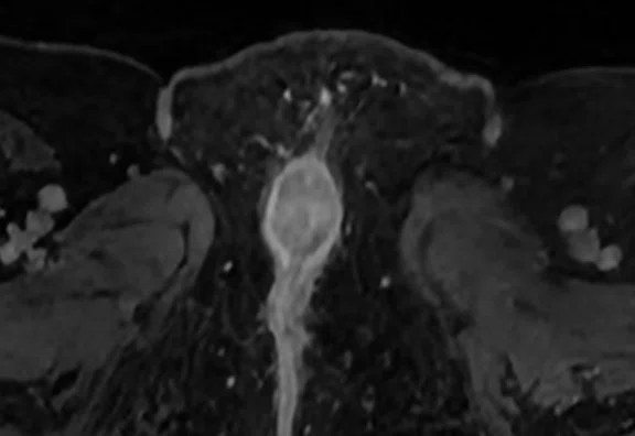
Figure 1.
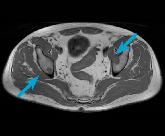
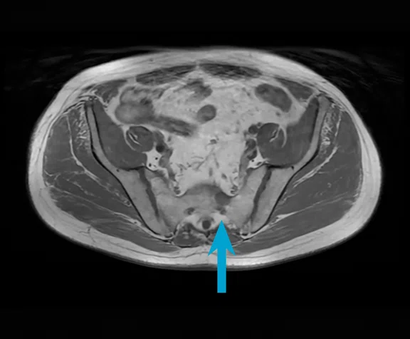
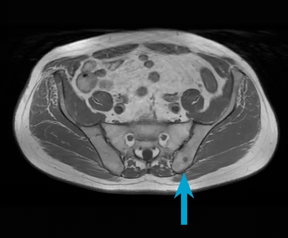
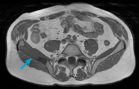
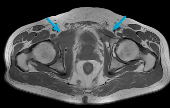
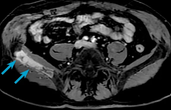
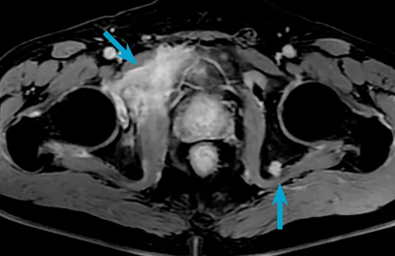
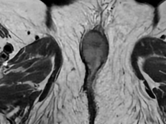
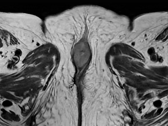
Multiple metastases from a primary tumor in the breast were found in the soft tissue of a 68-year-old man referred for a prostate exam. (A-E) Axial T1, 1 x 1.8 x 5 mm, 1:13 min. The use of AIR™ Recon DL reduced image noise for a clear depiction of the metastases.
B
Figure 1.
Multiple metastases from a primary tumor in the breast were found in the soft tissue of a 68-year-old man referred for a prostate exam. (A-E) Axial T1, 1 x 1.8 x 5 mm, 1:13 min. The use of AIR™ Recon DL reduced image noise for a clear depiction of the metastases.
C
Figure 1.
Multiple metastases from a primary tumor in the breast were found in the soft tissue of a 68-year-old man referred for a prostate exam. (A-E) Axial T1, 1 x 1.8 x 5 mm, 1:13 min. The use of AIR™ Recon DL reduced image noise for a clear depiction of the metastases.
D
Figure 1.
Multiple metastases from a primary tumor in the breast were found in the soft tissue of a 68-year-old man referred for a prostate exam. (A-E) Axial T1, 1 x 1.8 x 5 mm, 1:13 min. The use of AIR™ Recon DL reduced image noise for a clear depiction of the metastases.
E
Figure 1.
Multiple metastases from a primary tumor in the breast were found in the soft tissue of a 68-year-old man referred for a prostate exam. (A-E) Axial T1, 1 x 1.8 x 5 mm, 1:13 min. The use of AIR™ Recon DL reduced image noise for a clear depiction of the metastases.
A
Figure 2.
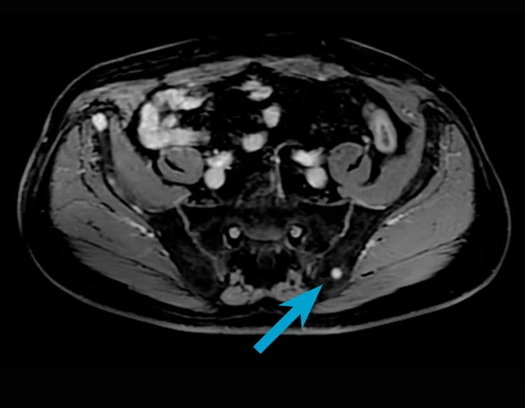
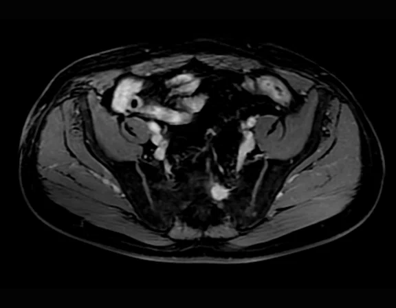
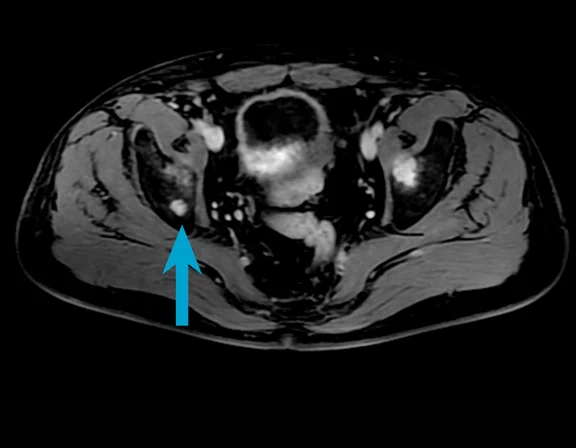
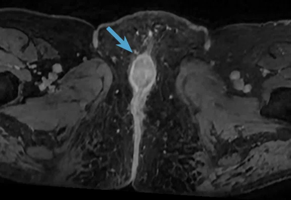
Same patient as Figure 1. (A-E) Axial LAVA Flex, 1.3 x 1.8 x 4 mm, 1:12 min.
B
Figure 2.
Same patient as Figure 1. (A-E) Axial LAVA Flex, 1.3 x 1.8 x 4 mm, 1:12 min.
C
Figure 2.
Same patient as Figure 1. (A-E) Axial LAVA Flex, 1.3 x 1.8 x 4 mm, 1:12 min.
D
Figure 2.
Same patient as Figure 1. (A-E) Axial LAVA Flex, 1.3 x 1.8 x 4 mm, 1:12 min.
E
Figure 2.
Same patient as Figure 1. (A-E) Axial LAVA Flex, 1.3 x 1.8 x 4 mm, 1:12 min.
A
Figure 3.
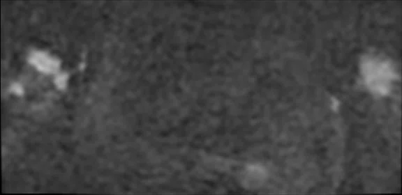
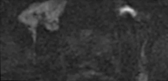
Same patient as Figure 1. (A, B) Axial Focus DWI with AIR™ Recon DL, b1500, 3 mm slice thickness, 37 slices for both b50 (not shown) and b1500, 5:40 min.
B
Figure 3.
Same patient as Figure 1. (A, B) Axial Focus DWI with AIR™ Recon DL, b1500, 3 mm slice thickness, 37 slices for both b50 (not shown) and b1500, 5:40 min.
A
Figure 4.
Same patient as Figure 1. (A-C) Axial 3D LAVA with AIR™ Recon DL, 1.3 x 1.3 x 1 mm, 1:57 min.
B
Figure 4.
Same patient as Figure 1. (A-C) Axial 3D LAVA with AIR™ Recon DL, 1.3 x 1.3 x 1 mm, 1:57 min.
C
Figure 4.
Same patient as Figure 1. (A-C) Axial 3D LAVA with AIR™ Recon DL, 1.3 x 1.3 x 1 mm, 1:57 min.
A
Figure 5.
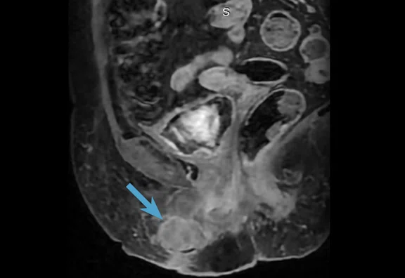
A 74-year-old female was referred with a diagnosis of suspected stage 3 vulvar cancer. AIR™ Recon DL 2D was utilized in each sequence. (A) Sagittal T2, 0.6 x 1.1 x 3.5 mm, 2:32 min.; (B) axial DWI, 2.7 x 2.7 x 5 mm, 1:50 min.; and (C) axial ADC.
B
Figure 5.
A 74-year-old female was referred with a diagnosis of suspected stage 3 vulvar cancer. AIR™ Recon DL 2D was utilized in each sequence. (A) Sagittal T2, 0.6 x 1.1 x 3.5 mm, 2:32 min.; (B) axial DWI, 2.7 x 2.7 x 5 mm, 1:50 min.; and (C) axial ADC.
C
Figure 5.
A 74-year-old female was referred with a diagnosis of suspected stage 3 vulvar cancer. AIR™ Recon DL 2D was utilized in each sequence. (A) Sagittal T2, 0.6 x 1.1 x 3.5 mm, 2:32 min.; (B) axial DWI, 2.7 x 2.7 x 5 mm, 1:50 min.; and (C) axial ADC.
A
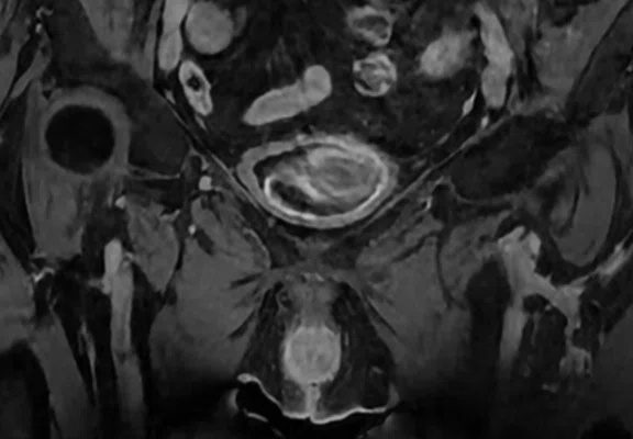
Figure 6.
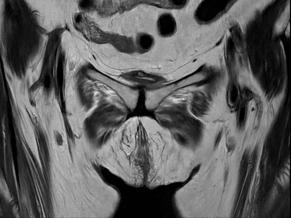
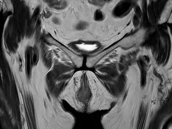
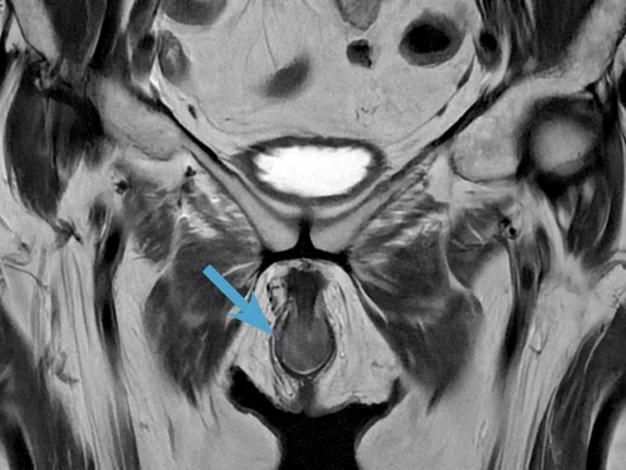
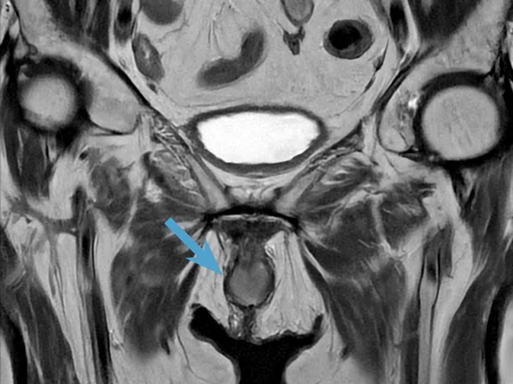
Same patient as Figure 5 with AIR™ Recon DL PROPELLER applied to the T2 acquisition. (A-D) Coronal T2 PROPELLER, 3.4 x 3.4 x 3 mm, 3:29 min.; (E, F) axial T2 PROPELLER, 0.8 x 0.8 x 3 mm, 3:11 min.
B
Figure 6.
Same patient as Figure 5 with AIR™ Recon DL PROPELLER applied to the T2 acquisition. (A-D) Coronal T2 PROPELLER, 3.4 x 3.4 x 3 mm, 3:29 min.; (E, F) axial T2 PROPELLER, 0.8 x 0.8 x 3 mm, 3:11 min.
C
Figure 6.
Same patient as Figure 5 with AIR™ Recon DL PROPELLER applied to the T2 acquisition. (A-D) Coronal T2 PROPELLER, 3.4 x 3.4 x 3 mm, 3:29 min.; (E, F) axial T2 PROPELLER, 0.8 x 0.8 x 3 mm, 3:11 min.
D
Figure 6.
Same patient as Figure 5 with AIR™ Recon DL PROPELLER applied to the T2 acquisition. (A-D) Coronal T2 PROPELLER, 3.4 x 3.4 x 3 mm, 3:29 min.; (E, F) axial T2 PROPELLER, 0.8 x 0.8 x 3 mm, 3:11 min.
E
Figure 6.
Same patient as Figure 5 with AIR™ Recon DL PROPELLER applied to the T2 acquisition. (A-D) Coronal T2 PROPELLER, 3.4 x 3.4 x 3 mm, 3:29 min.; (E, F) axial T2 PROPELLER, 0.8 x 0.8 x 3 mm, 3:11 min.
F
Figure 6.
Same patient as Figure 5 with AIR™ Recon DL PROPELLER applied to the T2 acquisition. (A-D) Coronal T2 PROPELLER, 3.4 x 3.4 x 3 mm, 3:29 min.; (E, F) axial T2 PROPELLER, 0.8 x 0.8 x 3 mm, 3:11 min.
A
Figure 7.
Same patient as Figure 5 with AIR™ Recon DL 3D applied to the LAVA sequence, which enabled reducing the number of sequences to one isotropic volumetric dataset with equal sharpness in all three planes, hence reducing total exam time. (A-D) 3D LAVA, 1.1 x 1.2 x 1.4 mm, 2:49 min.
B
Figure 7.
Same patient as Figure 5 with AIR™ Recon DL 3D applied to the LAVA sequence, which enabled reducing the number of sequences to one isotropic volumetric dataset with equal sharpness in all three planes, hence reducing total exam time. (A-D) 3D LAVA, 1.1 x 1.2 x 1.4 mm, 2:49 min.
C
Figure 7.
Same patient as Figure 5 with AIR™ Recon DL 3D applied to the LAVA sequence, which enabled reducing the number of sequences to one isotropic volumetric dataset with equal sharpness in all three planes, hence reducing total exam time. (A-D) 3D LAVA, 1.1 x 1.2 x 1.4 mm, 2:49 min.
D
Figure 7.
Same patient as Figure 5 with AIR™ Recon DL 3D applied to the LAVA sequence, which enabled reducing the number of sequences to one isotropic volumetric dataset with equal sharpness in all three planes, hence reducing total exam time. (A-D) 3D LAVA, 1.1 x 1.2 x 1.4 mm, 2:49 min.
result


PREVIOUS
${prev-page}
NEXT
${next-page}
Subscribe Now
Manage Subscription
FOLLOW US
Contact Us • Cookie Preferences • Privacy Policy • California Privacy PolicyDo Not Sell or Share My Personal Information • Terms & Conditions • Security
© 2024 GE HealthCare. GE is a trademark of General Electric Company. Used under trademark license.
Case Studies
Pushing the limits of oncologic pelvic MR imaging with 3D and motion-insensitive acquisitions
Pushing the limits of oncologic pelvic MR imaging with 3D and motion-insensitive acquisitions
By Lukasz Pruszynski, Lead Radiographer, Radom Oncology Center, Radom, Poland
The 2D PROPELLER technique is a widely recognized clinical application that relies on radial spin echo principles. It demonstrates resilience to motion interference and effectively eliminates pulsation artifacts. With this method, it is possible to obtain consistently sharp, motion-free images featuring the desired tissue contrast in a single scan.
While the use of 2D PROPELLER may serve as a secondary option in certain anatomical situations, it becomes the primary sequence choice in others, such as the female pelvis, shoulder examinations or in patients prone to agitation.
We recently upgraded our system to the SIGNA™ Explorer 1.5T with MR 30 for SIGNA™. This allows us to use AIR™ Recon DL 3D on prostate and other pelvic cases. It also enables us to use AIR™ Recon DL for PROPELLER motion-insensitive imaging and 3D LAVA, a 3D isotropic sequence, which shows considerable clinical benefit over conventional reconstruction with an improvement in image sharpness and SNR, along with the reduction in both motion artifacts and scan time. We also use 3D LAVA Flex.
Before the introduction of AIR™ Recon DL 3D, 3D sequences with isotropic properties often had suboptimal multiplanar reconstructions, especially when the technologist attempted to accelerate scan time. By incorporating AIR™ Recon DL into 3D sequences, we are able to enhance SNR and achieve equally sharp images in all three planes, irrespective of the scanning plane. In essence, we only need to scan one plane and then subsequently reformat it according to clinical preferences without sacrificing image quality or fine details.
Case #1
Patient history
A 68-year-old male with breast cancer was referred for an MR of the pelvis to assess possible metastases.
Results
Prostate scored PI-RADS® 1, showing no clinical indication of cancer. Metastases in the soft tissues and metastatic changes to the bones were observed.
The prostate gland measures 44/34/43 mm (33 ml). It appears normal in the peripheral, transitional and central zones, with smooth external outlines and without obvious features. The seminal vesicles were not enlarged and showed no signs of infiltration.
The bladder, well-filled with urine, showed no changes. The sigmoid colon presented numerous diverticula. Additionally, the rectum and sigmoid colon have shrunk, with no other changes visible.
The fatty tissue in the small pelvis showed no signs of infiltrative changes. There were no significantly enlarged lymph nodes in the pelvis, and there was no fluid present in the peritoneal cavity in the pelvis.
Osteolytic lesions in the examined bones measured approximately 45 mm x 70 mm, affecting mainly the upper part, the shaft of the right pubic bone and parts of the acetabulum of the right hip joint. Metastasis was observed in the plate of the right ilium (up to 85 mm), and smaller foci appeared in the shafts of the iliac bones and in the sacrum.
Case #2
Patient history
A 74-year-old female was referred with a diagnosis of suspected stage 3 vulvar cancer. An outpatient gynecological exam showed ulceration of the left labia with infiltration into the bone, and suspected infiltration of the urethra and anal sphincter.
She received radiotherapy on the infiltrative lesions of the vulva. After irradiation, partial regression of the infiltration was obtained with a reaction grade II. She was discharged home in good condition with the recommendation for an MR exam.
Results
AIR™ Recon DL was used for a dynamic study, as well as for an overview of the whole pelvis post-contrast (3D coronal LAVA isotropic). We also applied AIR™ Recon DL PROPELLER for small FOV axial and coronal T2 images, as well as LAVA and DWI.
The pelvic MR exam included FSE and T1 and T2 sequences, with fat saturation, DWI/ADC, and dynamic LAVA examination after administration of a paramagnetic contrast agent in transverse, coronal and sagittal sections.
A nodular mass emerging from the left labia was observed, measuring approximately 47 x 72 x 25 mm, infiltrating fatty tissue and likely infiltrating the opening of the urethra and vagina.
Infiltration of the anterior part of the anal sphincter could not be ruled out, but the assessment was uncertain. The uterus was rebuilt in a myomatous manner, with the largest fibroid measuring up to 50 mm. The normal uterus was not visualized.
Small atrophic ovaries were observed, and the bladder, rectum and sigmoid colon were unchanged. The lymph nodes did not have any features suspicious for metastasis. A structure was observed at the end of the coccyx measuring approximately 13-14 mm, which could be a small fistula. No free fluid was detected in the pelvis. The skeletal system was examined without any visible metastatic lesions.
Discussion
The prostate MR examination was performed with the SIGNA™ Explorer 1.5T and a surface coil. AIR™ Recon DL PROPELLER was used in all T2 images. The patient was anxious about the exam, so PROPELLER was used to guarantee we captured the images in one scan without repetitions. We applied LAVA isotropic imaging in the prostate region, and LAVA Flex for dynamic study and an overview of the entire pelvis post-contrast. We also used FOCUS DWI, as diffusion is critical in a multi-parametric MR prostate exam.
In the female pelvis case, AIR™ Recon DL 3D increased resolution and enabled us to reduce the number of sequences to one isotropic volumetric dataset with equal sharpness in all three planes, reducing total exam time. The use of AIR™ Recon DL PROPELLER reduced motion artifacts, allowing for the depiction of a small infiltration. The radiologist was also able to differentiate between residual changes and post-therapeutic changes and the degree of infiltration of adjacent structures, which was necessary for local staging of the disease. Further, the narrow connection of the dominant vulvar infiltration with a narrow pedicle within the vaginal vestibule and the distal part of the vagina was clearly captured in this MR examination. It can be difficult to visualize these anatomies with other imaging methods.
Upgrading to the SIGNA™ Explorer with MR 30 for SIGNA™ enabled us to ascend to a higher level of MR capabilities that were previously inaccessible within this modality. Of significant importance for us was the ease of use and a trouble-free experience with the new software. Our technologists have seamlessly integrated MR 30 for SIGNA™ features into their workflow.
In these cases, the fast scan times with the use of AIR™ Recon DL 3D and motion-insensitive imaging afforded by AIR™ Recon DL PROPELLER were essential to capturing diagnostic-quality images when patients were in significant pain and unable to hold still for long periods of time. These technological advancements in the MR 30 for SIGNA™ upgrade allowed us to address these issues and delivered crystal-clear images that made diagnosing and measuring lesions and other anomalies simple and accurate.
DOWNLOAD ARTICLE HERE


