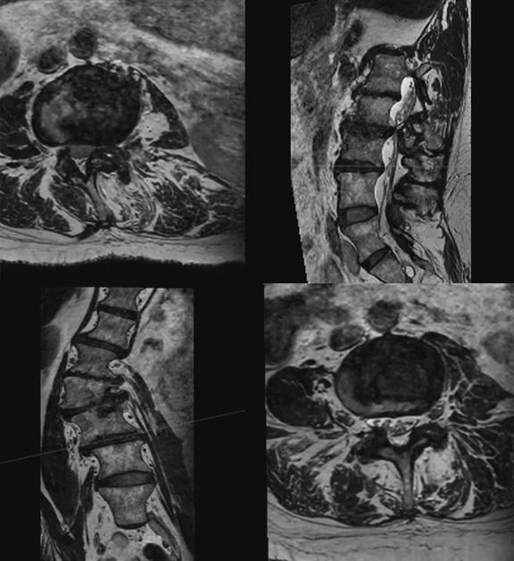
Figure 2.
3D sagittal acquisition in the lumbar spine with AIR™ Recon DL. All images were acquired with a research prototype. Images courtesy of Hospital for Special Surgery.
A
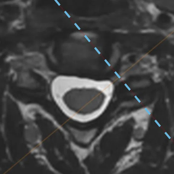
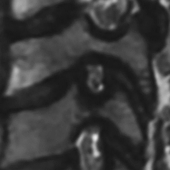
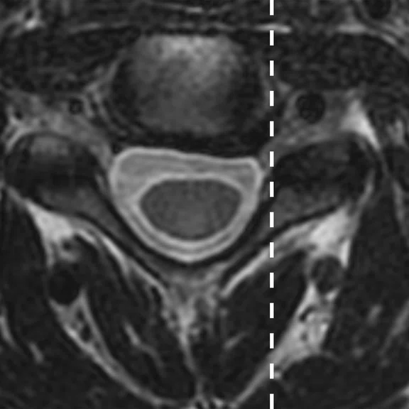
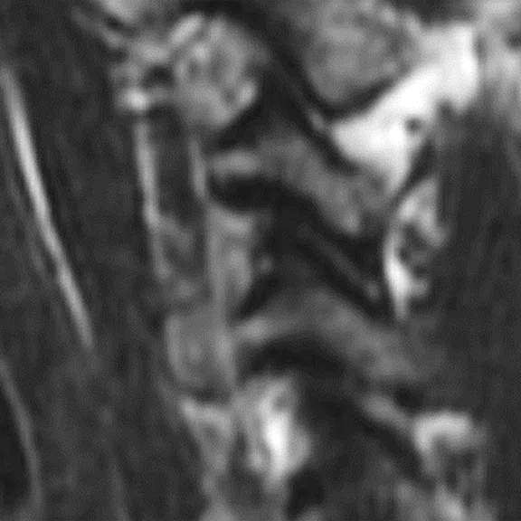
Figure 1.
Improved image quality from AIR™ Recon DL 3D combined with 3D multiplanar reformatting. (A) 3D axial reformat, (B) 3D orthogonal reformat, (C) 2D axial and (D) 2D sagittal. All images were acquired with a research prototype. Images courtesy of Hospital for Special Surgery.
B
Figure 1.
Improved image quality from AIR™ Recon DL 3D combined with 3D multiplanar reformatting. (A) 3D axial reformat, (B) 3D orthogonal reformat, (C) 2D axial and (D) 2D sagittal. All images were acquired with a research prototype. Images courtesy of Hospital for Special Surgery.
C
Figure 1.
Improved image quality from AIR™ Recon DL 3D combined with 3D multiplanar reformatting. (A) 3D axial reformat, (B) 3D orthogonal reformat, (C) 2D axial and (D) 2D sagittal. All images were acquired with a research prototype. Images courtesy of Hospital for Special Surgery.
D
Figure 1.
Improved image quality from AIR™ Recon DL 3D combined with 3D multiplanar reformatting. (A) 3D axial reformat, (B) 3D orthogonal reformat, (C) 2D axial and (D) 2D sagittal. All images were acquired with a research prototype. Images courtesy of Hospital for Special Surgery.
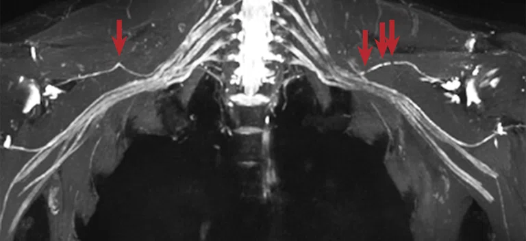
Figure 3.
A 43-year-old firefighter with bilateral Parsonage-Turner syndrome following firefight with suprascapular nerve constrictions (arrows). Acquired on SIGNA™ Premier with AIR™ Coils Multi-Purpose Coils, Large and Medium. MIP is from post-contrast Cube STIR reconstructed with AIR™ Recon DL 3D, 6:18 min. All images were acquired with a research prototype. Image courtesy of Hospital for Special Surgery.


result
A
Figure 4.
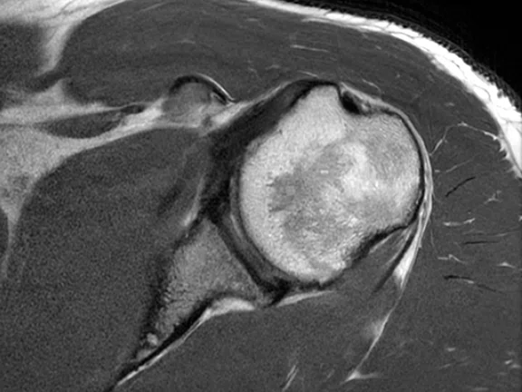
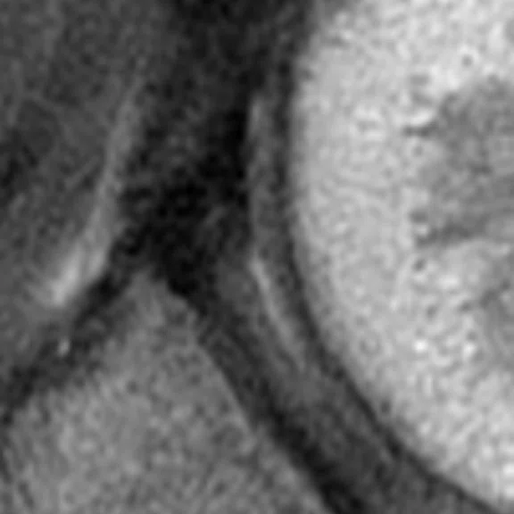
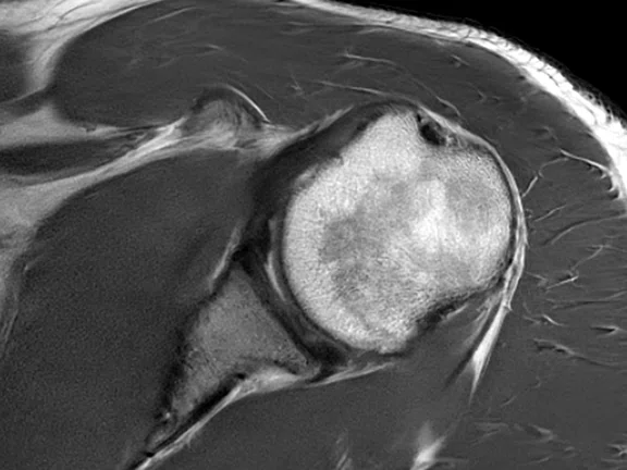
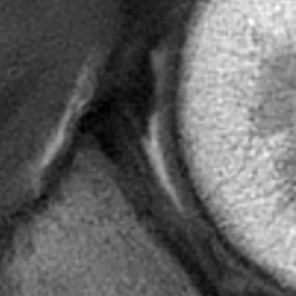
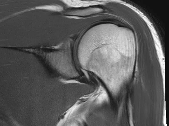
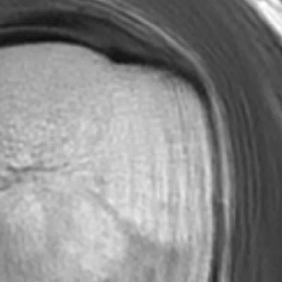
Shoulder exams demonstrating the value of AIR™ Recon DL PROPELLER. (A, B) Fissure at the base of labrum. (A) Axial 2D FSE PD with AIR™ Recon DL and (B) same acquisition with AIR™ Recon DL PROPELLER. (C, D) Thickening of bursae, (C) coronal 2D FSE PD with AIR™ Recon DL and (D) same acquisition with AIR™ Recon DL PROPELLER. All images were acquired with a research prototype. Images courtesy of Hospital for Special Surgery.
C
Figure 4.
Shoulder exams demonstrating the value of AIR™ Recon DL PROPELLER. (A, B) Fissure at the base of labrum. (A) Axial 2D FSE PD with AIR™ Recon DL and (B) same acquisition with AIR™ Recon DL PROPELLER. (C, D) Thickening of bursae, (C) coronal 2D FSE PD with AIR™ Recon DL and (D) same acquisition with AIR™ Recon DL PROPELLER. All images were acquired with a research prototype. Images courtesy of Hospital for Special Surgery.
B
Figure 4.
Shoulder exams demonstrating the value of AIR™ Recon DL PROPELLER. (A, B) Fissure at the base of labrum. (A) Axial 2D FSE PD with AIR™ Recon DL and (B) same acquisition with AIR™ Recon DL PROPELLER. (C, D) Thickening of bursae, (C) coronal 2D FSE PD with AIR™ Recon DL and (D) same acquisition with AIR™ Recon DL PROPELLER. All images were acquired with a research prototype. Images courtesy of Hospital for Special Surgery.
D
Figure 4.
Shoulder exams demonstrating the value of AIR™ Recon DL PROPELLER. (A, B) Fissure at the base of labrum. (A) Axial 2D FSE PD with AIR™ Recon DL and (B) same acquisition with AIR™ Recon DL PROPELLER. (C, D) Thickening of bursae, (C) coronal 2D FSE PD with AIR™ Recon DL and (D) same acquisition with AIR™ Recon DL PROPELLER. All images were acquired with a research prototype. Images courtesy of Hospital for Special Surgery.
A
Figure 4.
Shoulder exams demonstrating the value of AIR™ Recon DL PROPELLER. (A, B) Fissure at the base of labrum. (A) Axial 2D FSE PD with AIR™ Recon DL and (B) same acquisition with AIR™ Recon DL PROPELLER. (C, D) Thickening of bursae, (C) coronal 2D FSE PD with AIR™ Recon DL and (D) same acquisition with AIR™ Recon DL PROPELLER. All images were acquired with a research prototype. Images courtesy of Hospital for Special Surgery.
B
Figure 4.
Shoulder exams demonstrating the value of AIR™ Recon DL PROPELLER. (A, B) Fissure at the base of labrum. (A) Axial 2D FSE PD with AIR™ Recon DL and (B) same acquisition with AIR™ Recon DL PROPELLER. (C, D) Thickening of bursae, (C) coronal 2D FSE PD with AIR™ Recon DL and (D) same acquisition with AIR™ Recon DL PROPELLER. All images were acquired with a research prototype. Images courtesy of Hospital for Special Surgery.
A
Figure 4.
Shoulder exams demonstrating the value of AIR™ Recon DL PROPELLER. (A, B) Fissure at the base of labrum. (A) Axial 2D FSE PD with AIR™ Recon DL and (B) same acquisition with AIR™ Recon DL PROPELLER. (C, D) Thickening of bursae, (C) coronal 2D FSE PD with AIR™ Recon DL and (D) same acquisition with AIR™ Recon DL PROPELLER. All images were acquired with a research prototype. Images courtesy of Hospital for Special Surgery.
C
Figure 4.
Shoulder exams demonstrating the value of AIR™ Recon DL PROPELLER. (A, B) Fissure at the base of labrum. (A) Axial 2D FSE PD with AIR™ Recon DL and (B) same acquisition with AIR™ Recon DL PROPELLER. (C, D) Thickening of bursae, (C) coronal 2D FSE PD with AIR™ Recon DL and (D) same acquisition with AIR™ Recon DL PROPELLER. All images were acquired with a research prototype. Images courtesy of Hospital for Special Surgery.
D
Figure 4.
Shoulder exams demonstrating the value of AIR™ Recon DL PROPELLER. (A, B) Fissure at the base of labrum. (A) Axial 2D FSE PD with AIR™ Recon DL and (B) same acquisition with AIR™ Recon DL PROPELLER. (C, D) Thickening of bursae, (C) coronal 2D FSE PD with AIR™ Recon DL and (D) same acquisition with AIR™ Recon DL PROPELLER. All images were acquired with a research prototype. Images courtesy of Hospital for Special Surgery.
PREVIOUS
${prev-page}
NEXT
${next-page}
Subscribe Now
Manage Subscription
FOLLOW US
Contact Us • Cookie Preferences • Privacy Policy • California Privacy PolicyDo Not Sell or Share My Personal Information • Terms & Conditions • Security
© 2024 GE HealthCare. GE is a trademark of General Electric Company. Used under trademark license.
IN PRACTICE
Delivering on the promise of more deep-learning-based MR image reconstruction
Delivering on the promise of more deep-learning-based MR image reconstruction
With the recent FDA clearance of AIR™ Recon DL for 3D and PROPELLER imaging sequences, the benefits of AIR™ Recon DL are now extended to nearly all MR clinical procedures, covering all anatomies and enabling better image quality, shorter scan times and an enhanced patient experience. AIR™ Recon DL for 3D and PROPELLER are commercially available via the MR 30 for SIGNA™ software release.
With the recent FDA clearance of AIR™ Recon DL for 3D and PROPELLER imaging sequences, the benefits of AIR™ Recon DL are now extended to nearly all MR clinical procedures, covering all anatomies and enabling better image quality, shorter scan times and an enhanced patient experience. AIR™ Recon DL for 3D and PROPELLER are commercially available via the MR 30 for SIGNA™ software release.
Physicians have already realized the clinical benefit of improved SNR and sharpness that has fundamentally shifted the balance between image quality and scan time. Now they will be able to realize the same gains in image quality and shorter scan times in 3D imaging for greater clinical efficiency and to eliminate the need for multiple 2D acquisitions, potentially leading to faster diagnoses.
AIR™ Recon DL is now compatible with PROPELLER, a motion-insensitive imaging sequence particularly important for anatomies susceptible to motion, such as respiration, as well as pediatric, neurodegenerative, geriatric and claustrophobic patients who have difficulty remaining physically still for the duration of an MR scan. Often, a repeat scan is required when motion degrades the MR image, leading to longer exams times, less patient throughout and a poor patient experience.
“With AIR™ Recon DL 3D you can’t tell the difference between the acquisition plane and the reformatted planes when you have isotropic voxels,” says Tiron Pechet, MD, Assistant Medical Director at Shields Health. “This is a big improvement for Cube imaging, where the acquisition and reformatted planes were not equivalent without AIR™ Recon DL, even though voxels were isotropic.”
Specifically, Dr. Pechet expects that the reformats from
AIR™ Recon DL 3D will be sufficient to eliminate acquiring the same sequence in multiple planes, saving significant time by eliminating the need to acquire the same information multiple times. He also anticipates using AIR™ Recon DL 3D to improve imaging of the cervical and thoracic cord in multiple sclerosis patients for better lesion and plaque identification. Extending AIR™ Recon DL to 3D sequences is a crucial next step, he says.
“We already use it in 2D acquisitions where we’ve been able to improve image quality while saving time,” he continues. “And then, we have these examinations that have multiple 3D sequences. If we can apply AIR™ Recon DL across the board, to 3D as well as 2D pulse sequences, it’s a benefit for all patients and will improve their experience.”
In MR neurography, the ability to shorten lengthy 3D acquisition times is key for reducing motion artifacts when scanning anatomies such as the brachial plexus and lumbosacral plexus. At the Hospital for Special Surgery (HSS), Darryl Sneag, MD, Director of MRI Research and MR neurography, and his colleagues previously did not use acceleration for these exams because of the need to obtain high spatial resolution, 1 mm isotropic acquisitions. AIR™ Recon DL changes the game.
“I’m very excited about AIR™ Recon DL 3D. One of the challenges we’ve had for a very long time with 3D imaging is to obtain sufficient in-plane and through-plane resolution and to acquire isotropic voxels. Those elements are critical to avoid the appearance of averaging or distortion when the data is reformatted into arbitrary planes. At the same time,
AIR™ Recon DL allows us to acquire 3D sequences with dramatically reduced acquisition times.”
Now with AIR™ Recon DL, HSS can add parallel imaging and HyperSense to the MR neurography and more standard joint imaging protocols to further reduce scan times.
“A 3D acquisition in half the scan time that maintains image quality is tremendous,” Dr. Sneag adds. “The overall image sharpness is slightly better and these acquisitions without AIR™ Recon DL would otherwise be too noisy for adequate interpretation.”
In neuro imaging, the impact of AIR™ Recon DL is equally significant, particularly in centers such as the University of California San Francisco that have a high volume of 3D neuro imaging. Javier Villanueva-Meyer, MD, Associate Professor of Neuroradiology, specializes in brain tumor imaging. He estimates that 80% of brain MR imaging protocols are 3D at his institution.
“We have an immediate need to increase our capacity for MR neuro imaging in both the inpatient and outpatient settings,” Dr. Villanueva-Meyer says.
“The ability to accelerate imaging with
AIR™ Recon DL in neuro cases is going to enhance our turnaround times and ability to get quick answers to very targeted questions in our emergency department and observation units.”
Dr. Javier Villanueva-Meyer
Figure 3.
A 43-year-old firefighter with bilateral Parsonage-Turner syndrome following firefight with suprascapular nerve constrictions (arrows). Acquired on SIGNA™ Premier with AIR™ Coils Multi-Purpose Coils, Large and Medium. MIP is from post-contrast Cube STIR reconstructed with AIR™ Recon DL 3D, 6:18 min. All images were acquired with a research prototype. Image courtesy of Hospital for Special Surgery.
He has explored the use of 3D imaging in the pituitary gland, hippocampus and the superficial vessels for temporal arteritis. In infiltrated brain tumor cases, Dr. Villanueva-Miller and his colleagues rely on high-resolution 3D T2 FLAIR, while
3D PROMO is crucial for cases where motion correction is needed.
“Since realizing the promise of AIR™ Recon DL in 2D imaging, we always felt that there is one piece that was missing to unlock its full potential – and that was its use in 3D imaging,” Dr. Villanueva-Meyer adds. “With long wait times, the ability to reduce scan times in our most commonly acquired studies, such as brain MRI, will permit increased capacity across our healthcare system.”
In body and cardiac imaging, Melany Atkins, MD, Division Chief, Body/Cardiovascular Imaging and Medical Director at Fairfax MRI Center, says the ability to make thin section reformats for coronal and sagittal planes rather than acquire them with AIR™ Recon DL will deliver the added benefit of enabling the organization to perform more isotropic imaging with better resolution. Patients benefit, too, especially as the demand for MR imaging increases.
“The utilization of cardiac MR has skyrocketed over the last decade,” she says. “But it is such a labor-intensive and time-consuming study, especially for the patient who has to hold their breath. Anything we can do so the patient doesn’t have to hold their breath simply makes their experience better – and then we usually get a better quality exam, too.”
One of her goals is to provide patients with a free-breathing cardiac exam in less than 30 minutes. AIR™ Recon DL 3D is an important component that will help her achieve this.
Dr. Atkins also intends to work with her team to evaluate the use of PROPELLER in body imaging. What was once their fallback sequence in cases of excessive patient motion may now become the practice’s primary protocol.
“We will look at replacing many of our traditional high-resolution T2 sequences with PROPELLER because of the improvements in image quality with AIR™ Recon DL,” Dr. Atkins says. “It is just so motion robust, and we won’t have to repeat sequences.”
Figure 4.
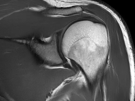
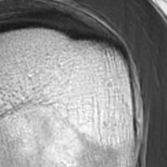
Shoulder exams demonstrating the value of AIR™ Recon DL PROPELLER. (A, B) Fissure at the base of labrum. (A) Axial 2D FSE PD with AIR™ Recon DL and (B) same acquisition with AIR™ Recon DL PROPELLER. (C, D) Thickening of bursae, (C) coronal 2D FSE PD with AIR™ Recon DL and (D) same acquisition with AIR™ Recon DL PROPELLER. All images were acquired with a research prototype. Images courtesy of Hospital for Special Surgery.
Ali Pirasteh, MD, Associate Chief of MRI and Assistant Professor at the University of Wisconsin-Madison, believes a large number of sequences in abdominal imaging can benefit from both AIR™ Recon DL 3D and PROPELLER, especially when acquisitions are long and motion is a consistent issue. Traditionally, obtaining both adequate SNR and anatomic detail within a reasonable scan time required a compromise, and sometimes was not even feasible.
“For prostate or rectal cancer patients, AIR™ Recon DL with PROPELLER enables high-resolution image acquisitions in a reasonable time, while mitigating peristaltic motion. This is an area where high-resolution/accurate delineation of the anatomy is important,” Dr. Pirasteh says.
“Patients with chronic liver disease or metastatic disease to the liver are often too sick to perform an adequate, or long enough, breath-hold,” he adds. “By acquiring noisier images through a faster acquisition, the breath-hold is more doable for the patient, while AIR™ Recon DL can provide a diagnostic-quality image through de-noising. This would not be feasible otherwise.”
Dr. Pirasteh sees several areas where AIR™ Recon DL for 3D
and/or PROPELLER can rescue an acquisition that may otherwise be suboptimal or non-diagnostic in difficult patient cases. For example, using AIR™ Recon DL PROPELLER when a Cartesian-based breath-hold in the liver fails, or using it to replace free-breathing or navigated high-resolution sequences when detecting liver metastases.
Dr. Pechet also envisions that his practice will maximize the benefit that AIR™ Recon DL PROPELLER brings to musculoskeletal (MSK) imaging. “Initially, we were using PROPELLER for the majority of our MSK examinations until AIR™ Recon DL FSE became available. Now, with AIR™ Recon DL PROPELLER looking so impressive, I suspect we’re going to be transitioning all 2D FSE scanning back to PROPELLER. We find that using PROPELLER imaging significantly decreases scan repeats for motion, and the absence of the motion artifacts leads to greater diagnostic confidence.”


