A
Figure 1.
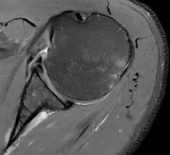
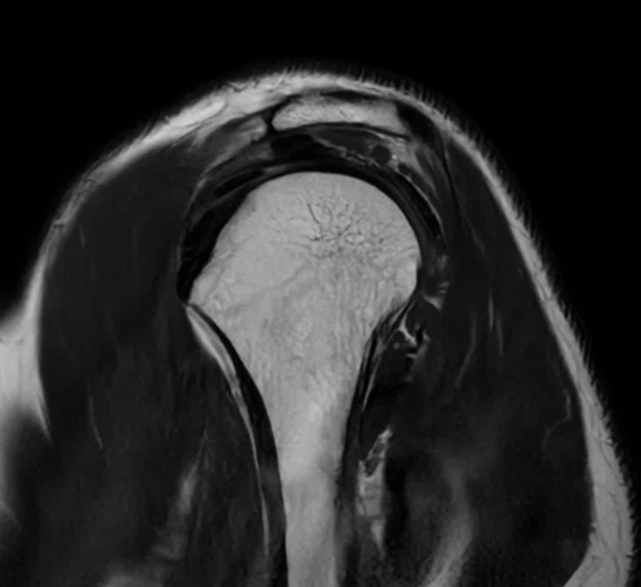
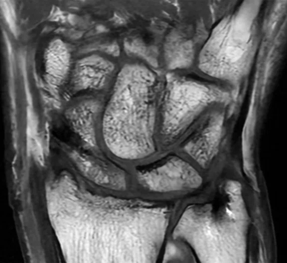
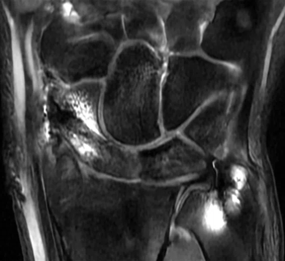
(A) Shoulder axial PD FatSat, 0.5 x 0.6 x 4.0 mm, 1:49 min.; (B) shoulder sagittal T2, 0.5 x 0.6 x 3.5 mm, 1:07 min.; (C) wrist coronal T1, 0.3 x 0.5 x 2.5 mm, 1:24 min.; and (D) wrist coronal STIR, 0.3 x 0.4 x 2.5 mm, 2:36 min.
B
Figure 1.
(A) Shoulder axial PD FatSat, 0.5 x 0.6 x 4.0 mm, 1:49 min.; (B) shoulder sagittal T2, 0.5 x 0.6 x 3.5 mm, 1:07 min.; (C) wrist coronal T1, 0.3 x 0.5 x 2.5 mm, 1:24 min.; and (D) wrist coronal STIR, 0.3 x 0.4 x 2.5 mm, 2:36 min.
C
Figure 1.
(A) Shoulder axial PD FatSat, 0.5 x 0.6 x 4.0 mm, 1:49 min.; (B) shoulder sagittal T2, 0.5 x 0.6 x 3.5 mm, 1:07 min.; (C) wrist coronal T1, 0.3 x 0.5 x 2.5 mm, 1:24 min.; and (D) wrist coronal STIR, 0.3 x 0.4 x 2.5 mm, 2:36 min.
D
Figure 1.
(A) Shoulder axial PD FatSat, 0.5 x 0.6 x 4.0 mm, 1:49 min.; (B) shoulder sagittal T2, 0.5 x 0.6 x 3.5 mm, 1:07 min.; (C) wrist coronal T1, 0.3 x 0.5 x 2.5 mm, 1:24 min.; and (D) wrist coronal STIR, 0.3 x 0.4 x 2.5 mm, 2:36 min.
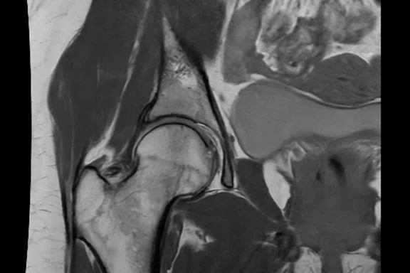
A
Figure 2.
All images with AIR™ Recon DL. (A) Hip coronal T1 post-contrast, 0.6 x 1.0 x 3.5 mm,
1:23 min.; (B) knee sagittal PD, 0.4 x 0.6 x 3.5 mm, 59 sec.; and (C) whole foot sagittal STIR, 0.4 x 0.6 x 3.0 mm, 2:28min.
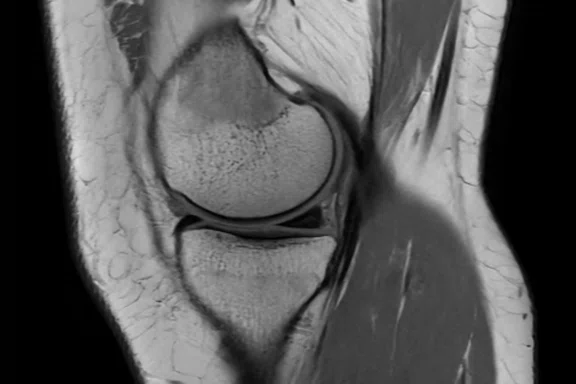
B
Figure 2.
All images with AIR™ Recon DL. (A) Hip coronal T1 post-contrast, 0.6 x 1.0 x 3.5 mm,
1:23 min.; (B) knee sagittal PD, 0.4 x 0.6 x 3.5 mm, 59 sec.; and (C) whole foot sagittal STIR, 0.4 x 0.6 x 3.0 mm, 2:28min.
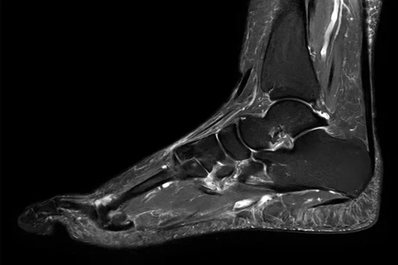
C
Figure 2.
All images with AIR™ Recon DL. (A) Hip coronal T1 post-contrast, 0.6 x 1.0 x 3.5 mm,
1:23 min.; (B) knee sagittal PD, 0.4 x 0.6 x 3.5 mm, 59 sec.; and (C) whole foot sagittal STIR, 0.4 x 0.6 x 3.0 mm, 2:28min.
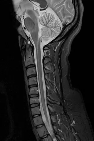
A
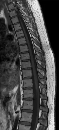
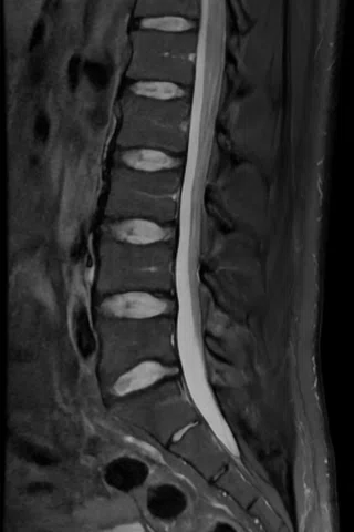
Figure 3.
All images with AIR™ Recon DL. (A) C-spine sagittal STIR, 0.8 x 1.0 x 3.0 mm, 2:20 min.;
(B) T-spine sagittal T1, 0.7 x 1.0 x 3.0 mm, 1:38 min.; and (C) L-spine sagittal T2 FatSat, 0.8 x 0.9 x 3.5 mm, 2:12 min.
B
Figure 3.
All images with AIR™ Recon DL. (A) C-spine sagittal STIR, 0.8 x 1.0 x 3.0 mm, 2:20 min.; (B) T-spine sagittal T1, 0.7 x 1.0 x 3.0 mm, 1:38 min.; and (C) L-spine sagittal T2 FatSat, 0.8 x 0.9 x 3.5 mm, 2:12 min.
C
Figure 3.
All images with AIR™ Recon DL. (A) C-spine sagittal STIR, 0.8 x 1.0 x 3.0 mm, 2:20 min.; (B) T-spine sagittal T1, 0.7 x 1.0 x 3.0 mm, 1:38 min.; and (C) L-spine sagittal T2 FatSat, 0.8 x 0.9 x 3.5 mm, 2:12 min.


result
PREVIOUS
${prev-page}
NEXT
${next-page}


Subscribe Now
Manage Subscription
FOLLOW US
Contact Us • Cookie Preferences • Privacy Policy • California Privacy PolicyDo Not Sell or Share My Personal Information • Terms & Conditions • Security
© 2024 GE HealthCare. GE is a trademark of General Electric Company. Used under trademark license.
IN PRACTICE
Fleury Group brings MR for everyone in Brazil
Fleury Group brings MR for everyone in Brazil
As one of the leading healthcare providers in Brazil, Fleury Group has invested in systems and solutions for imaging, laboratory, ophthalmology, reproductive medicine and more. Recently, the group installed one of the first SIGNA™ Prime systems worldwide.
As one of the leading healthcare providers in Brazil, Fleury Group has invested in systems and solutions for imaging, laboratory, ophthalmology, reproductive medicine and more. Recently, the group installed one of the first SIGNA™ Prime systems worldwide.
For 96 years, Fleury Group has delivered healthcare to residents throughout Brazil, from preventative medicine to tertiary care, with more than 300 sites in 10 states. According to Edgar Gil Rizzatti, MBA, MD, PhD, Executive Medical and Technical Director at Fleury Group, there is a long history of his organization working with GE Healthcare, culminating in a sizeable volume of imaging equipment from the company including MR, CT, X-ray and bone densitometry. Beyond equipment, he says that GE is recognized as a good service provider in the region, which further impacts the quality of imaging the Fleury Group provides. So, when one of the group’s centers in Tirol, in the Rio Grande do Norte State, needed a new MR system, he turned to GE and learned about a new system, SIGNA™ Prime.
SIGNA™ Prime, one of GE’s newest 1.5T MR systems, is ingeniously simple and environmentally friendly with future-ready capabilities. It was designed to be easy to set up, easy to use and easy to read – making the move to MR simple and seamless. Yet, it is a powerful system equipped with some of GE’s latest MR advancements, including AIR™ Recon DL, state-of-the-art Total Digital Imaging (TDI) 2.0 technology and a new, intuitive user interface.
“We saw the opportunity to bring new technology that adds significant value to our patients and physicians with SIGNA™ Prime,” Dr. Rizzatti says. “We acquired one of the first SIGNA™ Prime systems in the world – and certainly the first in Latin America – to continue our role as a pioneer in healthcare throughout our country and the region.”
MR for everyone
MR does not have to be difficult or just for the fully trained. It can be for everyone. This was the vision behind the development of SIGNA™ Prime, including the use of as much green technology in the design as possible, so that emerging markets could access MR. A key limitation in some countries can be the siting requirements for MR, including energy consumption. SIGNA™ Prime has a small footprint and, compared to other GE MR systems, a 25% lower power rating and up to 70% less helium consumption over its lifecycle, which means it will not require helium refills.
“Space is an issue in some of our centers, and it’s also about how we use that space to deliver the best services to our patients,” Dr. Rizzatti says.
Most impressive on SIGNA™ Prime is the new user interface that makes it easy for technologists to learn how to use it in just a matter of hours. It is very simple and intuitive, yet also very powerful. The user interface has all the key parameters well organized in key tabs for quick access by the technologist. An exam can be easily prescribed by following the guided workflow with step-by-step instructions.
“Because SIGNA™ Prime is so easy to operate, our technologists can give more attention to the patient.”
Dr. Edgar Rizzatti
“With fewer repetitive tasks, our technologists are more consistent across exams. Because of this, we believe the imaging is better and we are experiencing fewer patient callbacks.”
That ease of operation and focus on the patient leads to a better experience for patients, who may be anxious undergoing an MR due to claustrophobia or magnet noise.
SIGNA™ Prime has one of the simplest and easiest user interfaces that I’ve worked with,” says Onilda Duarte de Souza Cavalcante, BSc, Lead Technologist at the Fleury Group. “Even a junior technologist can learn to use it quickly and efficiently, thanks to the express mode.”
“The patient experience is very important to us and we place a lot of value on the connection between the patient and the physician. This really differentiates our services,” Dr. Rizzatti adds. “In fact, this is one of our organizational pillars, to closely support the patient and establish good relationships with our client physicians by delivering efficient and high-quality service.”
AI-infused technologies
Simplicity did not compromise quality. SIGNA™ Prime uses artificial intelligence to improve the user experience and shorten the learning curve. It includes AIR Touch™, a workflow application that automates coil element selection and landmarking, further accelerating the scanning process as soon as the patient is on the table. SIGNA™ Prime comes with SIGNA™Works, GE’s fully customizable productivity platform that hosts a suite of applications to enhance imaging accuracy and consistency, even for the most challenging clinical cases. In addition, it includes AIR™ Recon DL for improved SNR, which can be used to enhance image quality, shorten scan times or achieve a combination of both.
Shorter scan times with AIR™ Recon DL is an added benefit that Dr. Rizzatti did not anticipate with SIGNA™ Prime. Overall, the Fleury Group has shortened MSK exam times by at least 30% at the site, including ankle and elbow exams by 50%, hand exams by 40% and arm exams by 30%. While this technology will allow Fleury Group to increase productivity and patient volume, shorter scans also enhance the patient experience.
When he looks at the quality of the images in these shorter exams, he is further impressed with the potential of AIR™ Recon DL and plans to implement it across anatomies and compatible sequences.
“We intend to implement AIR™ Recon DL in as many GE MR systems as we can after seeing the impressive results.”
Dr. Edgar Rizzatti
“Of course, it will be a key consideration to have in any additional MR systems we purchase. SIGNA™ Prime is definitely a system that we would buy again and recommend to others, as well.”


