A
Figure 1.
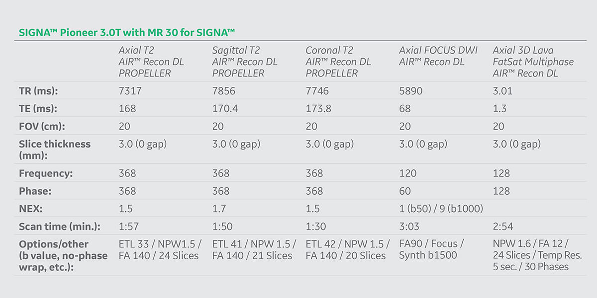
High-resolution mpMRI of the prostate with AIR™ Recon DL and AIR™ Recon DL PROPELLER, total exam time 11:14 min. (A) Axial T2 PROPELLER, 0.5 x 0.5 x 3.0 mm, 1:57 min.; (B) sagittal T2 PROPELLER, 0.5 x 0.5 x 3.0 mm, 1:50 min.; (C) coronal T2 PROPELLER, 0.5 x 0.5 x 3.0 mm, 1:30 min.; (D-F) axial FOCUS DWI, 1.7 x 1.7 x 3.0 mm, 3:03 min., with (D) b1000, (E) synthetic b1500 and (F) ADC; and (G) axial 3D LAVA FatSat multi-phase, 1.7 x 1.7 x 3.0 mm, 30 phases, 2:54 min.
B
Figure 1.
High-resolution mpMRI of the prostate with AIR™ Recon DL and AIR™ Recon DL PROPELLER, total exam time 11:14 min. (A) Axial T2 PROPELLER, 0.5 x 0.5 x 3.0 mm, 1:57 min.; (B) sagittal T2 PROPELLER, 0.5 x 0.5 x 3.0 mm, 1:50 min.; (C) coronal T2 PROPELLER, 0.5 x 0.5 x 3.0 mm, 1:30 min.; (D-F) axial FOCUS DWI, 1.7 x 1.7 x 3.0 mm, 3:03 min., with (D) b1000, (E) synthetic b1500 and (F) ADC; and (G) axial 3D LAVA FatSat multi-phase, 1.7 x 1.7 x 3.0 mm, 30 phases, 2:54 min.
C
Figure 1.
High-resolution mpMRI of the prostate with AIR™ Recon DL and AIR™ Recon DL PROPELLER, total exam time 11:14 min. (A) Axial T2 PROPELLER, 0.5 x 0.5 x 3.0 mm, 1:57 min.; (B) sagittal T2 PROPELLER, 0.5 x 0.5 x 3.0 mm, 1:50 min.; (C) coronal T2 PROPELLER, 0.5 x 0.5 x 3.0 mm, 1:30 min.; (D-F) axial FOCUS DWI, 1.7 x 1.7 x 3.0 mm, 3:03 min., with (D) b1000, (E) synthetic b1500 and (F) ADC; and (G) axial 3D LAVA FatSat multi-phase, 1.7 x 1.7 x 3.0 mm, 30 phases, 2:54 min.
D
Figure 1.
High-resolution mpMRI of the prostate with AIR™ Recon DL and AIR™ Recon DL PROPELLER, total exam time 11:14 min. (A) Axial T2 PROPELLER, 0.5 x 0.5 x 3.0 mm, 1:57 min.; (B) sagittal T2 PROPELLER, 0.5 x 0.5 x 3.0 mm, 1:50 min.; (C) coronal T2 PROPELLER, 0.5 x 0.5 x 3.0 mm, 1:30 min.; (D-F) axial FOCUS DWI, 1.7 x 1.7 x 3.0 mm, 3:03 min., with (D) b1000, (E) synthetic b1500 and (F) ADC; and (G) axial 3D LAVA FatSat multi-phase, 1.7 x 1.7 x 3.0 mm, 30 phases, 2:54 min.
E
Figure 1.
High-resolution mpMRI of the prostate with AIR™ Recon DL and AIR™ Recon DL PROPELLER, total exam time 11:14 min. (A) Axial T2 PROPELLER, 0.5 x 0.5 x 3.0 mm, 1:57 min.; (B) sagittal T2 PROPELLER, 0.5 x 0.5 x 3.0 mm, 1:50 min.; (C) coronal T2 PROPELLER, 0.5 x 0.5 x 3.0 mm, 1:30 min.; (D-F) axial FOCUS DWI, 1.7 x 1.7 x 3.0 mm, 3:03 min., with (D) b1000, (E) synthetic b1500 and (F) ADC; and (G) axial 3D LAVA FatSat multi-phase, 1.7 x 1.7 x 3.0 mm, 30 phases, 2:54 min.
F
Figure 1.
High-resolution mpMRI of the prostate with AIR™ Recon DL and AIR™ Recon DL PROPELLER, total exam time 11:14 min. (A) Axial T2 PROPELLER, 0.5 x 0.5 x 3.0 mm, 1:57 min.; (B) sagittal T2 PROPELLER, 0.5 x 0.5 x 3.0 mm, 1:50 min.; (C) coronal T2 PROPELLER, 0.5 x 0.5 x 3.0 mm, 1:30 min.; (D-F) axial FOCUS DWI, 1.7 x 1.7 x 3.0 mm, 3:03 min., with (D) b1000, (E) synthetic b1500 and (F) ADC; and (G) axial 3D LAVA FatSat multi-phase, 1.7 x 1.7 x 3.0 mm, 30 phases, 2:54 min.
G
Figure 1.
High-resolution mpMRI of the prostate with AIR™ Recon DL and AIR™ Recon DL PROPELLER, total exam time 11:14 min. (A) Axial T2 PROPELLER, 0.5 x 0.5 x 3.0 mm, 1:57 min.; (B) sagittal T2 PROPELLER, 0.5 x 0.5 x 3.0 mm, 1:50 min.; (C) coronal T2 PROPELLER, 0.5 x 0.5 x 3.0 mm, 1:30 min.; (D-F) axial FOCUS DWI, 1.7 x 1.7 x 3.0 mm, 3:03 min., with (D) b1000, (E) synthetic b1500 and (F) ADC; and (G) axial 3D LAVA FatSat multi-phase, 1.7 x 1.7 x 3.0 mm, 30 phases, 2:54 min.


result
PREVIOUS
${prev-page}
NEXT
${next-page}
Subscribe Now
Manage Subscription
FOLLOW US
Contact Us • Cookie Preferences • Privacy Policy • California Privacy PolicyDo Not Sell or Share My Personal Information • Terms & Conditions • Security
© 2024 GE HealthCare. GE is a trademark of General Electric Company. Used under trademark license.

Christopher Ahlers, MD
radiomed
Wiesbaden, Germany
CASE STUDIES
DL-based 3D and motion-insensitive reconstruction advances multiparametric MR prostate imaging
DL-based 3D and motion-insensitive reconstruction advances multiparametric MR prostate imaging
By Christopher Ahlers, MD, Managing Partner, radiomed, Weisbaden, Germany
Christopher Ahlers, MD
radiomed
Wiesbaden, Germany
The multiparametric prostate MR (mpMRI) exam allows for the reliable representation of clinically relevant tumors. In our center, the examination protocol for a prostate MR is specified by the guidelines of the European Society for Urogenital Radiology (ESUR). This includes an assessment using a scoring system and structured diagnosis and documentation (PI-RADS® v2.1). The 2D FSE-based sequences are considered the gold standard acquisition. To achieve good image quality in these cases, it is recommended to administer Buscopan® intravenously to the patient before the exam. Elevated intraocular pressure or severe cardiac arrhythmias are Phasescontraindications for the administration of Buscopan®. In addition, patients should avoid operating a motor vehicle for five hours after administering the medicine. If these contraindications exist, as in the case presented below, then the technologists and clinician must choose between a likely poor image quality of the prostate with the 2D FSE sequence or a longer examination using the PROPELLER acquisition.
This patient case is, in many aspects, an exciting clinical case to present. First, Buscopan® could not be administered to the patient, which means that constant bowel motion would have a potentially negative impact on the findings of the examination. Secondly, AIR™ Recon DL can now be used for 100% of all sequences that are prescribed by the ESUR and PI-RADS® guidelines, including 3D. This applies in particular to the T2-weighted sequences, e.g., AIR™ Recon DL PROPELLER, the FOCUS Diffusion (b50-1000/synthetic b1500) and the Dynamic T1 FatSat GRE (3D LAVA FatSat multiphase). Further, the compromise that previously had to be made with PROPELLER, such as longer examination times and the slight loss of spatial sharpness, can now be compensated for with AIR™ Recon DL PROPELLER.
With the complete use of AIR™ Recon DL, the examination time was 11:14 minutes with adherence to the resolution guidelines.
Patient history
A 63-year-old male presenting with a suspicious palpation and a PSA of 10.9 ng/ml was referred for an MR exam of the prostate.

A

B

C
D
E
F
G
Figure 1.
High-resolution mpMRI of the prostate with AIR™ Recon DL and AIR™ Recon DL PROPELLER, total exam time 11:14 min. (A) Axial T2 PROPELLER, 0.5 x 0.5 x 3.0 mm, 1:57 min.; (B) sagittal T2 PROPELLER, 0.5 x 0.5 x 3.0 mm, 1:50 min.; (C) coronal T2 PROPELLER, 0.5 x 0.5 x 3.0 mm, 1:30 min.; (D-F) axial FOCUS DWI, 1.7 x 1.7 x 3.0 mm, 3:03 min., with (D) b1000, (E) synthetic b1500 and (F) ADC; and (G) axial 3D LAVA FatSat multi-phase, 1.7 x 1.7 x 3.0 mm, 30 phases, 2:54 min.
Results
Size: The prostate measures approximately 5.3 x 4.7 x 5.7 cm (right left x anteroposterior x craniocaudal diameters). This corresponds to a prostate volume of approximately 74 ml.
Peripheral zone: Oval, diffusionrestricted lesion of the PZpm paramedian left center with a size of 11 mm and circumscribed roundish lesion with diffusion restriction of the PZpm/ pl apical right with a size of 6 mm. Wedge-shaped T2-weighted and ADC hypointensities in the peripheral zone, most likely in the context of (chronic) prostatitis.
Transition zone: Pronounced stromal adenomatous change in the transitional zone with a significant cystic regressive component and prolapsing benign prostatic hyperplasia (BPH) nodes in the bladder floor.
A recommendation for further treatment by a urologist was also defined.
Shape: Largely unilateral configuration.
Peripheral zone: Oval, diffusionrestricted lesion of the PZpm paramedian left center with a size of 11 mm and circumscribed roundish lesion with diffusion restriction of the PZpm/ pl apical right with a size of 6 mm. Wedge-shaped T2-weighted and ADC hypointensities in the peripheral zone, most likely in the context of (chronic) prostatitis.
Transition zone: Pronounced stromal adenomatous change in the transitional zone with a significant cystic regressive component and prolapsing benign prostatic hyperplasia (BPH) nodes in the bladder floor.
Conclusion
PI-RADS® 4 lesion of the PZpm paramedian left center with a size of 11 mm and the PZpm/pl apical right with a size of 6 mm. Targeted biopsy recommended.
Discussion
In mpMRI of the prostate, it is very important for the patient’s outcome to always achieve a high spatial resolution and detailed diagnosis. The PI-RADS® guidelines help ensure this. If there is any deviation in the examination due to individual characteristics (contraindications, claustrophobia, obesity, etc.), the quality of the diagnosis and examination should not suffer.
The ability to use AIR™ Recon DL for all required sequences resulted in a robust imaging exam. Despite the fact that the examination protocol deviated from the standard, a very thorough and quick examination was completed, which led to a high-quality, confident diagnosis. The utilization of AIR™ Recon DL PROPELLER helped overcome bowel motion due to the non-administration of Buscopan®, limiting any disadvantage or negative impact on image quality for the radiologist’s review and ultimately the patient diagnosis. In addition, an increase in spatial resolution with AIR™ Recon DL for diffusion imaging and dynamic contrast examination meant that the differentiation of the lesion using the PI-RADS® 2.1 scheme was significantly better.