A
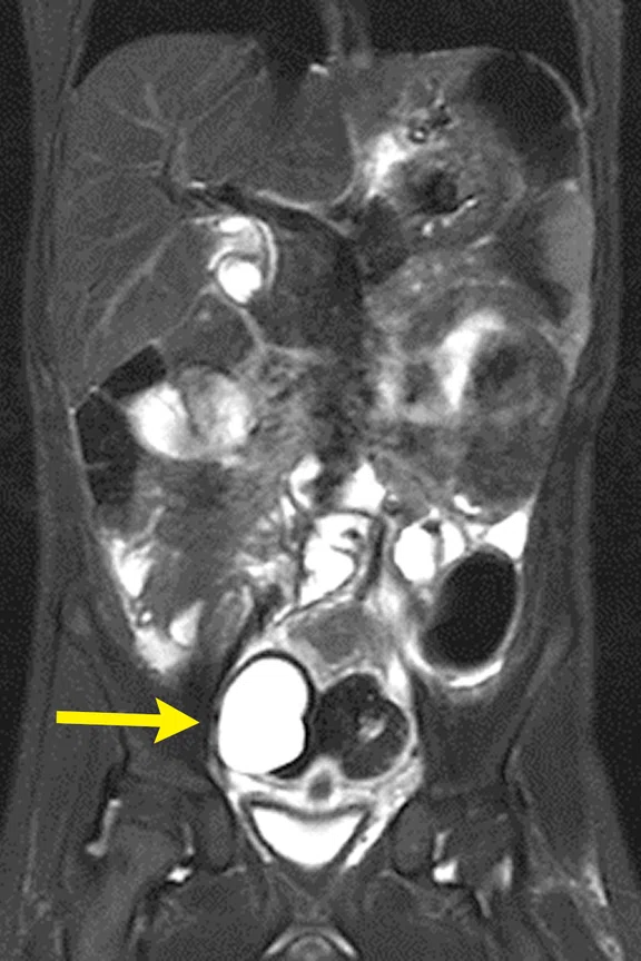
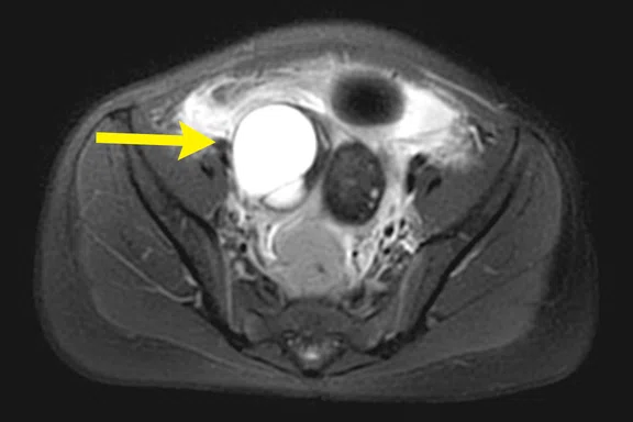
Figure 1.
Coronal T2 HyperCube STIR with HyperSense and Auto Navigator, 0.9 x 1.1 x 3 mm and 0.9 x 1.1 x 1.5 mm, ARC 2 x 1, HyperSense 1.2, 4:24 min.
A
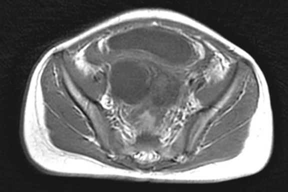
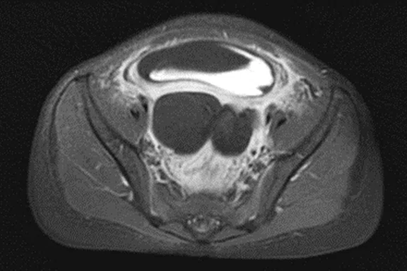
Figure 2.
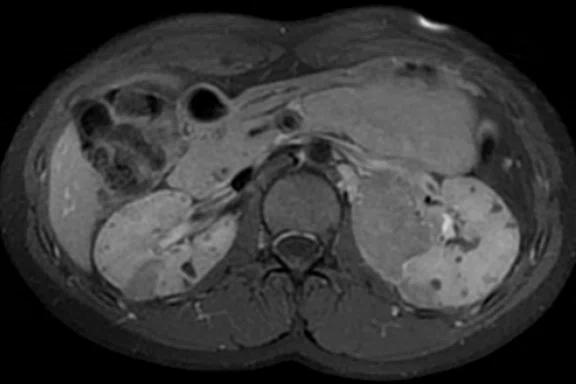
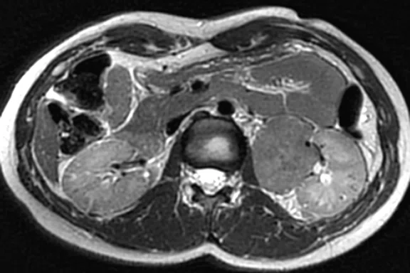
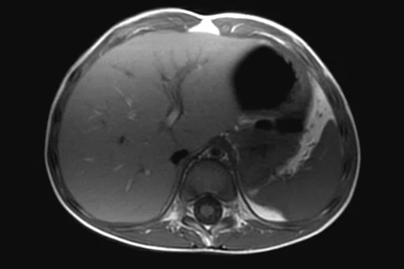
Axial PROPELLER MB with RTr T2 FatSat and free-breathing T1. (A) Axial PROPELLER T2 FatSat RTr FOV 24, 300 x 300, 4:39 min. (B) Axial PROPELLER T1 free breathing, 0.8 x 0.8 x 5 mm, 4:17 min. (C) Axial PROPELLER T1 FatSat post-contrast free breathing, 0.9 x 0.9 x 5 mm, 4:34 min.
B
Figure 2.
Axial PROPELLER MB with RTr T2 FatSat and free-breathing T1. (A) Axial PROPELLER T2 FatSat RTr FOV 24, 300 x 300, 4:39 min. (B) Axial PROPELLER T1 free breathing, 0.8 x 0.8 x 5 mm, 4:17 min. (C) Axial PROPELLER T1 FatSat post-contrast free breathing, 0.9 x 0.9 x 5 mm, 4:34 min.
C
Figure 2.
Axial PROPELLER MB with RTr T2 FatSat and free-breathing T1. (A) Axial PROPELLER T2 FatSat RTr FOV 24, 300 x 300, 4:39 min. (B) Axial PROPELLER T1 free breathing, 0.8 x 0.8 x 5 mm, 4:17 min. (C) Axial PROPELLER T1 FatSat post-contrast free breathing, 0.9 x 0.9 x 5 mm, 4:34 min.
A
Figure 4.
Free-breathing coronal HyperCube STIR with HyperSense and Auto Navigator, 0.8 x 1.0 x 3 mm, ARC 2x1, HyperSense 1.2, 2:05 min.
B
Figure 4.
Free-breathing coronal HyperCube STIR with HyperSense and Auto Navigator, 0.8 x 1.0 x 3 mm, ARC 2x1, HyperSense 1.2, 2:05 min.
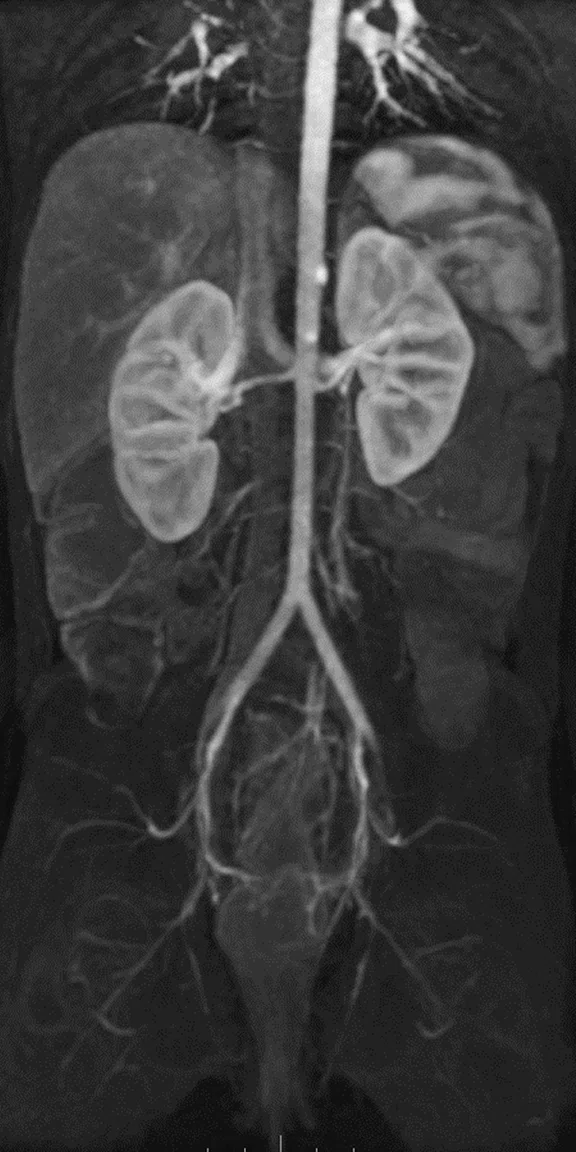
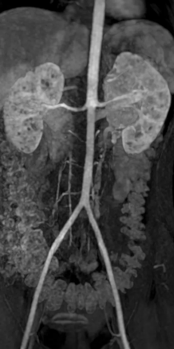
A
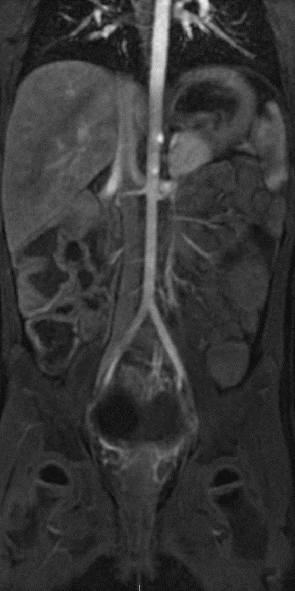
Figure 3.
Free-breathing DCE imaging with DISCO and Auto Navigator, (A) coronal 3 mm/1.5 mm arterial and (B) derived MRA, arterial thick MIP.
B
Figure 3.
Free-breathing DCE imaging with DISCO and Auto Navigator, (A) coronal 3 mm/1.5 mm arterial and (B) derived MRA, arterial thick MIP.
A
Figure 5.
PROPELLER MB, free-breathing T1 and RTr T2 FatSat. (A) Coronal PROPELLER T1 free breathing, 1.0 x 1.0 x 5 mm, 3:40 min. (B) Axial PROPELLER T1 free breathing, 1.0 x 1.0 x 5 mm, 3:09 min. (C) Axial PROPELLER T2 FatSat RTr, 1.0 x 1.0 x 5 mm, 3:32 min.
B
Figure 5.
PROPELLER MB, free-breathing T1 and RTr T2 FatSat. (A) Coronal PROPELLER T1 free breathing, 1.0 x 1.0 x 5 mm, 3:40 min. (B) Axial PROPELLER T1 free breathing, 1.0 x 1.0 x 5 mm, 3:09 min. (C) Axial PROPELLER T2 FatSat RTr, 1.0 x 1.0 x 5 mm, 3:32 min.
C
Figure 5.
PROPELLER MB, free-breathing T1 and RTr T2 FatSat. (A) Coronal PROPELLER T1 free breathing, 1.0 x 1.0 x 5 mm, 3:40 min. (B) Axial PROPELLER T1 free breathing, 1.0 x 1.0 x 5 mm, 3:09 min. (C) Axial PROPELLER T2 FatSat RTr, 1.0 x 1.0 x 5 mm, 3:32 min.
A
Figure 6.
DWI and PROPELLER MB. (A) Axial PROPELLER T1 FatSat post-contrast free breathing, 1.0 x 1.0 x 5 mm, 3:29 min. (B) Axial PROPELLER T2 RTr, 1.0 x 1.0 x 3 mm, 3:46 min. (C-E) Axial DWI RTCF RTr, 2.1 x 2.0 x 5 mm, with (C) b50, (D) b1500 and (E) ADC.
B
Figure 6.
DWI and PROPELLER MB. (A) Axial PROPELLER T1 FatSat post-contrast free breathing, 1.0 x 1.0 x 5 mm, 3:29 min. (B) Axial PROPELLER T2 RTr, 1.0 x 1.0 x 3 mm, 3:46 min. (C-E) Axial DWI RTCF RTr, 2.1 x 2.0 x 5 mm, with (C) b50, (D) b1500 and (E) ADC.
A
Figure 8.
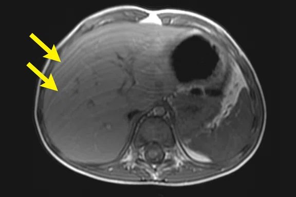
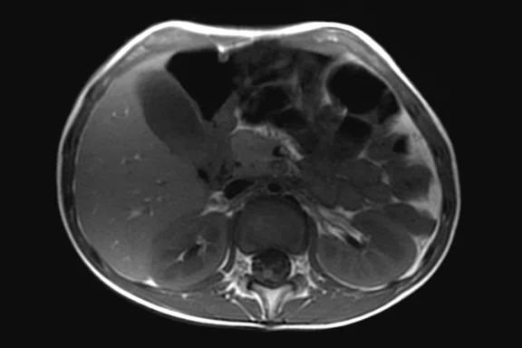
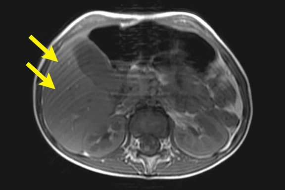
T1 PROPELLER MB vs. T1 SE respiratory compensation (RC). Note how PROPELLER MB T1 suppresses breathing motion artifacts compared to conventional T1 SE acquisition with remaining motion artifacts (yellow arrows). (A, C) Axial T1 PROPELLER MB free-breathing, 0.8 x 0.8 x 5 mm, 3:40 min., (B, D) Axial T1 SE RC free-breathing, 1.0 x 1.4 x 5 mm, 4:08 min.
B
Figure 8.
T1 PROPELLER MB vs. T1 SE respiratory compensation (RC). Note how PROPELLER MB T1 suppresses breathing motion artifacts compared to conventional T1 SE acquisition with remaining motion artifacts (yellow arrows). (A, C) Axial T1 PROPELLER MB free-breathing, 0.8 x 0.8 x 5 mm, 3:40 min., (B, D) Axial T1 SE RC free-breathing, 1.0 x 1.4 x 5 mm, 4:08 min.
C
Figure 8.
T1 PROPELLER MB vs. T1 SE respiratory compensation (RC). Note how PROPELLER MB T1 suppresses breathing motion artifacts compared to conventional T1 SE acquisition with remaining motion artifacts (yellow arrows). (A, C) Axial T1 PROPELLER MB free-breathing, 0.8 x 0.8 x 5 mm, 3:40 min., (B, D) Axial T1 SE RC free-breathing, 1.0 x 1.4 x 5 mm, 4:08 min.
D
Figure 8.
T1 PROPELLER MB vs. T1 SE respiratory compensation (RC). Note how PROPELLER MB T1 suppresses breathing motion artifacts compared to conventional T1 SE acquisition with remaining motion artifacts (yellow arrows). (A, C) Axial T1 PROPELLER MB free-breathing, 0.8 x 0.8 x 5 mm, 3:40 min., (B, D) Axial T1 SE RC free-breathing, 1.0 x 1.4 x 5 mm, 4:08 min.
C
Figure 6.
DWI and PROPELLER MB. (A) Axial PROPELLER T1 FatSat post-contrast free breathing, 1.0 x 1.0 x 5 mm, 3:29 min. (B) Axial PROPELLER T2 RTr, 1.0 x 1.0 x 3 mm, 3:46 min. (C-E) Axial DWI RTCF RTr, 2.1 x 2.0 x 5 mm, with (C) b50, (D) b1500 and (E) ADC.
D
Figure 6.
DWI and PROPELLER MB. (A) Axial PROPELLER T1 FatSat post-contrast free breathing, 1.0 x 1.0 x 5 mm, 3:29 min. (B) Axial PROPELLER T2 RTr, 1.0 x 1.0 x 3 mm, 3:46 min. (C-E) Axial DWI RTCF RTr, 2.1 x 2.0 x 5 mm, with (C) b50, (D) b1500 and (E) ADC.
E
Figure 6.
DWI and PROPELLER MB. (A) Axial PROPELLER T1 FatSat post-contrast free breathing, 1.0 x 1.0 x 5 mm, 3:29 min. (B) Axial PROPELLER T2 RTr, 1.0 x 1.0 x 3 mm, 3:46 min. (C-E) Axial DWI RTCF RTr, 2.1 x 2.0 x 5 mm, with (C) b50, (D) b1500 and (E) ADC.
A
Figure 7.
DCE free-breathing imaging with DISCO and Auto Navigator. (A-D) Coronal DISCO Flex and Auto Navigator 3.4 mm/1.7 mm, (A) arterial, (B) portal-venous and (C-D) derived MRA – arterial thick MIP.
B
Figure 7.
DCE free-breathing imaging with DISCO and Auto Navigator. (A-D) Coronal DISCO Flex and Auto Navigator 3.4 mm/1.7 mm, (A) arterial, (B) portal-venous and (C-D) derived MRA – arterial thick MIP.
C
Figure 7.
DCE free-breathing imaging with DISCO and Auto Navigator. (A-D) Coronal DISCO Flex and Auto Navigator 3.4 mm/1.7 mm, (A) arterial, (B) portal-venous and (C-D) derived MRA – arterial thick MIP.
D
Figure 7.
DCE free-breathing imaging with DISCO and Auto Navigator. (A-D) Coronal DISCO Flex and Auto Navigator 3.4 mm/1.7 mm, (A) arterial, (B) portal-venous and (C-D) derived MRA – arterial thick MIP.
D
Figure 4.
Free-breathing coronal HyperCube STIR with HyperSense and Auto Navigator, 0.8 x 1.0 x 3 mm, ARC 2x1, HyperSense 1.2, 2:05 min.
C
Figure 4.
Free-breathing coronal HyperCube STIR with HyperSense and Auto Navigator, 0.8 x 1.0 x 3 mm, ARC 2x1, HyperSense 1.2, 2:05 min.
A
Figure 5.
PROPELLER MB, free-breathing T1 and RTr T2 FatSat. (A) Coronal PROPELLER T1 free breathing, 1.0 x 1.0 x 5 mm, 3:40 min. (B) Axial PROPELLER T1 free breathing, 1.0 x 1.0 x 5 mm, 3:09 min. (C) Axial PROPELLER T2 FatSat RTr, 1.0 x 1.0 x 5 mm, 3:32 min.
B
Figure 5.
PROPELLER MB, free-breathing T1 and RTr T2 FatSat. (A) Coronal PROPELLER T1 free breathing, 1.0 x 1.0 x 5 mm, 3:40 min. (B) Axial PROPELLER T1 free breathing, 1.0 x 1.0 x 5 mm, 3:09 min. (C) Axial PROPELLER T2 FatSat RTr, 1.0 x 1.0 x 5 mm, 3:32 min.
C
Figure 5.
PROPELLER MB, free-breathing T1 and RTr T2 FatSat. (A) Coronal PROPELLER T1 free breathing, 1.0 x 1.0 x 5 mm, 3:40 min. (B) Axial PROPELLER T1 free breathing, 1.0 x 1.0 x 5 mm, 3:09 min. (C) Axial PROPELLER T2 FatSat RTr, 1.0 x 1.0 x 5 mm, 3:32 min.
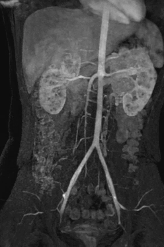
A
Figure 7.
DCE free-breathing imaging with DISCO and Auto Navigator. (A-D) Coronal DISCO Flex and Auto Navigator 3.4 mm/1.7 mm, (A) arterial, (B) portal-venous and (C-D) derived MRA – arterial thick MIP.
B
Figure 7.
DCE free-breathing imaging with DISCO and Auto Navigator. (A-D) Coronal DISCO Flex and Auto Navigator 3.4 mm/1.7 mm, (A) arterial, (B) portal-venous and (C-D) derived MRA – arterial thick MIP.
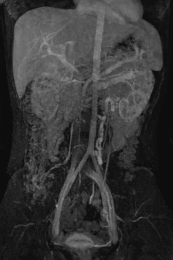
C
Figure 7.
DCE free-breathing imaging with DISCO and Auto Navigator. (A-D) Coronal DISCO Flex and Auto Navigator 3.4 mm/1.7 mm, (A) arterial, (B) portal-venous and (C-D) derived MRA – arterial thick MIP.
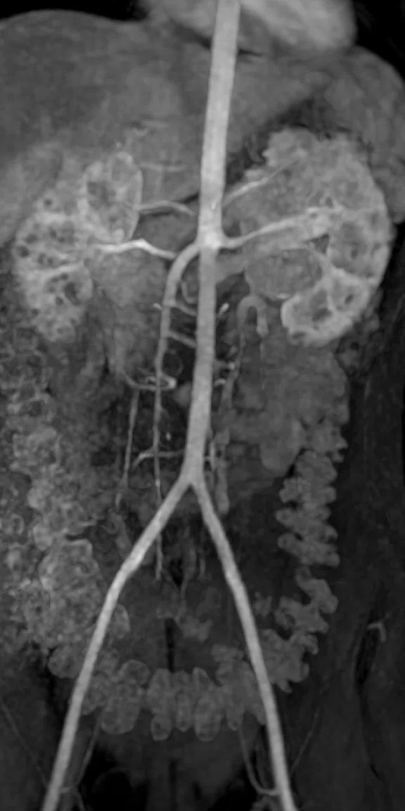
D
Figure 7.
DCE free-breathing imaging with DISCO and Auto Navigator. (A-D) Coronal DISCO Flex and Auto Navigator 3.4 mm/1.7 mm, (A) arterial, (B) portal-venous and (C-D) derived MRA – arterial thick MIP.
A
Figure 8.
T1 PROPELLER MB vs. T1 SE respiratory compensation (RC). Note how PROPELLER MB T1 suppresses breathing motion artifacts compared to conventional T1 SE acquisition with remaining motion artifacts (yellow arrows). (A, C) Axial T1 PROPELLER MB free-breathing, 0.8 x 0.8 x 5 mm, 3:40 min., (B, D) Axial T1 SE RC free-breathing, 1.0 x 1.4 x 5 mm, 4:08 min.
B
Figure 8.
T1 PROPELLER MB vs. T1 SE respiratory compensation (RC). Note how PROPELLER MB T1 suppresses breathing motion artifacts compared to conventional T1 SE acquisition with remaining motion artifacts (yellow arrows). (A, C) Axial T1 PROPELLER MB free-breathing, 0.8 x 0.8 x 5 mm, 3:40 min., (B, D) Axial T1 SE RC free-breathing, 1.0 x 1.4 x 5 mm, 4:08 min.
C
Figure 8.
T1 PROPELLER MB vs. T1 SE respiratory compensation (RC). Note how PROPELLER MB T1 suppresses breathing motion artifacts compared to conventional T1 SE acquisition with remaining motion artifacts (yellow arrows). (A, C) Axial T1 PROPELLER MB free-breathing, 0.8 x 0.8 x 5 mm, 3:40 min., (B, D) Axial T1 SE RC free-breathing, 1.0 x 1.4 x 5 mm, 4:08 min.
D
Figure 8.
T1 PROPELLER MB vs. T1 SE respiratory compensation (RC). Note how PROPELLER MB T1 suppresses breathing motion artifacts compared to conventional T1 SE acquisition with remaining motion artifacts (yellow arrows). (A, C) Axial T1 PROPELLER MB free-breathing, 0.8 x 0.8 x 5 mm, 3:40 min., (B, D) Axial T1 SE RC free-breathing, 1.0 x 1.4 x 5 mm, 4:08 min.
result


PREVIOUS
${prev-page}
NEXT
${next-page}
Subscribe Now
Manage Subscription
FOLLOW US
Contact Us • Cookie Preferences • Privacy Policy • California Privacy PolicyDo Not Sell or Share My Personal Information • Terms & Conditions • Security
© 2024 GE HealthCare. GE is a trademark of General Electric Company. Used under trademark license.
CASE STUDIES
Pediatric free-breathing abdominal imaging
Pediatric free-breathing abdominal imaging
by Christian Kellenberger, MD, Radiologist-in-Chief, University Children’s Hospital Zürich, Zürich, Switzerland
Abdominal MR imaging is challenging in young children because they often cannot hold their breath and may be unable to follow breathing instructions. Thus, having the ability to perform fast and completely free-breathing examinations is crucial to obtain high-quality images and ensure an accurate diagnosis.
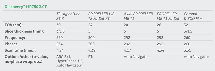
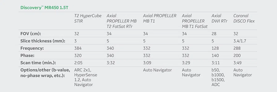
After updating our software to SIGNA™Works on the Discovery™ MR750 3.0T and Discovery™ MR450 1.5T systems, we now have access to acceleration techniques such as HyperSense and HyperCube that are compatible with Auto Navigator and Respiratory Triggering (RTr) methods. These capabilities have helped to significantly reduce the acquisition time of 3D Cube sequences for abdominal imaging while increasing spatial resolution. We are routinely using Auto Navigator in our free-breathing protocols, as it allows for automatic diaphragm detection and tracker placement without user interaction, which simplifies the workflow.
Additionally, the latest generation of PROPELLER Multi-Blade (MB) allows us to achieve T1 and T1 FatSat FSE contrasts without respiratory motion artifacts. Without the use of respiratory gating or triggering, PROPELLER MB provides us with high-quality and artifact-free FSE T1-weighted images in free-breathing children, which is essential for the assessment of abdominal organs. Image quality is clearly improved and the acquisition is faster when compared to prior approaches for compensating respiratory motion, such as signal averaging by increasing the number of excitations or the use of respiratory compensation (RT) for T1-weighted SE.
Finally, the 3D DISCO sequence combined with Auto Navigator allows us to do free-breathing dynamic contrast enhanced (DCE) imaging without compromising spatial resolution and with a temporal resolution between 10-20 ms per phase, depending on the patient’s respiration rate. It can be done in any plane and is compatible with Dixon techniques for robust fat saturation in every case.
Case 1
Patient history
A three-year-old child with a pelvic cystic mass.
Technique
Performed a complete free-breathing exam on the Discovery™ MR750 using PROPELLER MB, T1 and RTr T2 FatSat, HyperCube, 3D DISCO and Auto Navigator (Figures 1-3).
Case 2
Patient history
A 16-year-old child with suspicion of angiomyolipoma or kidney cell carcinoma.
Technique
Performed a complete free-breathing exam on the Discovery™ MR450 using PROPELLER MB, T1 and RTr T2 FatSat, HyperCube STIR, 3D DISCO, DWI and Auto Navigator (Figures 4-6).
Discussion
The latest sequence options from GE Healthcare are a real asset for achieving robust abdominal imaging in free-breathing children, adolescents and adults who are unable to hold their breath. In particular, PROPELLER MB T1 efficiently suppresses breathing motion artifacts compared to respiratory compensation techniques (Figure 8).
Figure 8.
T1 PROPELLER MB vs. T1 SE respiratory compensation (RC). Note how PROPELLER MB T1 suppresses breathing motion artifacts compared to conventional T1 SE acquisition with remaining motion artifacts (yellow arrows). (A, C) Axial T1 PROPELLER MB free-breathing, 0.8 x 0.8 x 5 mm, 3:40 min., (B, D) Axial T1 SE RC free-breathing, 1.0 x 1.4 x 5 mm, 4:08 min.


