A
Figure 2.
A 69-year-old female with shoulder pain due to trauma and suspected rotator cuff tear. All images were processed with AIR™ Recon DL PROPELLER and acquired at 0.5 x 0.5 x 3.0 mm. (A) Coronal PD FatSat PROPELLER, 1:22 min.; (B) coronal T1 PROPELLER, 1:58 min.; (C) sagittal T2 PROPELLER, 1:40 min.; and (D) axial PD FatSat PROPELLER, 1:43 min. Total exam time was 6:43 min with a 16ch Flex medium coil.
B
Figure 2.
A 69-year-old female with shoulder pain due to trauma and suspected rotator cuff tear. All images were processed with AIR™ Recon DL PROPELLER and acquired at 0.5 x 0.5 x 3.0 mm. (A) Coronal PD FatSat PROPELLER, 1:22 min.; (B) coronal T1 PROPELLER, 1:58 min.; (C) sagittal T2 PROPELLER, 1:40 min.; and (D) axial PD FatSat PROPELLER, 1:43 min. Total exam time was 6:43 min with a 16ch Flex medium coil.
C
Figure 2.
A 69-year-old female with shoulder pain due to trauma and suspected rotator cuff tear. All images were processed with AIR™ Recon DL PROPELLER and acquired at 0.5 x 0.5 x 3.0 mm. (A) Coronal PD FatSat PROPELLER, 1:22 min.; (B) coronal T1 PROPELLER, 1:58 min.; (C) sagittal T2 PROPELLER, 1:40 min.; and (D) axial PD FatSat PROPELLER, 1:43 min. Total exam time was 6:43 min with a 16ch Flex medium coil.
D
Figure 2.
A 69-year-old female with shoulder pain due to trauma and suspected rotator cuff tear. All images were processed with AIR™ Recon DL PROPELLER and acquired at 0.5 x 0.5 x 3.0 mm. (A) Coronal PD FatSat PROPELLER, 1:22 min.; (B) coronal T1 PROPELLER, 1:58 min.; (C) sagittal T2 PROPELLER, 1:40 min.; and (D) axial PD FatSat PROPELLER, 1:43 min. Total exam time was 6:43 min with a 16ch Flex medium coil.
A
Figure 1.
A 45-year-old female suffered trauma to her finger eight days prior. This case was processed with both conventional and AIR™ Recon DL PROPELLER. A 16ch Flex small coil was used for this case. (A, B) Sagittal PD FatSat PROPELLER, 0.4 x 0.4 x 1.0 mm, 3:32 min., (A) with conventional reconstruction and (B) AIR™ Recon DL PROPELLER. (C-E) Axial PD FatSat PROPELLER, 0.4 x 0.4 x 1.0 mm, 3:15 min., (C) with conventional reconstruction and (D, E) AIR™ Recon DL PROPELLER. (F, G) Coronal PD FatSat 0.4 x 0.4 x 1.0 mm, 3:21 min. with AIR™ Recon DL PROPELLER.
B
Figure 1.
A 45-year-old female suffered trauma to her finger eight days prior. This case was processed with both conventional and AIR™ Recon DL PROPELLER. A 16ch Flex small coil was used for this case. (A, B) Sagittal PD FatSat PROPELLER, 0.4 x 0.4 x 1.0 mm, 3:32 min., (A) with conventional reconstruction and (B) AIR™ Recon DL PROPELLER. (C-E) Axial PD FatSat PROPELLER, 0.4 x 0.4 x 1.0 mm, 3:15 min., (C) with conventional reconstruction and (D, E) AIR™ Recon DL PROPELLER. (F, G) Coronal PD FatSat 0.4 x 0.4 x 1.0 mm, 3:21 min. with AIR™ Recon DL PROPELLER.
C
Figure 1.
A 45-year-old female suffered trauma to her finger eight days prior. This case was processed with both conventional and AIR™ Recon DL PROPELLER. A 16ch Flex small coil was used for this case. (A, B) Sagittal PD FatSat PROPELLER, 0.4 x 0.4 x 1.0 mm, 3:32 min., (A) with conventional reconstruction and (B) AIR™ Recon DL PROPELLER. (C-E) Axial PD FatSat PROPELLER, 0.4 x 0.4 x 1.0 mm, 3:15 min., (C) with conventional reconstruction and (D, E) AIR™ Recon DL PROPELLER. (F, G) Coronal PD FatSat 0.4 x 0.4 x 1.0 mm, 3:21 min. with AIR™ Recon DL PROPELLER.
D
Figure 1.
A 45-year-old female suffered trauma to her finger eight days prior. This case was processed with both conventional and AIR™ Recon DL PROPELLER. A 16ch Flex small coil was used for this case. (A, B) Sagittal PD FatSat PROPELLER, 0.4 x 0.4 x 1.0 mm, 3:32 min., (A) with conventional reconstruction and (B) AIR™ Recon DL PROPELLER. (C-E) Axial PD FatSat PROPELLER, 0.4 x 0.4 x 1.0 mm, 3:15 min., (C) with conventional reconstruction and (D, E) AIR™ Recon DL PROPELLER. (F, G) Coronal PD FatSat 0.4 x 0.4 x 1.0 mm, 3:21 min. with AIR™ Recon DL PROPELLER.
E
Figure 1.
A 45-year-old female suffered trauma to her finger eight days prior. This case was processed with both conventional and AIR™ Recon DL PROPELLER. A 16ch Flex small coil was used for this case. (A, B) Sagittal PD FatSat PROPELLER, 0.4 x 0.4 x 1.0 mm, 3:32 min., (A) with conventional reconstruction and (B) AIR™ Recon DL PROPELLER. (C-E) Axial PD FatSat PROPELLER, 0.4 x 0.4 x 1.0 mm, 3:15 min., (C) with conventional reconstruction and (D, E) AIR™ Recon DL PROPELLER. (F, G) Coronal PD FatSat 0.4 x 0.4 x 1.0 mm, 3:21 min. with AIR™ Recon DL PROPELLER.
F
Figure 1.
A 45-year-old female suffered trauma to her finger eight days prior. This case was processed with both conventional and AIR™ Recon DL PROPELLER. A 16ch Flex small coil was used for this case. (A, B) Sagittal PD FatSat PROPELLER, 0.4 x 0.4 x 1.0 mm, 3:32 min., (A) with conventional reconstruction and (B) AIR™ Recon DL PROPELLER. (C-E) Axial PD FatSat PROPELLER, 0.4 x 0.4 x 1.0 mm, 3:15 min., (C) with conventional reconstruction and (D, E) AIR™ Recon DL PROPELLER. (F, G) Coronal PD FatSat 0.4 x 0.4 x 1.0 mm, 3:21 min. with AIR™ Recon DL PROPELLER.
G
Figure 1.
A 45-year-old female suffered trauma to her finger eight days prior. This case was processed with both conventional and AIR™ Recon DL PROPELLER. A 16ch Flex small coil was used for this case. (A, B) Sagittal PD FatSat PROPELLER, 0.4 x 0.4 x 1.0 mm, 3:32 min., (A) with conventional reconstruction and (B) AIR™ Recon DL PROPELLER. (C-E) Axial PD FatSat PROPELLER, 0.4 x 0.4 x 1.0 mm, 3:15 min., (C) with conventional reconstruction and (D, E) AIR™ Recon DL PROPELLER. (F, G) Coronal PD FatSat 0.4 x 0.4 x 1.0 mm, 3:21 min. with AIR™ Recon DL PROPELLER.
result


PREVIOUS
${prev-page}
NEXT
${next-page}


Subscribe Now
Manage Subscription
FOLLOW US
Contact Us • Cookie Preferences • Privacy Policy • California Privacy PolicyDo Not Sell or Share My Personal Information • Terms & Conditions • Security
© 2024 GE HealthCare. GE is a trademark of General Electric Company. Used under trademark license.
CASE STUDIES
Rapid, motion-insensitive imaging in musculoskeletal exams
Rapid, motion-insensitive imaging in musculoskeletal exams
By Christopher Ahlers, MD, Managing Partner, radiomed, Weisbaden, Germany
Small joints, such as those in the fingers, are very sensitive to movement, often due to the uncomfortable position that the patient must attain. There is also the issue of peripheral nerve stimulation. The small field-of-view (FOV) also causes issues in the chemical shift, which can only be remedied by a high bandwidth that leads to a loss of signal. In general, these factors all lead to a high risk of annefact and/or artifacts. In addition, high in-plane resolution often leads to long scan times, further promoting the influence of flow and motion artifacts.
Shoulder examinations are also very sensitive to movement, which can be addressed with the use of the 2D PROPELLER technique to obtain sharp and high-resolution images. As this is a complex joint, a robust and high-resolution exam is important for a good diagnosis. However, the use of the PROPELLER technique to address motion artifacts usually results in grainier images and longer scan times than 2D FSE.
The introduction of AIR™ Recon DL PROPELLER eliminates the disadvantages described above and, as a result, delivers robust and high-resolution images. The compromise that previously had to be made with the PROPELLER technology, such as longer examination times and the slight loss of spatial sharpness, can now be compensated for with AIR™ Recon DL PROPELLER.
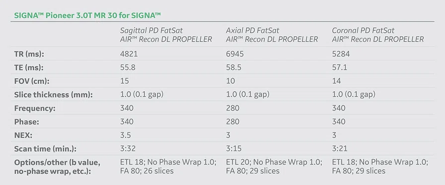
In the finger exam (Case 1), we used an extraordinarily high in-plane resolution and a very thin slice. The aim was to show the new possibilities of the SIGNA™ Pioneer 3.0T MR system with MR 30 for SIGNA™ and AIR™ Recon DL PROPELLER. With a 2D FSE sequence, movement and flow artifacts would certainly have occurred with such long scan times, which is not the case with the 2D PROPELLER technology. This technology enables very high in-plane resolution in combination with very thin slice thickness without any problems. In addition, we compared conventionally reconstructed images to the AIR™ Recon DL reconstructed images.
The shoulder exam (Case 2) is particularly interesting because the patient was claustrophobic and could not tolerate a long exam. It was also very important to not lose small anatomical details due to the pathology. The complete high-resolution and robust examination took only 6:43 minutes.
Case 1
Patient history
A 45-year-old obese female suffered trauma to her finger eight days prior. Patient had suspected capsule rupture/collateral ligament rupture on the left thumb. X-ray image suggested secondary findings of suspected enchondroma.
Figure 1.
A 45-year-old female suffered trauma to her finger eight days prior. This case was processed with both conventional and AIR™ Recon DL PROPELLER. A 16ch Flex small coil was used for this case. (A, B) Sagittal PD FatSat PROPELLER, 0.4 x 0.4 x 1.0 mm, 3:32 min., (A) with conventional reconstruction and (B) AIR™ Recon DL PROPELLER. (C-E) Axial PD FatSat PROPELLER, 0.4 x 0.4 x 1.0 mm, 3:15 min., (C) with conventional reconstruction and (D, E) AIR™ Recon DL PROPELLER. (F, G) Coronal PD FatSat 0.4 x 0.4 x 1.0 mm, 3:21 min. with AIR™ Recon DL PROPELLER.
Results
Slight soft tissue swelling at the metacarpophalangeal joint on the flexor side in the vicinity of the flexor tendon, ulnar accentuated with discrete capsule strain and no evidence of a collateral ligament rupture. Intact palmar plate. No joint effusion with proper articulation. Also, slight soft tissue swelling at the thumb saddle joint and discrete patchy bone marrow edema on the joint side, as well as bone marrow edema in the flexor side sesamoid. Adjacent hyperintense signal alteration of the joint capsule on the flexor and extensor sides, although the ligaments are also intact. No relevant joint effusion. The extensor and flexor tendons are regularly depicted, no peritendinitis. In the phalanx of the thumb, popcorn-like hyperintense signal alterations PD-weighted with a size of about 0.9 x 0.8 cm, also morphologically consistent with an enchondroma on MR.
Conclusion
No evidence of a collateral ligament rupture of the thumb. There is capsule strain on the flexor side and enchondroma in the basal segment. Moderate rhizarthrosis in the thumb saddle joint with bone marrow edema on the joint side and edematous changes in the sesamoid bone and immediate capsulitis, indicating differential diagnosis possibly due to this trauma. Extensor and flexor tendons are intact.
Case 2
Patient history
A 69-year-old claustrophic female with shoulder pain due to trauma; suspected rotator cuff tear.
Figure 2.
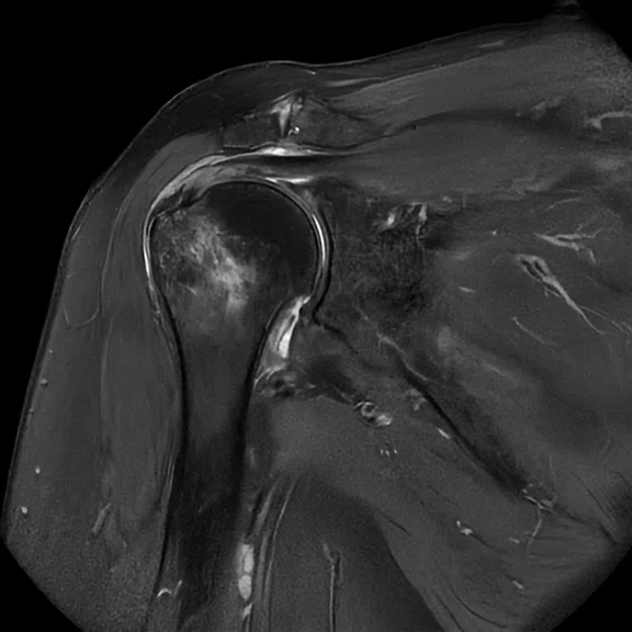
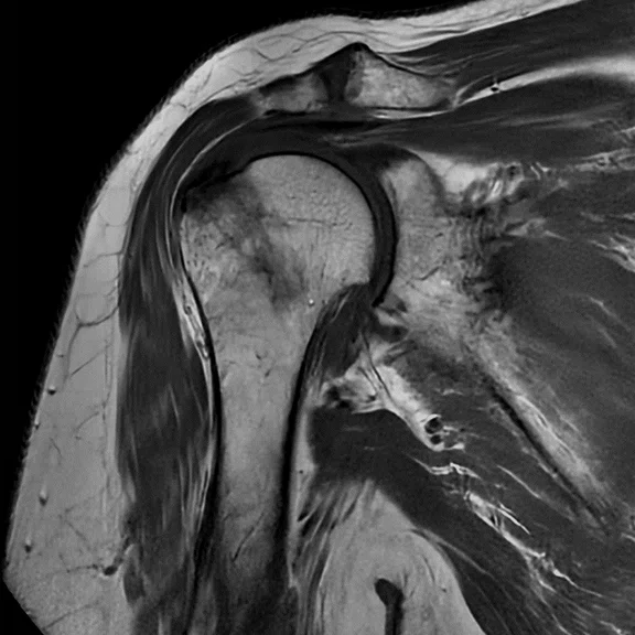
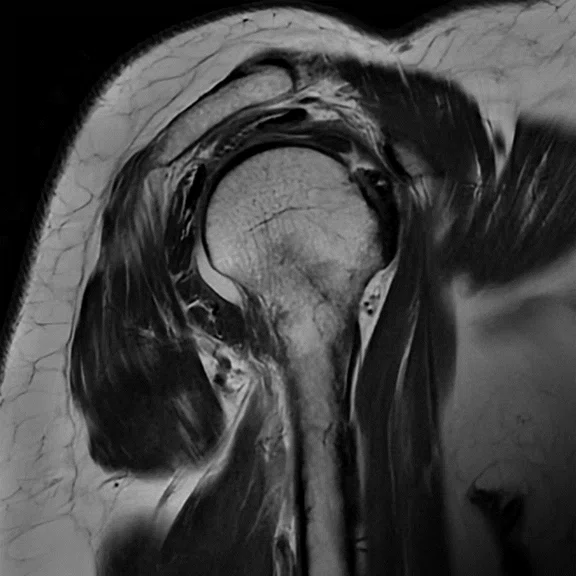
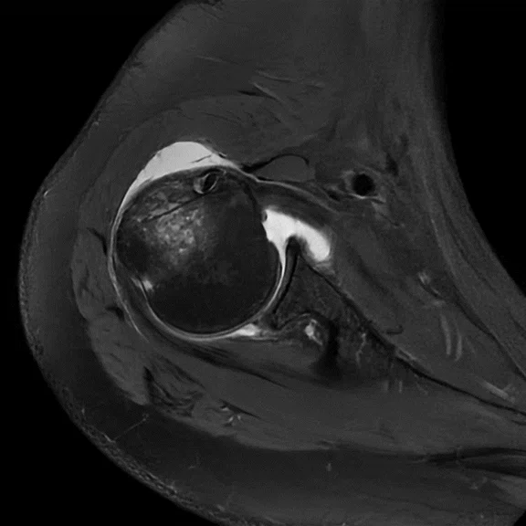
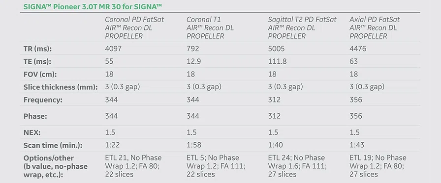
A 69-year-old female with shoulder pain due to trauma and suspected rotator cuff tear. All images were processed with AIR™ Recon DL PROPELLER and acquired at 0.5 x 0.5 x 3.0 mm. (A) Coronal PD FatSat PROPELLER, 1:22 min.; (B) coronal T1 PROPELLER, 1:58 min.; (C) sagittal T2 PROPELLER, 1:40 min.; and (D) axial PD FatSat PROPELLER, 1:43 min. Total exam time was 6:43 min with a 16ch Flex medium coil.
Results
Cutis and subcutis were unremarkable. Slightly activated acromioclavicular (AC) joint arthrosis with discrete joint effusion and small patchy bone marrow edema on the joint side were observed. The subacromial space was moderately narrowed at 0.6 cm. Images showed fluid collection in the subdeltoid/subacromial bursa and humeroglenoidal joint effusion. The glenoid was intact. On the head of the humerus, small cortical impression on the greater tuberosity with adjacent strong bone marrow edema and smallest avulsion fragment in the distal course of the supraspinatus tendon were present. The supraspinatus tendon hyperintensely signaled distally with no evidence of rupture. Infraspinatus tendon, tendon of the musculus teres minor and subscapularis tendon were all intact. Correct course and caliber of the long bicep tendon in the sulcus intertubercularis is also intact. No labrum lesion and no relevant chondropathy depicted.
Conclusion
There is a compression fracture at the greater tuberosity with small bony avulsion and strain of the distal supraspinatus tendon without evidence of a rotator cuff tear. Subdeltoid bursitis/subacromial and joint effusion. Mildly activated AC joint osteoarthritis.
Discussion
The smallest structures play an important role in small joints and, therefore, must be optimally imaged with MR. In the thumb case, diagnosis with the conventional reconstruction technology (without AIR™ Recon DL) would have been difficult due to the poor SNR. The MR finger examination on the SIGNA™ Pioneer with the combination of the 16-channel Flex small coil and AIR™ Recon DL PROPELLER was very helpful in order to make a confident diagnosis.
Robust and artifact-free imaging is important in patients with a suspected rotator cuff tear. In this particular case, since the patient was in pain and claustrophobic, the situation was particularly difficult. The MR shoulder examination on the SIGNA™ Pioneer with the combination of the 16-channel Flex medium and AIR™ Recon DL PROPELLER helped greatly to make the diagnosis.
In both of these cases, the acquired sequences are used for optimal findings, which is why each sequence must have the highest possible contrast, resolution and homogeneity.
The 2D FSE sequence is typically the first choice for high-resolution MSK examinations, as it provides sharp and low-noise images. However, this is not always the case in difficult patient populations, such as those who are in pain, obese or claustrophobic. In shoulder MR imaging, PROPELLER sequences were only utilized as a backup for restless patients when motion artifacts degraded the 2D FSE acquisition.
Since the implementation of MR 30 for SIGNA™ at our site, many small joints, such as those in the finger, and all shoulder protocols have been converted from 2D FSE to AIR™ Recon DL PROPELLER, allowing for the consistent acquisition of robust and sharp MR images. As demonstrated in the small joint (finger) case, even the smallest enchondromas can be defined in their size. In the shoulder case with the direct comparison of the 2D FSE and AIR™ Recon DL PROPELLER acquisitions, the “conventional” 2D FSE imaging technique did not provide a reliable diagnosis, whereas the AIR™ Recon DL PROPELLER delivered the image quality required for a confident diagnosis.


