1. Laurent V, Trausch G, Bruot O, Oliver P, Felblinger J, Regent D. Comparative study of two whole-body imaging techniques in the case of melanoma metastases: advantages of multi-contrast MRI examination including a diffusion-weighted sequence in comparison with PET-CT. Eur J Radiol. 2010 Sep;75(3):376-83.
1. Laurent V, Trausch G, Bruot O, Oliver P, Felblinger J, Regent D. Comparative study of two whole-body imaging techniques in the case of melanoma metastases: advantages of multi-contrast MRI examination including a diffusion-weighted sequence in comparison with PET-CT. Eur J Radiol. 2010 Sep;75(3):376-83.
A
Figure 1.
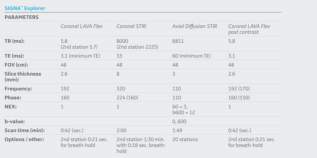
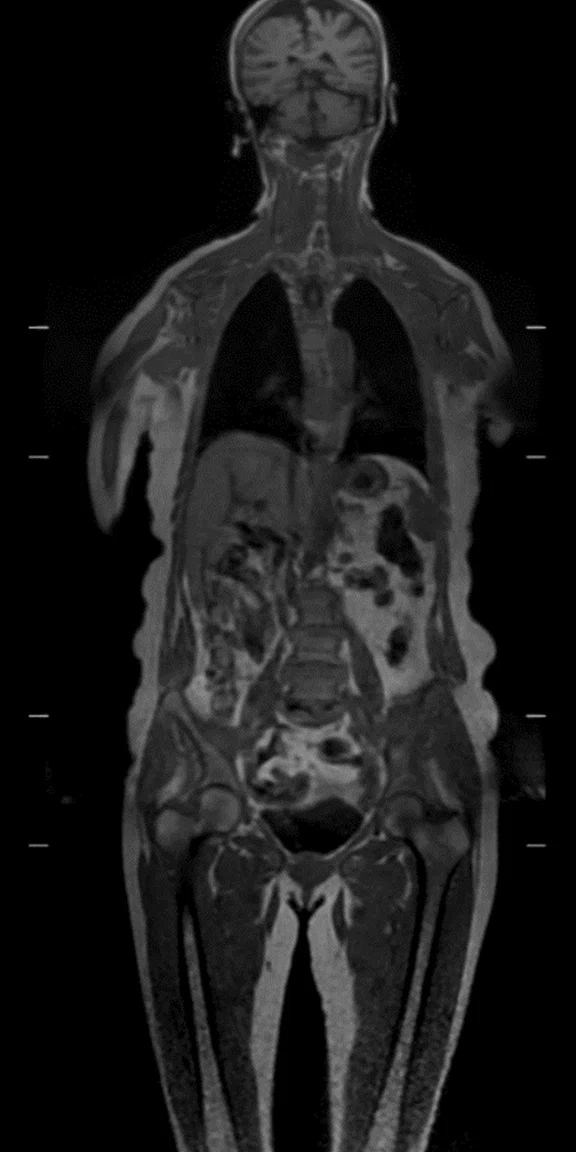
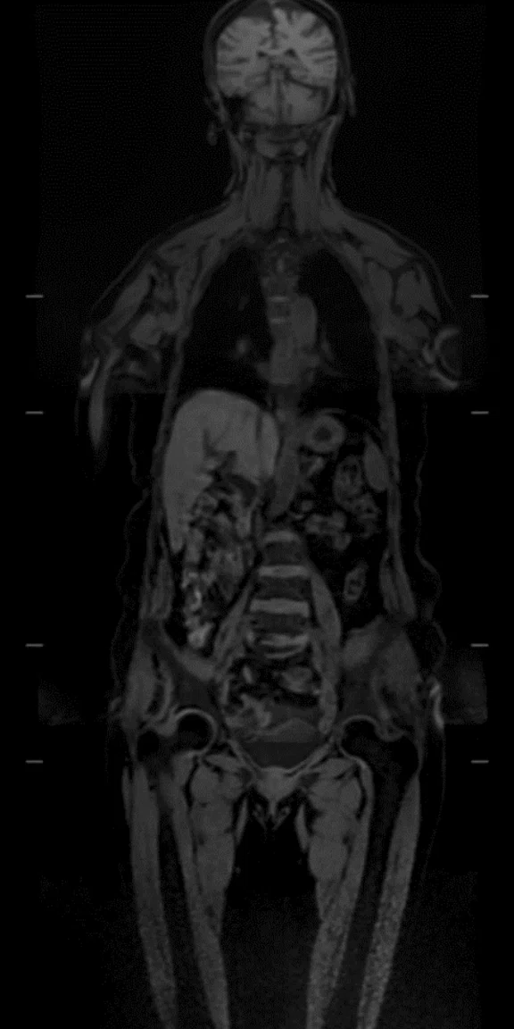
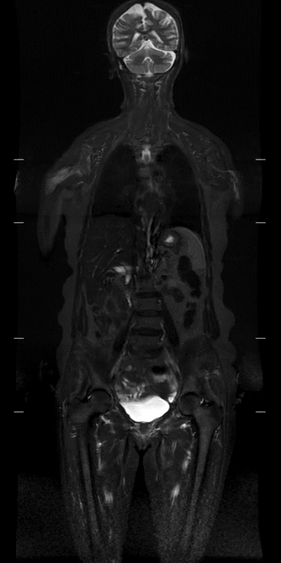
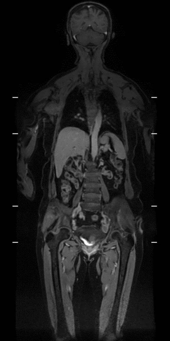
Whole-body MR imaging with approximately 120 cm coverage. (A-B) Coronal in-phase and water LAVA Flex; 2.5 x 3 x 2.6 mm, 3 stations, 1:45 min.; (C) Coronal STIR, 1.5 x 2.1 x 8 mm, 3 stations, 5:30 min.; and (D) Coronal water LAVA Flex, post contrast, 2.5 x 3 x 2.6 mm, 3 stations, 1:45 min.
B
Figure 1.
Whole-body MR imaging with approximately 120 cm coverage. (A-B) Coronal in-phase and water LAVA Flex; 2.5 x 3 x 2.6 mm, 3 stations, 1:45 min.; (C) Coronal STIR, 1.5 x 2.1 x 8 mm, 3 stations, 5:30 min.; and (D) Coronal water LAVA Flex, post contrast, 2.5 x 3 x 2.6 mm, 3 stations, 1:45 min.
C
Figure 1.
Whole-body MR imaging with approximately 120 cm coverage. (A-B) Coronal in-phase and water LAVA Flex; 2.5 x 3 x 2.6 mm, 3 stations, 1:45 min.; (C) Coronal STIR, 1.5 x 2.1 x 8 mm, 3 stations, 5:30 min.; and (D) Coronal water LAVA Flex, post contrast, 2.5 x 3 x 2.6 mm, 3 stations, 1:45 min.
D
Figure 1.
Whole-body MR imaging with approximately 120 cm coverage. (A-B) Coronal in-phase and water LAVA Flex; 2.5 x 3 x 2.6 mm, 3 stations, 1:45 min.; (C) Coronal STIR, 1.5 x 2.1 x 8 mm, 3 stations, 5:30 min.; and (D) Coronal water LAVA Flex, post contrast, 2.5 x 3 x 2.6 mm, 3 stations, 1:45 min.
A
Figure 2.
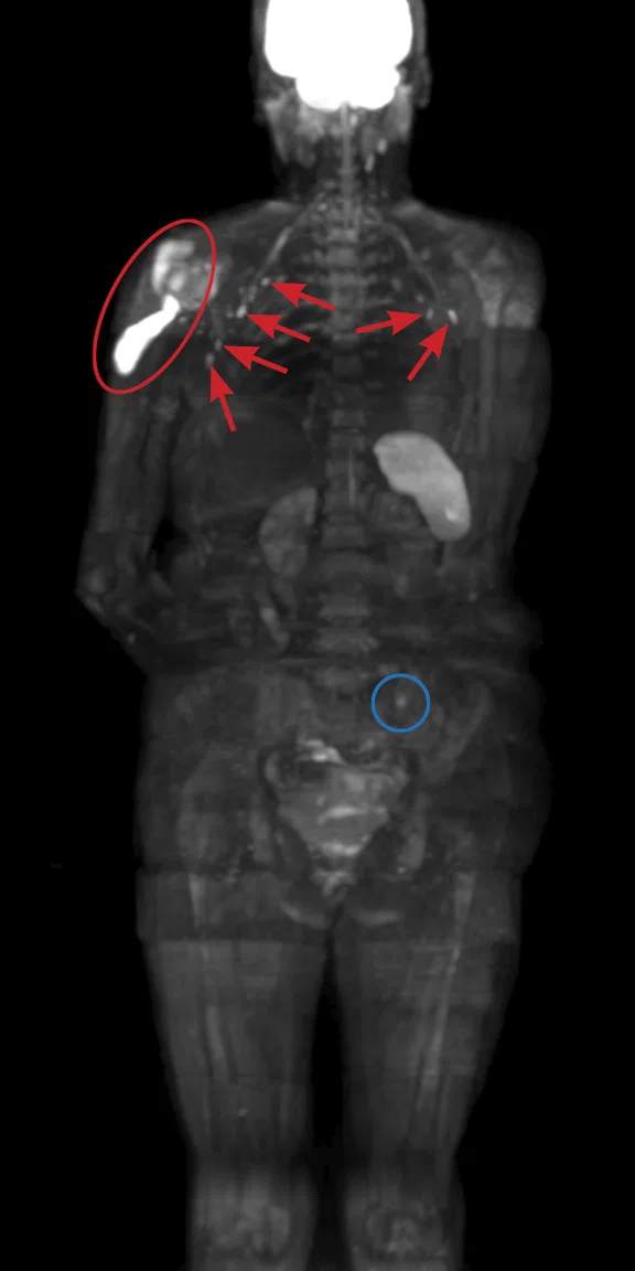
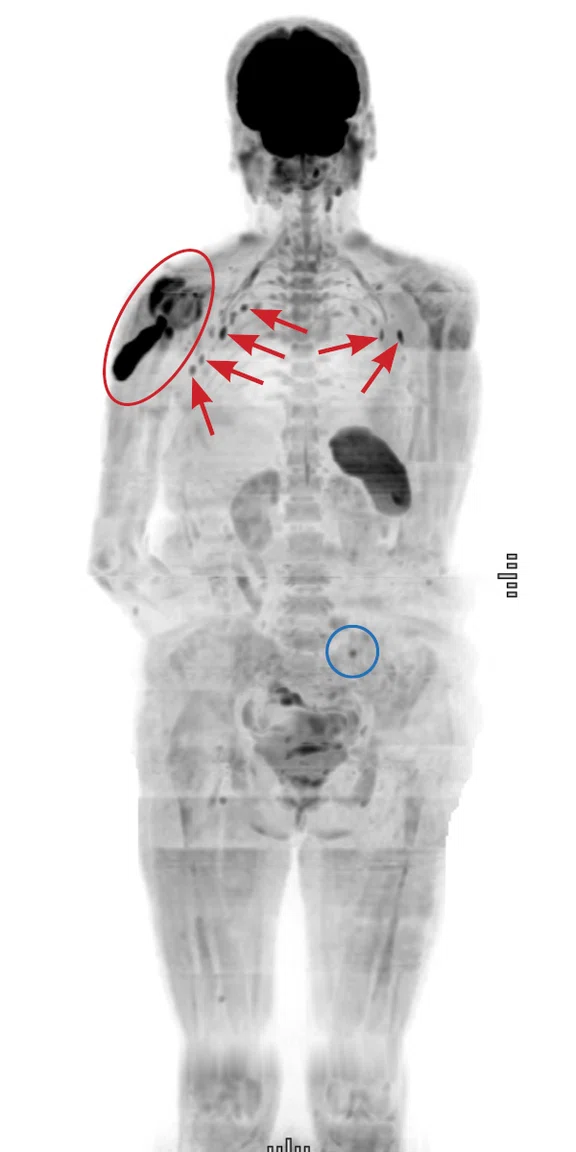
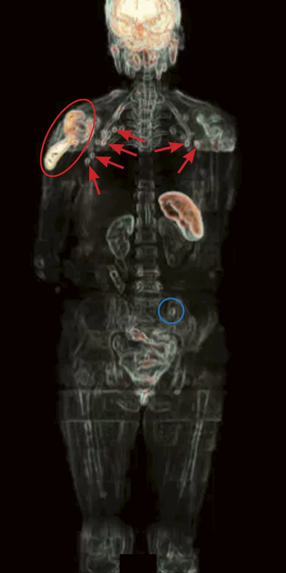
Whole-body MR imaging with approximately 120 cm coverage. Binded Axial diffusion STIR 3-in-1, b600, 4.4 x 4.4 x 3 mm, 20 stations, 36:20 min. (A-B) MIP projections; and (C) volume rendered image. Red circle depicts the effusion on the glenohumeral joint of the right shoulder. Red arrows show the bilateral swollen axillary lymph nodes. Blue circle denotes the lesion on the left iliac joint.
B
Figure 2.
Whole-body MR imaging with approximately 120 cm coverage. Binded Axial diffusion STIR 3-in-1, b600, 4.4 x 4.4 x 3 mm, 20 stations, 36:20 min. (A-B) MIP projections; and (C) volume rendered image. Red circle depicts the effusion on the glenohumeral joint of the right shoulder. Red arrows show the bilateral swollen axillary lymph nodes. Blue circle denotes the lesion on the left iliac joint.
C
Figure 2.
Whole-body MR imaging with approximately 120 cm coverage. Binded Axial diffusion STIR 3-in-1, b600, 4.4 x 4.4 x 3 mm, 20 stations, 36:20 min. (A-B) MIP projections; and (C) volume rendered image. Red circle depicts the effusion on the glenohumeral joint of the right shoulder. Red arrows show the bilateral swollen axillary lymph nodes. Blue circle denotes the lesion on the left iliac joint.
A
Figure 3.
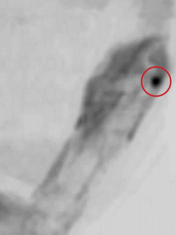
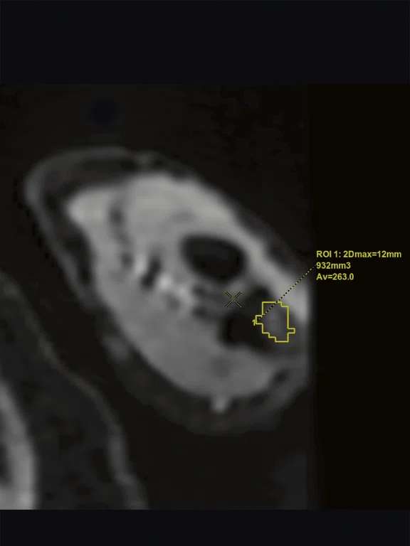
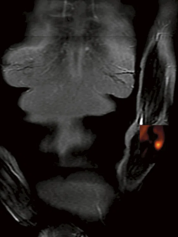
(A) Coronal STIR, 1.5 x 2.1 x 8 mm; (B) same image with color map; (C-D) diffusion STIR 3-in-1 b600, 4.4 x 4.4 x 3 mm; and (E-F) Coronal water LAVA Flex post contrast, 2.5 x 3 x 2.6 mm and Axial MPR. Lesion located on the proximal metaphysis of the ulna of the left arm (red circle), suspected to be metastatic.
C
Figure 3.
(A) Coronal STIR, 1.5 x 2.1 x 8 mm; (B) same image with color map; (C-D) diffusion STIR 3-in-1 b600, 4.4 x 4.4 x 3 mm; and (E-F) Coronal water LAVA Flex post contrast, 2.5 x 3 x 2.6 mm and Axial MPR. Lesion located on the proximal metaphysis of the ulna of the left arm (red circle), suspected to be metastatic.
E
Figure 3.
(A) Coronal STIR, 1.5 x 2.1 x 8 mm; (B) same image with color map; (C-D) diffusion STIR 3-in-1 b600, 4.4 x 4.4 x 3 mm; and (E-F) Coronal water LAVA Flex post contrast, 2.5 x 3 x 2.6 mm and Axial MPR. Lesion located on the proximal metaphysis of the ulna of the left arm (red circle), suspected to be metastatic.
B
Figure 3.
(A) Coronal STIR, 1.5 x 2.1 x 8 mm; (B) same image with color map; (C-D) diffusion STIR 3-in-1 b600, 4.4 x 4.4 x 3 mm; and (E-F) Coronal water LAVA Flex post contrast, 2.5 x 3 x 2.6 mm and Axial MPR. Lesion located on the proximal metaphysis of the ulna of the left arm (red circle), suspected to be metastatic.
D
Figure 3.
(A) Coronal STIR, 1.5 x 2.1 x 8 mm; (B) same image with color map; (C-D) diffusion STIR 3-in-1 b600, 4.4 x 4.4 x 3 mm; and (E-F) Coronal water LAVA Flex post contrast, 2.5 x 3 x 2.6 mm and Axial MPR. Lesion located on the proximal metaphysis of the ulna of the left arm (red circle), suspected to be metastatic.
F
Figure 3.
(A) Coronal STIR, 1.5 x 2.1 x 8 mm; (B) same image with color map; (C-D) diffusion STIR 3-in-1 b600, 4.4 x 4.4 x 3 mm; and (E-F) Coronal water LAVA Flex post contrast, 2.5 x 3 x 2.6 mm and Axial MPR. Lesion located on the proximal metaphysis of the ulna of the left arm (red circle), suspected to be metastatic.
A
Figure 4.
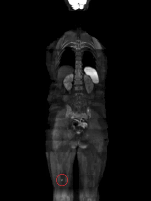
(A) Fusion STIR + diffusion STIR 3-in-1 b600; (B) diffusion STIR b600, 4.4 x 4.4 x 3 mm. Small nodule was located on the soft tissues of the right thigh (red circle).
B
Figure 4.
(A) Fusion STIR + diffusion STIR 3-in-1 b600; (B) diffusion STIR b600, 4.4 x 4.4 x 3 mm. Small nodule was located on the soft tissues of the right thigh (red circle).
2. Skeletal Radiol. 2010 Apr;39(4):333-43. doi: 10.1007/s00256-009-0789-4. Gutzeit A, Doert A, Froehlich JM, et al. Comparison of diffusion-weighted whole-body MRI and skeletal scintigraphy for the detection of bone metastases in patients with prostate or breast carcinoma. Skeletal Radiol. 2010 Apr;39(4):333-43.
A
Figure 1.
Whole-body MR imaging with approximately 120 cm coverage. (A-B) Coronal in-phase and water LAVA Flex; 2.5 x 3 x 2.6 mm, 3 stations, 1:45 min.; (C) Coronal STIR, 1.5 x 2.1 x 8 mm, 3 stations, 5:30 min.; and (D) Coronal water LAVA Flex, post contrast, 2.5 x 3 x 2.6 mm, 3 stations, 1:45 min.
B
Figure 1.
Whole-body MR imaging with approximately 120 cm coverage. (A-B) Coronal in-phase and water LAVA Flex; 2.5 x 3 x 2.6 mm, 3 stations, 1:45 min.; (C) Coronal STIR, 1.5 x 2.1 x 8 mm, 3 stations, 5:30 min.; and (D) Coronal water LAVA Flex, post contrast, 2.5 x 3 x 2.6 mm, 3 stations, 1:45 min.
C
Figure 1.
Whole-body MR imaging with approximately 120 cm coverage. (A-B) Coronal in-phase and water LAVA Flex; 2.5 x 3 x 2.6 mm, 3 stations, 1:45 min.; (C) Coronal STIR, 1.5 x 2.1 x 8 mm, 3 stations, 5:30 min.; and (D) Coronal water LAVA Flex, post contrast, 2.5 x 3 x 2.6 mm, 3 stations, 1:45 min.
D
Figure 1.
Whole-body MR imaging with approximately 120 cm coverage. (A-B) Coronal in-phase and water LAVA Flex; 2.5 x 3 x 2.6 mm, 3 stations, 1:45 min.; (C) Coronal STIR, 1.5 x 2.1 x 8 mm, 3 stations, 5:30 min.; and (D) Coronal water LAVA Flex, post contrast, 2.5 x 3 x 2.6 mm, 3 stations, 1:45 min.
result


PREVIOUS
${prev-page}
NEXT
${next-page}
Subscribe Now
Manage Subscription
FOLLOW US
Contact Us • Cookie Preferences • Privacy Policy • California Privacy PolicyDo Not Sell or Share My Personal Information • Terms & Conditions • Security
© 2024 GE HealthCare. GE is a trademark of General Electric Company. Used under trademark license.
SPOTLIGHT
Whole-body diffusion for evaluation of metastatic lesions
Whole-body diffusion for evaluation of metastatic lesions
By Abdelhamid Derriche, MD, site radiologist, and Orkia Ferdagha, MR technologist, PRIISM, EHP Kara
Cancer patients are often referred for whole-body imaging to evaluate the presence of secondary or metastatic lesions, which can change the course of patient treatment. In our institution, whole-body diffusion MR is our preferred modality for this type of study.
Diffusion-weighted imaging (DWI) on MR has shown excellent sensitivity (82%) and specificity (97%) compared to PET/CT (sensitivity of 72% and specificity of 92%) in whole-body imaging in melanoma metastases.1 Additionally, DWI MR has been shown to be the most accurate for detecting metastases in the bone, liver, subcutaneous and intra-peritoneal sites compared to PET/CT.1
However, non-optimal DWI MR acquisitions can produce scintigraphy-like images that may produce false positive results. Combining DWI MR with an inversion time technique can help overcome this issue by allowing for a better suppression of fat signal, which helps suppress background body signal. Further, utilizing an MR system that has strong magnet homogeneity allows for the acquisition of large field-of-view (FOV) images and helps reduce distortions in areas of off-center imaging, such as the extremities. DWI STIR with a "3-in-1" technique provides an excellent compromise between acquisition time and signal-to-noise ratio (SNR), allowing acquisition of thin slices that may help further enhance sensitivity of the DWI sequence.
Patient history
A 75-year-old female with known melanoma was referred for whole-body DWI MR for evaluation of suspected metastatic (secondary) lesions on her extremities. Post-contrast whole-body MR with LAVA Flex was also used to evaluate areas with suspicious contrast uptake.
MR findings
- Important effusion on the glenohumeral joint of the right shoulder, to be investigated
- Several bilateral swollen axillary lymph nodes, predominant in number and size at the right axillary region
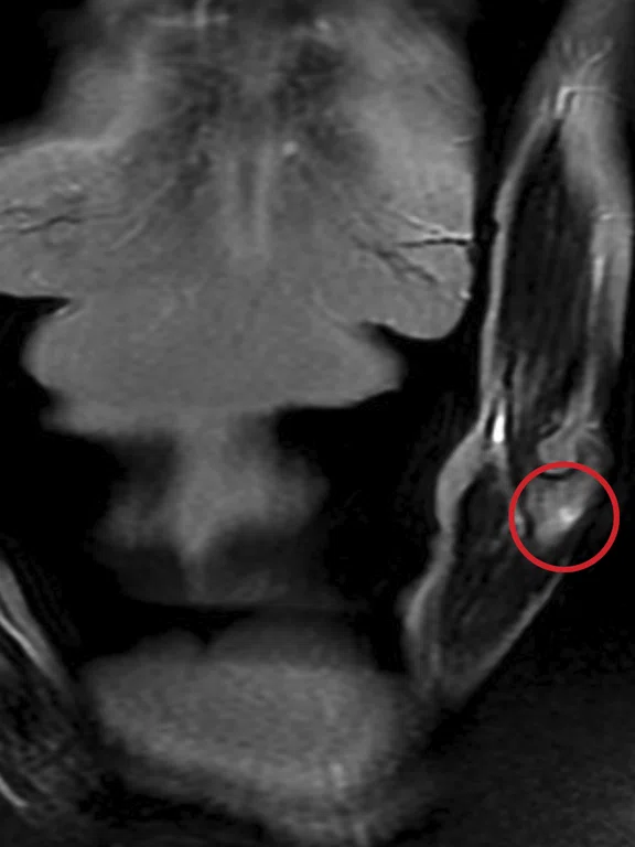
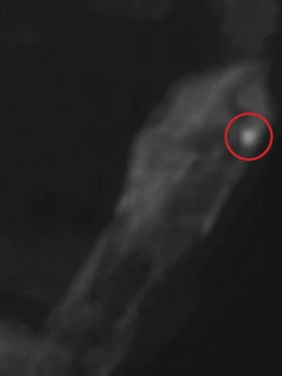
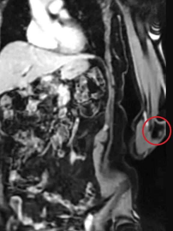
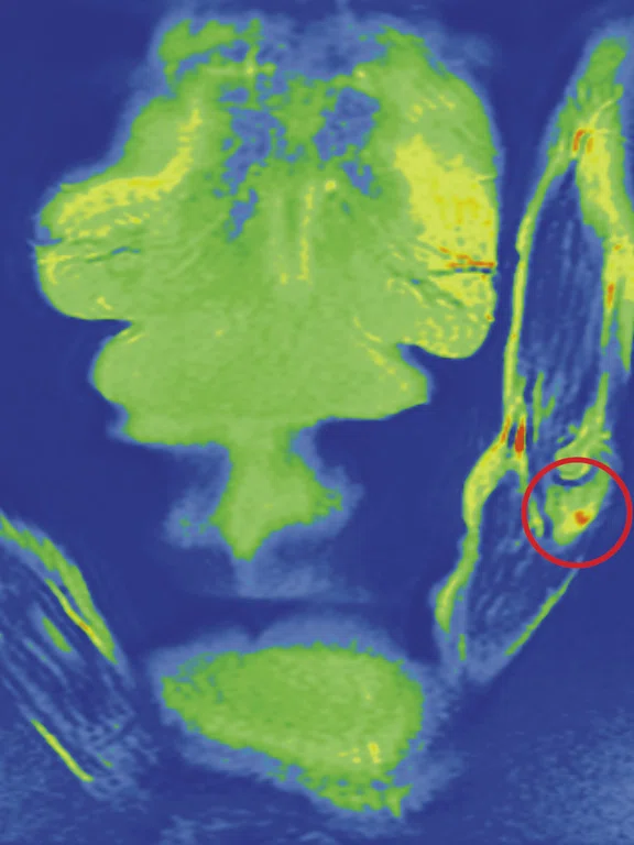
- Lesion located on the proximal metaphysis of the ulna of the left arm and suspected to be metastatic, measuring 13 mm from its long axis, hyperintense in diffusion, hyperintense in STIR and gadolinium-enhanced on post-contrast T1 sequence
- Second suspicious lesion with similar aspect, measuring 9 mm and located on the distal diaphysis of the radius of the right arm
- Lesion on the left iliac joint, measuring 6 mm, hyperintense in diffusion, with no aspects on the other sequences
- Small nodule of 15 mm situated on the soft tissues of the right thigh, at the inferior part located within the posterior muscular compartment, hyperintense in diffusion.
Figure 1.
Whole-body MR imaging with approximately 120 cm coverage. (A-B) Coronal in-phase and water LAVA Flex; 2.5 x 3 x 2.6 mm, 3 stations, 1:45 min.; (C) Coronal STIR, 1.5 x 2.1 x 8 mm, 3 stations, 5:30 min.; and (D) Coronal water LAVA Flex, post contrast, 2.5 x 3 x 2.6 mm, 3 stations, 1:45 min.
Figure 1.
Whole-body MR imaging with approximately 120 cm coverage. (A-B) Coronal in-phase and water LAVA Flex; 2.5 x 3 x 2.6 mm, 3 stations, 1:45 min.; (C) Coronal STIR, 1.5 x 2.1 x 8 mm, 3 stations, 5:30 min.; and (D) Coronal water LAVA Flex, post contrast, 2.5 x 3 x 2.6 mm, 3 stations, 1:45 min.
Figure 2.
Whole-body MR imaging with approximately 120 cm coverage. Binded Axial diffusion STIR 3-in-1, b600, 4.4 x 4.4 x 3 mm, 20 stations, 36:20 min. (A-B) MIP projections; and (C) volume rendered image. Red circle depicts the effusion on the glenohumeral joint of the right shoulder. Red arrows show the bilateral swollen axillary lymph nodes. Blue circle denotes the lesion on the left iliac joint.
Figure 3.
(A) Coronal STIR, 1.5 x 2.1 x 8 mm; (B) same image with color map; (C-D) diffusion STIR 3-in-1 b600, 4.4 x 4.4 x 3 mm; and (E-F) Coronal water LAVA Flex post contrast, 2.5 x 3 x 2.6 mm and Axial MPR. Lesion located on the proximal metaphysis of the ulna of the left arm (red circle), suspected to be metastatic.
Discussion
The patient has several small lesions suspected to be secondary bone lesions from her primary cancer, melanoma. Having the ability to detect these lesions on the patient’s arms, iliac joint and thigh will enable the oncologist to re-stage the cancer and adapt therapy.
Whole-body DWI is an excellent option for evaluation of metastatic bone lesions compared to PET/CT and scintigraphy. It provides good image quality and provides the information clinicians need for patient management.
Even if PET/CT were recommended, this type of system is not available in all regions and is often more expensive than MR. Scintigraphy exposes the patient to additional radiation and MR has been found to provide similar sensitivity and specificity for patients with a small number of bone lesions, 58% sensitivity/33% specificity for whole-body DWI MR versus 67% sensitivity/58% specificity for scintigraphy; and patients with multiple bone lesions, 97% sensitivity/91% specificity for whole-body DWI MR versus 48% sensitivity/42% specificity for scintigraphy.2
With MR, we can avoid patient exposure to additional radiation dose and achieve a high sensitivity and specificity for detection of metastatic lesions. Additionally, the strong magnet homogeneity on the SIGNA™Explorer helps to decrease distortion when imaging large FOVs and "3-in-1" diffusion STIR provides the thin-slice data needed for a confident diagnosis in shorter scan times.


