A
Figure 2.
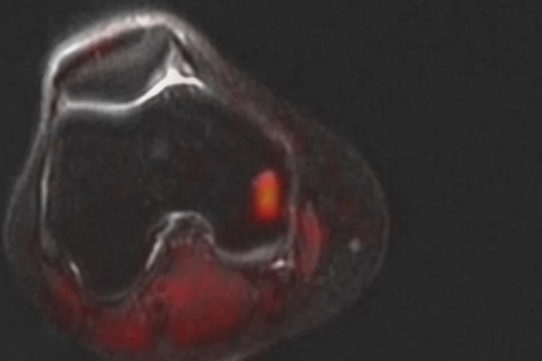
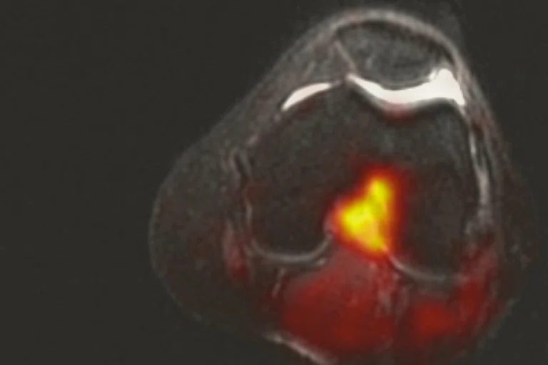
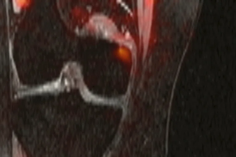
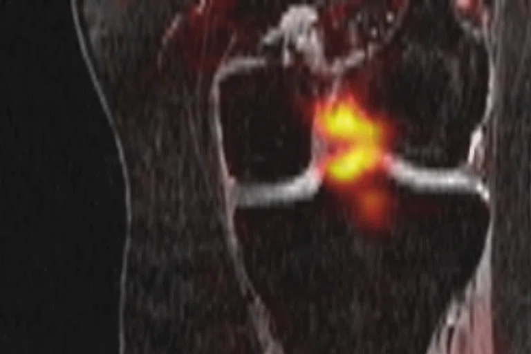
Simultaneous PET/MR with novel sigma-1 receptor-specific radiotracer identifies source of pain (an intraarticular inflamed synovial lipoma) in patient with chronic knee pain of previously unclear origin. Patient reported 8-10/10 left knee pain for years. Presented for S1R PET/MR which showed a “hotspot” in the center of knee, which was subsequently removed arthroscopically and completely resolved the patient’s pain. (A-D) Fused PET/MR images; (E) pathology; and (F) MR image depicting lesion.
B
Figure 2.
Simultaneous PET/MR with novel sigma-1 receptor-specific radiotracer identifies source of pain (an intraarticular inflamed synovial lipoma) in patient with chronic knee pain of previously unclear origin. Patient reported 8-10/10 left knee pain for years. Presented for S1R PET/MR which showed a “hotspot” in the center of knee, which was subsequently removed arthroscopically and completely resolved the patient’s pain. (A-D) Fused PET/MR images; (E) pathology; and (F) MR image depicting lesion.
C
Figure 2.
Simultaneous PET/MR with novel sigma-1 receptor-specific radiotracer identifies source of pain (an intraarticular inflamed synovial lipoma) in patient with chronic knee pain of previously unclear origin. Patient reported 8-10/10 left knee pain for years. Presented for S1R PET/MR which showed a “hotspot” in the center of knee, which was subsequently removed arthroscopically and completely resolved the patient’s pain. (A-D) Fused PET/MR images; (E) pathology; and (F) MR image depicting lesion.
D
Figure 2.
Simultaneous PET/MR with novel sigma-1 receptor-specific radiotracer identifies source of pain (an intraarticular inflamed synovial lipoma) in patient with chronic knee pain of previously unclear origin. Patient reported 8-10/10 left knee pain for years. Presented for S1R PET/MR which showed a “hotspot” in the center of knee, which was subsequently removed arthroscopically and completely resolved the patient’s pain. (A-D) Fused PET/MR images; (E) pathology; and (F) MR image depicting lesion.
1 Institute of Medicine Report from the Committee on Advancing Pain Research, Care, and Education: Relieving Pain in America, A Blueprint for Transforming Prevention, Care, Education and Research. The National Academies Press, 2011. Accessed October 18, 2019.
2 Goldberg DS, McGee SJ. Pain as a global public health priority. BMC Public Health. 2011;11:770.
1 Institute of Medicine Report from the Committee on Advancing Pain Research, Care, and Education: Relieving Pain in America, A Blueprint for Transforming Prevention, Care, Education and Research. The National Academies Press, 2011. Accessed October 18, 2019.
3 Englund M, Guermazi A, Gale D, et al. Incidental meniscal findings on knee MRI in middle-aged and elderly persons. N Engl J Med. 2008 Sep 11;359(11):1108-15.
3 Englund M, Guermazi A, Gale D, et al. Incidental meniscal
findings on knee MRI in middle-aged and elderly persons.
N Engl J Med. 2008 Sep 11;359(11):1108-15.
Figure 1.
In the US alone, the total cost to society and government for chronic pain is nearly $800 billion.
5 Cipriano PW, Lee SW, Yoon D, et al. Successful treatment of chronic knee pain following localization by a sigma-1 receptor radioligand and Pet/MRI: a case report. J Pain Res. 2018 Oct 12;11:2353-2357.
6 Institute for Chronic Pain. Sciatica. Available at: https://www.instituteforchronicpain.org/common-conditions/sciatica. Accessed October 18, 2019.
7 Cipriano PW, Yoon D, Gandhi H, et al. 18F-FDG PET/MRI in Chronic Sciatica: Early Results Revealing Spinal and Nonspinal Abnormalities. J Nucl Med. 2018 Jun;59(6):967-972.
E
Figure 2.
Simultaneous PET/MR with novel sigma-1 receptor-specific radiotracer identifies source of pain (an intraarticular inflamed synovial lipoma) in patient with chronic knee pain of previously unclear origin. Patient reported 8-10/10 left knee pain for years. Presented for S1R PET/MR which showed a “hotspot” in the center of knee, which was subsequently removed arthroscopically and completely resolved the patient’s pain. (A-D) Fused PET/MR images; (E) pathology; and (F) MR image depicting lesion.
F
Figure 2.
Simultaneous PET/MR with novel sigma-1 receptor-specific radiotracer identifies source of pain (an intraarticular inflamed synovial lipoma) in patient with chronic knee pain of previously unclear origin. Patient reported 8-10/10 left knee pain for years. Presented for S1R PET/MR which showed a “hotspot” in the center of knee, which was subsequently removed arthroscopically and completely resolved the patient’s pain. (A-D) Fused PET/MR images; (E) pathology; and (F) MR image depicting lesion.
‡18F-S1R is not cleared or approved by the US FDA or any other global regulator.
4 Beyer T, Tellmann L, Nickel I, Pietrzyk U. On the Use of Positioning Aids to Reduce Misregistration in the Head and Neck in Whole-Body PET/CT Studies. J Nucl Med. 2005 Apr;46(4):596-602.
result


PREVIOUS
${prev-page}
NEXT
${next-page}
Subscribe Now
Manage Subscription
FOLLOW US
Contact Us • Cookie Preferences • Privacy Policy • California Privacy PolicyDo Not Sell or Share My Personal Information • Terms & Conditions • Security
© 2024 GE HealthCare. GE is a trademark of General Electric Company. Used under trademark license.
IN PRACTICE
Understanding the underlying molecular underpinnings and pathology of pain using PET/MR
Understanding the underlying molecular underpinnings and pathology of pain using PET/MR
The numbers are staggering. In the US alone, nearly one-third of adults report suffering from at least one type of chronic pain (e.g., neck, back, leg, knee or other kind of pain).1 Estimates for the global incidence rate of chronic pain sufferers are currently one in five people, and one in 10 adults are newly diagnosed with chronic pain each year.2 Moreover, the Institute of Medicine estimates that pain costs society between $560 and $635 billion per year while federal and state costs add another $100 billion annually, making the total burden nearly $800 billion each year.1 These facts highlight that chronic pain is a significant public health issue.
The numbers are staggering. In the US alone, nearly one-third of adults report suffering from at least one type of chronic pain (e.g., neck, back, leg, knee or other kind of pain).1 Estimates for the global incidence rate of chronic pain sufferers are currently one in five people, and one in 10 adults are newly diagnosed with chronic pain each year.2 Moreover, the Institute of Medicine estimates that pain costs society between $560 and $635 billion per year while federal and state costs add another $100 billion annually, making the total burden nearly $800 billion each year.1 These facts highlight that chronic pain is a significant public health issue.
Sandip Biswal, MD, a co-founder of SiteOne Therapeutics, Inc., and PainSeek, has spent the last 20 years pioneering a better way to accurately identify pain generators in patients with chronic pain.
“The ugly truth is that current methods to diagnose pain are not always accurate or based on objective methods,” he explains. “Instead, it’s based on empirical testing, often without getting to the root of the problem. In a large number of patients, we are prescribing treatments that merely mask pain without understanding its etiology. It’s no wonder current pain treatments are estimated to be only 30 percent effective. It is also no surprise we have an opioid epidemic since physicians resort to alleviating the symptoms of pain without understanding the cause of one’s pain since physicians do not have any good tools to pinpoint the site of pain generation.”
Although MR is widely utilized for diagnosing the source of pain, the findings from an MR exam may not be related to the patient’s pain. One of the most famous examples of this is what is known as the Framingham knee study, which used MR imaging of the knee in patients with an unclear cause of knee pain. The study found that 61 percent of subjects had meniscal tears in their knee yet did not have pain, aching or stiffness during the previous month. Further, only 63 percent of subjects experiencing pain, aching or stiffness in their knee actually had a meniscus tear.3
“We have found that conventional MR techniques either have limited sensitivity or find a number of abnormalities that may or may not be diagnosed as the pain generator,” Dr. Biswal explains. This leads Dr. Biswal to question whether the pathology visualized is related to the patient’s pain.
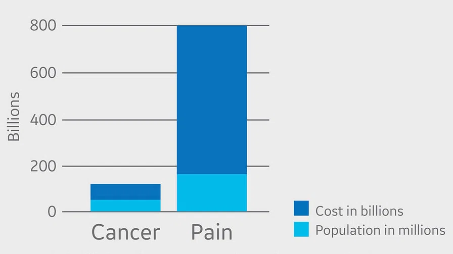
Figure 1.
In the US alone, the total cost to society and government for chronic pain is nearly $800 billion.
This is where the combination of simultaneous PET/MR can help. With PET/MR, Dr. Biswal can acquire both MR and PET image data at the same time — both cameras are on — and then register them together as one unified image. He interprets the PET images in real-time to identify abnormal FDG uptake throughout the body, using PET imaging findings as a guide to perform high-resolution multiparametric MR imaging to evaluate tissues and structures for the identification of pain generators and a specific diagnosis.
“The simultaneous acquisition in PET/MR gives me more confidence because fusing two studies acquired at different times, such as those acquired with PET/CT, may not be precise,” he says.
He references a paper that showed fusing a separate PET and CT in the head led to a difference in spatial registration of 7 mm between the two data sets using a standard PET head holder.4 That difference can introduce uncertainties that may result in different diagnoses and therapies when evaluating small structures such as nerve roots, joints and surrounding tissue.
The simultaneously acquired PET and MR image data led to much higher fidelity of co-registered data sets, which is critical for Dr. Biswal when making treatment management decisions based on small structures in complex anatomic areas. Additionally, the time-of-flight PET and Q.Clear image reconstruction algorithm available on the SIGNA™ PET/MR system allows him to accurately detect small abnormalities with very high sensitivity and low noise.
Whole-body PET/MR imaging provides further insight into the manifestation of pain in its evaluation of both central and peripheral pain generators. This whole-body view ensures that all potential causes of pain will be captured since pain can be referred from locations remote to the site of symptoms. This is in contrast to current conventional methods to pain diagnosis where the standard of care is limited to the examination of only the site of pain (e.g., when someone presents with knee pain, only the knee is scanned). With this new approach, the entire body is studied, including the brain and spine, thereby significantly increasing the likelihood the pain generator will be identified.
By using PET/MR, Dr. Biswal can see more than just the anatomic abnormalities that may give rise to pain. He can also visualize the cellular and molecular makeup of the injury or inflammation and understand the underlying biology and pathology of pain. He is also examining nociception and neuronal inflammation as a means of improving objective, image-guided diagnosis and treatment of chronic pain disorders.
Using a novel tracer, 18F-S1R‡, Dr. Biswal has imaged sigma-1 receptors that are overexpressed in injured or inflamed tissues and are thought to be more specific than FDG. In a published case report, the tracer was used to identify the source of refractory left knee pain of unknown origin. The patient was thought to have peroneal neuropathy, however, the PET showed uptake in an intercondylar notch that corresponded to an intra articular synovial lipoma detected in the simultaneously acquired MR exam.5
Another study that Dr. Biswal co-authored examined the use of whole-body FDG PET/MR as a diagnostic tool to visualize pathology and hypermetabolism in patients with chronic sciatica, a pain that starts in the lower back. Identifying the cause of sciatica can be difficult — it can result from disc herniations or other degenerative changes in the lower spine and it is not uncommon for the findings of an MR exam to be inconsistent with the patient symptoms.6 With FDG PET/MR, Dr. Biswal and co authors were able to locate the source of sciatica in nine patients. In five patients, the source of pain was a herniated disc. However, in four patients different lesions were detected in the peripheral nerve, leg muscles, pars and facet joints where the FDG uptake was abnormally high on PET, yet the MR demonstrated mild or no abnormalities in these areas.7
E
F
Figure 2.
Simultaneous PET/MR with novel sigma-1 receptor-specific radiotracer identifies source of pain (an intraarticular inflamed synovial lipoma) in patient with chronic knee pain of previously unclear origin. Patient reported 8-10/10 left knee pain for years. Presented for S1R PET/MR which showed a “hotspot” in the center of knee, which was subsequently removed arthroscopically and completely resolved the patient’s pain. (A-D) Fused PET/MR images; (E) pathology; and (F) MR image depicting lesion.
Addressing this need to identify pain generators is why Dr. Biswal cofounded PainSeek along with Michelle James, PhD, and others. According to Dr. Biswal, the company will be dedicated to developing pain diagnostics as well as new therapies for pain, including the development of PET tracers to identify certain molecules and cells specific to pain. They hope to develop objective molecular measurements of pain that highlight changes visualized in pain-sensing nerves.
“Even referring physicians are amazed at the initial results of imaging pain patients with PET/MR. It’s one thing to provide high-quality imaging, but another to help improve patient outcomes by figuring out what is really wrong.”
Dr. Sandip Biswal


