result
1. Iskurt A, Becerkli Y, Mahmutyazicioglu K. Automatic Identification of Landmarks for Standard Slice Positioning in Brain MRI. JMRI, 2011: 34(3);499-510.
1. Iskurt A, Becerkli Y, Mahmutyazicioglu K. Automatic Identification of Landmarks for Standard Slice Positioning in Brain MRI. JMRI, 2011: 34(3);499-510.
A
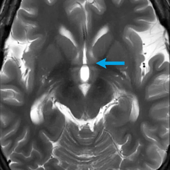
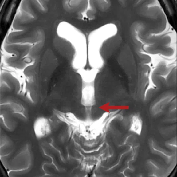
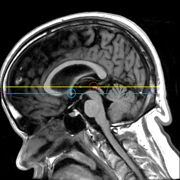
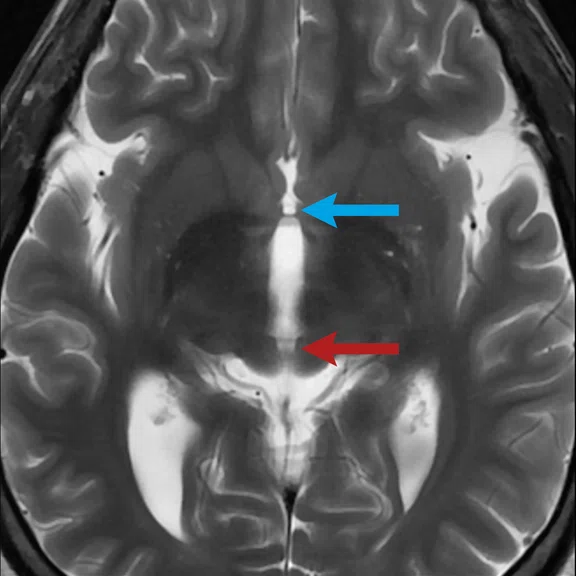
Figure 1.
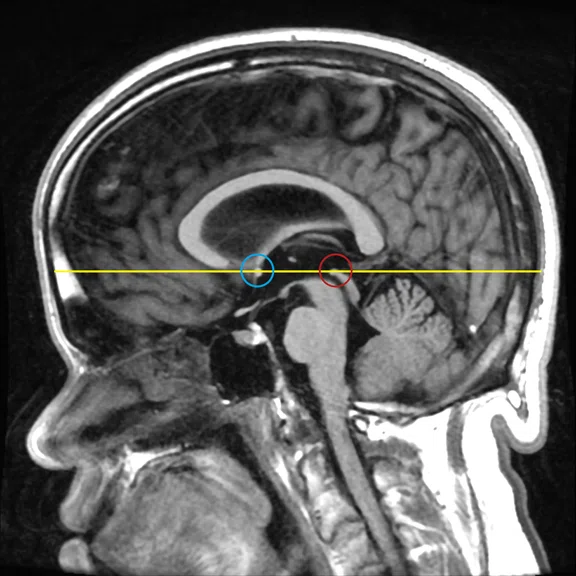
Atlas-based auto slice prescription compared to AIR x™ with AC-PC line, the most commonly used plane for planning a brain MR exam. (A-C) Atlas-based slices acquire the AC-PC plane, which varies between patients, on different slices in order to visualize the AC-PC in the same plane. (D, E) AIR x™ aligns to the AC-PC on the same plane and slice regardless of the head's position. AC=arrow/circle, PC=red arrow/circle.
B
Figure 1.
Atlas-based auto slice prescription compared to AIR x™ with AC-PC line, the most commonly used plane for planning a brain MR exam. (A-C) Atlas-based slices acquire the AC-PC plane, which varies between patients, on different slices in order to visualize the AC-PC in the same plane. (D, E) AIR x™ aligns to the AC-PC on the same plane and slice regardless of the head's position. AC=arrow/circle, PC=red arrow/circle.
C
Figure 1.
Atlas-based auto slice prescription compared to AIR x™ with AC-PC line, the most commonly used plane for planning a brain MR exam. (A-C) Atlas-based slices acquire the AC-PC plane, which varies between patients, on different slices in order to visualize the AC-PC in the same plane. (D, E) AIR x™ aligns to the AC-PC on the same plane and slice regardless of the head's position. AC=arrow/circle, PC=red arrow/circle.
D
Figure 1.
Atlas-based auto slice prescription compared to AIR x™ with AC-PC line, the most commonly used plane for planning a brain MR exam. (A-C) Atlas-based slices acquire the AC-PC plane, which varies between patients, on different slices in order to visualize the AC-PC in the same plane. (D, E) AIR x™ aligns to the AC-PC on the same plane and slice regardless of the head's position. AC=arrow/circle, PC=red arrow/circle.
E
Figure 1.
Atlas-based auto slice prescription compared to AIR x™ with AC-PC line, the most commonly used plane for planning a brain MR exam. (A-C) Atlas-based slices acquire the AC-PC plane, which varies between patients, on different slices in order to visualize the AC-PC in the same plane. (D, E) AIR x™ aligns to the AC-PC on the same plane and slice regardless of the head's position. AC=arrow/circle, PC=red arrow/circle.
A
Figure 3.
AIR x™ Knee enables consistent and true planes for critical scan planes of the knee such as the (A) ACL, (B) PCL, (C) meniscus and (D) patella.
B
Figure 3.
AIR x™ Knee enables consistent and true planes for critical scan planes of the knee such as the (A) ACL, (B) PCL, (C) meniscus and (D) patella.
C
Figure 3.
AIR x™ Knee enables consistent and true planes for critical scan planes of the knee such as the (A) ACL, (B) PCL, (C) meniscus and (D) patella.
D
Figure 3.
AIR x™ Knee enables consistent and true planes for critical scan planes of the knee such as the (A) ACL, (B) PCL, (C) meniscus and (D) patella.
2. IMV 2020 MR Market Outlook Report. Available at www.imvinfo.com.
2. IMV 2020 MR Market Outlook Report. Available at www.imvinfo.com.
1. Iskurt A, Becerkli Y, Mahmutyazicioglu K. Automatic Identification of Landmarks for Standard Slice Positioning in Brain MRI. JMRI, 2011: 34(3);499-510.
A
Figure 2.
Anomalous brain scanned using AIR x™ on three different SIGNA™ MR scanners at different time points. AIR x™ enables consistent anatomical slice planes regardless of the patient's head position. (A-C) Images were captured on various SIGNA™ MR systems delivering the same result. (D-F) Even in cases of extreme rotation, AIR xTM delivers consistent alignment as shown in A-C.
B
Figure 2.
Anomalous brain scanned using AIR x™ on three different SIGNA™ MR scanners at different time points. AIR x™ enables consistent anatomical slice planes regardless of the patient's head position. (A-C) Images were captured on various SIGNA™ MR systems delivering the same result. (D-F) Even in cases of extreme rotation, AIR xTM delivers consistent alignment as shown in A-C.
C
Figure 2.
Anomalous brain scanned using AIR x™ on three different SIGNA™ MR scanners at different time points. AIR x™ enables consistent anatomical slice planes regardless of the patient's head position. (A-C) Images were captured on various SIGNA™ MR systems delivering the same result. (D-F) Even in cases of extreme rotation, AIR xTM delivers consistent alignment as shown in A-C.
D
Figure 2.
Anomalous brain scanned using AIR x™ on three different SIGNA™ MR scanners at different time points. AIR x™ enables consistent anatomical slice planes regardless of the patient's head position. (A-C) Images were captured on various SIGNA™ MR systems delivering the same result. (D-F) Even in cases of extreme rotation, AIR xTM delivers consistent alignment as shown in A-C.
E
Figure 2.
Anomalous brain scanned using AIR x™ on three different SIGNA™ MR scanners at different time points. AIR x™ enables consistent anatomical slice planes regardless of the patient's head position. (A-C) Images were captured on various SIGNA™ MR systems delivering the same result. (D-F) Even in cases of extreme rotation, AIR xTM delivers consistent alignment as shown in A-C.
F
Figure 2.
Anomalous brain scanned using AIR x™ on three different SIGNA™ MR scanners at different time points. AIR x™ enables consistent anatomical slice planes regardless of the patient's head position. (A-C) Images were captured on various SIGNA™ MR systems delivering the same result. (D-F) Even in cases of extreme rotation, AIR xTM delivers consistent alignment as shown in A-C.


PREVIOUS
${prev-page}
NEXT
${next-page}
Subscribe Now
Manage Subscription
FOLLOW US
Contact Us • Cookie Preferences • Privacy Policy • California Privacy PolicyDo Not Sell or Share My Personal Information • Terms & Conditions • Security
© 2024 GE HealthCare. GE is a trademark of General Electric Company. Used under trademark license.
TECH TRENDS
Using DL to plan a scan with fewer clicks and more consistent results
Using DL to plan a scan with fewer clicks and more consistent results
by Heide Harris, RT(R)(MR), Global Product Marketing Director, MR Applications & Visualization and Steve Lawson, RT(R)(MR), Global MR Clinical Marketing Manager, GE Healthcare
Consistency in MR imaging results is important in the diagnosis and treatment of musculoskeletal injuries of the knee as well as in brain imaging, particularly for tumors and neurodegenerative disease patients. At GE Healthcare, artificial intelligence (AI) and deep-learning (DL) technologies are being utilized to highlight anatomical structures and deliver consistency in MR imaging results. AIR x™ uses a DL model trained on more than 200,000 MR images spanning across different populations – ages and ethnicities – and various pathologies. In the AIR x™ workflow, extra localizers are not required for set up. The exam can be planned on the three-plane localizer to identify anatomical structures. AIR x™ streamlines scan setup and determines the anatomical position for slices while improving workflow. The technologist does not have to wait for the first image to continue planning the scan.
While auto planning is not new to MR, AIR x™ is the first solution developed with DL for the most precise prescription set up. AIR x™ delivers graphic localization of slice placement regardless of the patient position, is reproducible scan after scan and is independent of the sequence, plane or whether it is a 2D or 3D acquisition. It provides consistently accurate imaging results so each subsequent scan looks like the prior ones, which is critical when patients come back for follow-up exams or longitudinal pharma studies.
Previous technologies that used atlas-based planning are not adequate solutions and historically had challenges. Atlas-based templates may not be the same and they do not uniquely define the slicing angles or provide recognition, thus resulting in improper angulation of slices. Often, high deviations are observed particularly in the pitch and yaw angles in intrasubject and intersubject scans1. It has been shown that this variability limits reproducibility and can lead to inconsistent measures for comparison1. Additionally, every patient's anatomy is slightly different, especially in cases of changing pathology such as tumor shape and size. This inconsistency and lack of reproducibility can result in misleading metrics for longitudinal studies and in monitoring disease1.
Figure 1.
Atlas-based auto slice prescription compared to AIR x™ with AC-PC line, the most commonly used plane for planning a brain MR exam. (A-C) Atlas-based slices acquire the AC-PC plane, which varies between patients, on different slices in order to visualize the AC-PC in the same plane. (D, E) AIR x™ aligns to the AC-PC on the same plane and slice regardless of the head's position. AC=arrow/circle, PC=red arrow/circle.
MR is a highly user-dependent imaging modality. There can be variability in the technologist’s technique across patients, scans and systems. Currently, neuro exams comprise 33% of all MR procedures2. In an internal GE study, five technologists with an average of 17 years of MR experience manually planned a brain scan as they normally would without the assistance of AIR x™ on the same volunteer. Each of these experienced technologists prescribed the common slice planes as they normally would, which turned out to be slightly different after identifying the anterior commissure – posterior commissure (AC-PC) line, in MR exams of the IAC, pituitary, temporal lobe and angiography of the brain. This is likely the result of how the technologist was taught and trained. However, one thing remained constant: it took each technologist an average of 95 clicks to achieve the scan set-up that AIR x™ does automatically in one to two steps. The accuracy of the slice prescriptions across the different technologists was widely variable when performed manually, yet they consistently matched when AIR x™ was utilized.
Figure 2.
Anomalous brain scanned using AIR x™ on three different SIGNA™ MR scanners at different time points. AIR x™ enables consistent anatomical slice planes regardless of the patient's head position. (A-C) Images were captured on various SIGNA™ MR systems delivering the same result. (D-F) Even in cases of extreme rotation, AIR x™ delivers consistent alignment as shown in A-C.
Patient positioning with the coil can also vary. Beyond the obvious differences across patients due to ethnicity and age, even the same patient can have changes in morphology, especially after surgery or oncology treatments, and they may not lie in the same position each time. This is also true for patients that need to get up in the middle of an exam for a break or to use the restroom.
This inconsistency and variability across scans can change the way pathology is depicted in the MR image, impacting accuracy and quantification. AIR x™ solves this problem. It reduces variability from patient to patient or technologist to technologist, saving set-up time, optimizing efficiency and providing more consistent results. With AIR x™, users experience 5x faster exam set-up time and 4x fewer mouse clicks. This consistency and efficiency is particularly valuable for more complex imaging studies of the optic nerve, IAC, hippocampus and pituitary, as well as in 3D TOF.
AIR x™ is now available for knee imaging, where variability and inconsistency can impact not just the diagnosis but also treatment decisions. Approximately 21% of all MR procedures are of the extremities2. A traumatic knee MR exam typically includes specialized views and very thin slices of the ACL, PCL, meniscus and patella (Figure 3). Proper set up and alignment is crucial for a clinician to evaluate knee injuries or degeneration of the bone, meniscus, cartilage, tendons or ligaments. AIR x™ delivers these specialized views of anatomical landmarks in the knee so that the MR exam can provide the clinician with the information needed for surgical planning and regenerative medicine therapy.
The consistency and repeatability in MR imaging enabled by AIR x™ may also benefit multi-center clinical trials and pharma, where evaluating new surgical techniques or biologics in the knee, or emerging drugs for neurodegenerative diseases, requires precision and comparable results. Even on different scanners and at different time points, AIR x™ will compensate and deliver the same scan prescription, limiting the need to have repeat patient exams performed on the same scanner with the same technologist.
AIR x™ Brain is compatible with GE software versions SIGNA™Works AIR™ Edition (MR28), and AIR x™ Knee has been recently introduced with SIGNA™Works AIR™ IQ Edition (MR29) across GE's MR portfolio.


