A
Figure 1.
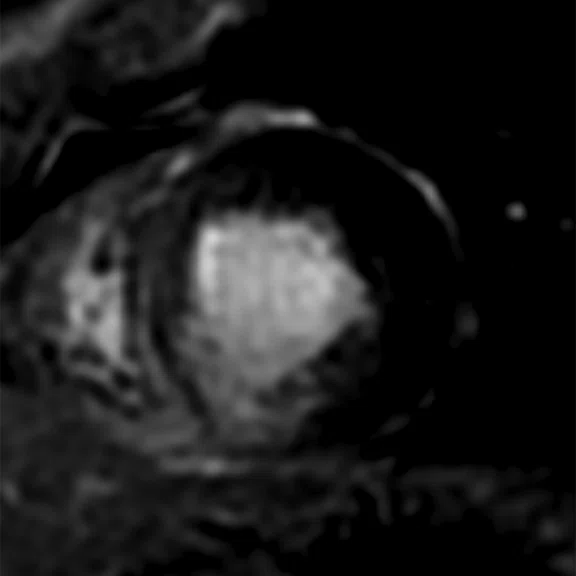
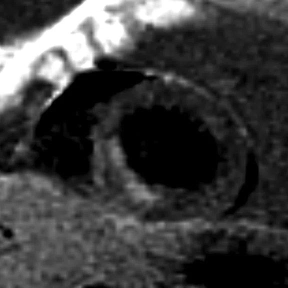
A 53-year-old patient with acute myocardial infarction on territory of right coronary artery. (A) Short axis shows subendocardial late gadolinium enhancement of inferior and inferoseptal myocardial segments. The findings were confirmed on (B) BB MDE images.
B
Figure 1.
A 53-year-old patient with acute myocardial infarction on territory of right coronary artery. (A) Short axis shows subendocardial late gadolinium enhancement of inferior and inferoseptal myocardial segments. The findings were confirmed on (B) BB MDE images.
‡Technology in development that represents ongoing research and development efforts. These technologies are not products and may never become products. Not for sale. Not cleared or approved by the US FDA or any other global regulator for commercial availability.
result


PREVIOUS
${prev-page}
NEXT
${next-page}
Subscribe Now
Manage Subscription
FOLLOW US
Contact Us • Cookie Preferences • Privacy Policy • California Privacy PolicyDo Not Sell or Share My Personal Information • Terms & Conditions • Security
© 2024 GE HealthCare. GE is a trademark of General Electric Company. Used under trademark license.
CASE STUDIES
Assessing damaged myocardium using a novel black blood MDE sequence
Assessing damaged myocardium using a novel black blood MDE sequence
By Giuseppe Muscogiuri, MD, PhD, FSCCT, radiologist, and Gianluca Pontone, MD, PhD, FESC, FEACVI, FSCCT, FISC, cardiologist and radiologist, Department of Cardiovascular Imaging, Centro Cardiologico Monzino, Milan, Italy
Cardiac MR enables both anatomical and functional assessment of the heart. Black blood (BB), T2 STIR and fat suppression are commonly used for the detection of areas at risk for infarction and scar. In some cases it is difficult to differentiate between scar and blood pool using late gadolinium enhancement (LGE) sequences. In particular, factors such as the volume of gadolinium-based contrast, clearance rate and the timing of the LGE acquisition contribute to the similar signal intensity of the blood pool and the area of infarct. To overcome this, usually we compare cine images and LGE images in the same cardiac phase of acquisition.
The availability of BB LGE sequences makes it easier to depict subendocardial infarction, as well as involvement of the papillary muscles with higher contrast-to-noise ratios. We evaluated GE Healthcare’s BB myocardial delayed enhancement (MDE)‡ sequence for research purposes in 73 patients with known or suspected coronary artery disease who underwent a cardiac MR exam.
Patient history
A 53-year-old patient weighing 52 kg (115 lbs.) for evaluation of acute myocardial infarction.
MR findings
Cardiac MR shows a subendocardial myocardial infarction with 50-100 percent of transmurality in inferoseptal, inferior segment of the middle-ventricle myocardium and apical septum (Figure 1A). The findings were confirmed on BB MDE images (Figure 1B).
Discussion
The BB MDE sequence enabled us to depict the subendocardial enhancement and involvement of papillary muscles, and clearly distinguish the damaged myocardium from the blood pool activity. This capability may be especially useful in cases where the standard LGE sequence is non-diagnostic.
In particular, the possibility to have a dark blood in both ventricles with the BB MDE sequence is extremely promising. Overall, the image quality was good, providing excellent diagnostic capability.


