A
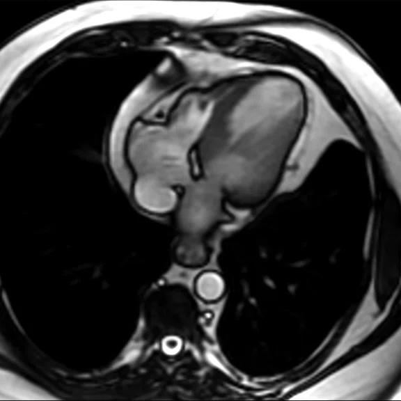
Figure 1.
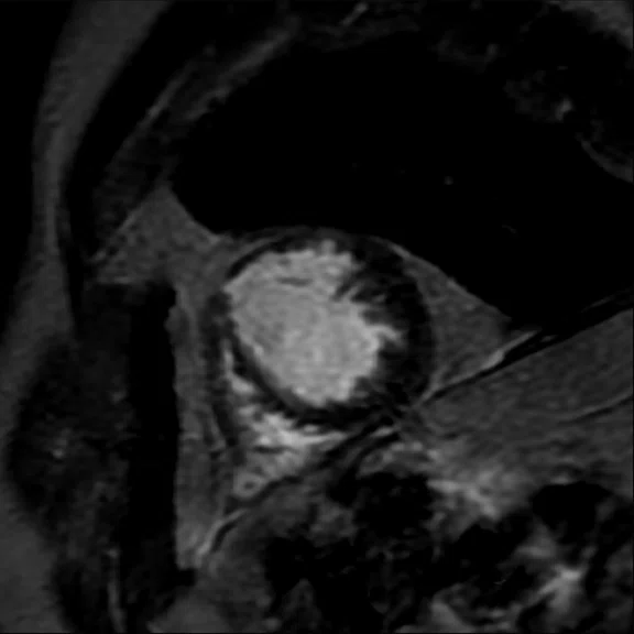
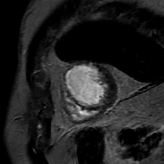
(A, B) Axial FIESTA, 2.9 x 2.4 x 7 mm, 15 sec.
B
Figure 1.
(A, B) Axial FIESTA, 2.9 x 2.4 x 7 mm, 15 sec.
2. Islam FM, Wu J, Jansson J, Wilson DP. Relative risk of cardiovascular disease among people living with HIV: A systematic review and meta-analysis. HIV Med. 2012;13(8):453-468.
1. The Heart and Stroke Foundation South Africa. Cardiovascular Disease Statistics Reference Document. Available at: http://www.heartfoundation.co.za/wp-content/uploads/2017/10/CVD-Stats-Reference-Document-2016-FOR-MEDIA-1.pdf.
A
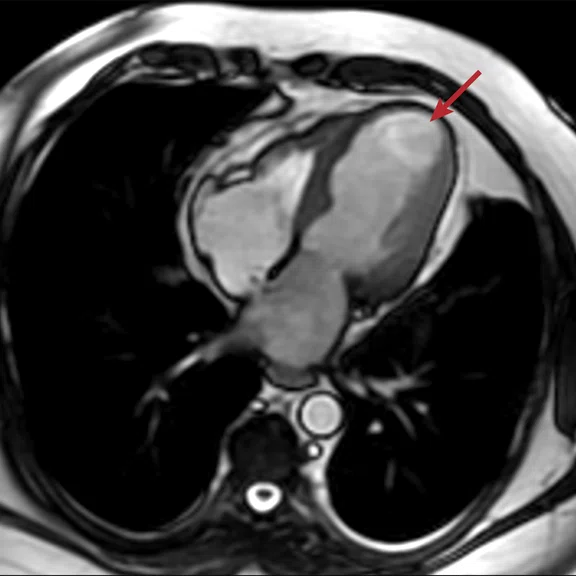
Figure 2.
Aneurysm dilation; note the thinning myocardium (arrow). (A) Long axis Cine FIESTA, 2.2 x 1.8 x 6 mm, 10 sec.; (B) 4-chamber Cine function, 2.2 x 2.0 x 6 mm, 7 sec.; and (C) short-axis Cine stack, 2.3 x 1.7 x 8 mm, 7 sec.
B
Figure 2.
Aneurysm dilation; note the thinning myocardium (arrow). (A) Long axis Cine FIESTA, 2.2 x 1.8 x 6 mm, 10 sec.; (B) 4-chamber Cine function, 2.2 x 2.0 x 6 mm, 7 sec.; and (C) short-axis Cine stack, 2.3 x 1.7 x 8 mm, 7 sec.
C
Figure 2.
Aneurysm dilation; note the thinning myocardium (arrow). (A) Long axis Cine FIESTA, 2.2 x 1.8 x 6 mm, 10 sec.; (B) 4-chamber Cine function, 2.2 x 2.0 x 6 mm, 7 sec.; and (C) short-axis Cine stack, 2.3 x 1.7 x 8 mm, 7 sec.
A
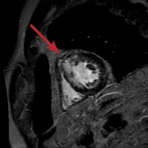
Figure 3.
(A-C) Short-axis PS MDE acquisition demonstrates subendocardial enhancement of the apex and apical anterior segment of the left ventricle with 50 percent involvement (arrow). Parameters: 1.7 x 2.4 x 8 mm, 9 sec.
B
Figure 3.
(A-C) Short-axis PS MDE acquisition demonstrates subendocardial enhancement of the apex and apical anterior segment of the left ventricle with 50 percent involvement (arrow). Parameters: 1.7 x 2.4 x 8 mm, 9 sec.
C
Figure 3.
(A-C) Short-axis PS MDE acquisition demonstrates subendocardial enhancement of the apex and apical anterior segment of the left ventricle with 50 percent involvement (arrow). Parameters: 1.7 x 2.4 x 8 mm, 9 sec.
result


PREVIOUS
${prev-page}
NEXT
${next-page}
Subscribe Now
Manage Subscription
FOLLOW US
Contact Us • Cookie Preferences • Privacy Policy • California Privacy PolicyDo Not Sell or Share My Personal Information • Terms & Conditions • Security
© 2024 GE HealthCare. GE is a trademark of General Electric Company. Used under trademark license.
CASE STUDIES
CMR for assessing coronary structure and function after myocardial infarction
CMR for assessing coronary structure and function after myocardial infarction
By Tinus Malan, MBChB, FC Rad Diag (SA), radiologist, and Celesté Pretorius, MSc Rad (UK), PhD (SA), Head MR Radiographer, De Beer De Jager Radiologists, Pretoria, South Africa
Cardiovascular disease (CVD) is a national health issue in South Africa, with cardiovascular disease being the country’s second-leading cause of death behind HIV and AIDS. Patients living with HIV are at an increased risk for CVD with antiretroviral therapies, and in particular protease inhibitor-based regimens, further increasing their risk1.
Cardiac MR (CMR) is now regarded as a very important tool in the diagnosis and management of CVD in South Africa2 as a result of efforts led by the Radiological Society of South Africa, including the development of the South African Cardiac Imaging Society subcommittee. CMR provides high spatial resolution, image contrast and tissue characterization, making it indispensable for evaluating ventricular size and function of the left and right ventricles. It can also help clinicians differentiate ischemic heart disease and non-ischemic cardiomyopathies.
Proper diagnosis of CVD with CMR not only enhances the quality of care, but it can help the clinician determine prognosis, the need for additional tests and appropriate treatment or therapeutic procedures.
Recent advances in CMR have led to the development of techniques for non-invasive assessment of cardiovascular structure and function. In particular, myocardial delayed enhancement (MDE) and phase-sensitive (PS) MDE are invaluable sequences. Cine IR is used to determine the optimal inversion recovery time (TI). With PS MDE, the TI evolution image can help us detect certain tissue T1 abnormalities such as myocardial viability, cardiomyopathy, myocarditis and other infiltrative myocardial processes.
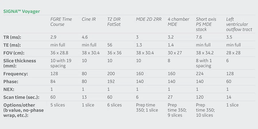
In late 2017, our facility upgraded its Optima™ MR360 system to SIGNA™ Voyager, which has a 70 cm bore, Total Digital Imaging (TDI) technology, high performance gradients with 36 mT/m amplitude and a 150 T/m/s slew rate.
Patient history
A 56-year-old male suffered an anterior myocardial infarction. PET and myocardial perfusion scintigraphy studies provided conflicting results. In the PET exam, a large perfusion defect was noted in the apex, anterior wall and apical aspect of the inferior wall; the apex of the left ventricle had an aneurysm appearance and questionable myocardium viability. The scintigraphy exam demonstrated a large fixed perfusion defect involving the anterior wall, apex and inferoapical aspect of the left ventricle; results indicated the possibility of an aneurysm and dilated cardiomyopathy.
The patient was referred to CMR to address the conflicting results from the PET and scintigraphy studies and assess the viability of the myocardium.
MR findings
CMR showed aneurysm dilatation and ballooning of the apex with thinning of the left ventricle myocardium (5-6 mm) and decreased perfusion of the left ventricle during rest (Figure 2). Post-contrast subendocardial enhancement of the apex and anterior segment of the left ventricle with 50 percent involvement (Figure 3). Enhancement is most likely due to subendocardial ischemia in the left anterior descending artery territory with a viable myocardium. Functional analysis showed an ejection fraction of 33 percent indicating left ventricle function impairment.
Discussion
The SIGNA™ Voyager provided the image quality needed so we could visualize the difference between normal and diseased tissue. The 2D FIESTA/FGRE Cine pulse sequence provides excellent blood-to-myocardium contrast. Short TR/TE allows for the acquisition of high-quality cardiac images that are less sensitive to turbulent blood flow and off-resonance artifacts. The FGRE Time Course (TC) performs fast, robust cardiac studies that enhance our clinical confidence. FGRE TC also delivers excellent temporal and spatial resolution and T1 contrast to aid in a more confident diagnosis of the myocardium. 2D/3D MDE helps us to determine myocardial tissue viability fast, simply and reliably, and assess fibrosis by improving the contrast-to-noise ratio between an infarct and normal myocardium.
With CMR, we can provide a non-invasive technique to evaluate the structures and function of the heart to help guide patient management, therapy and interventions.
References
3. Scholtz, L. Cardiovascular imaging in South Africa: Is the heartache easing? SA Journal of Radiology. 2016; (2), 1-2.


